Pediatric Femur Fracture Management in Cerebral Palsy with Osteopenia: Case 37

17 Apr 2026 18 min read 136 Views
Illustration of children with cerebral - Dr. Mohammed Hutaif

Key Takeaway

Managing femur fractures in children with GMFCS IV cerebral palsy and osteopenia presents unique challenges. Orthopedic specialists must address weakened, osteopenic bone, pre-existing spasticity, and contractures that complicate alignment and implant stability. Detailed imaging like CT is crucial for surgical planning, ensuring adequate fixation and considering long-term bone health strategies for these complex pediatric patients.
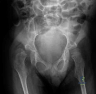
Patient Presentation and History

Mechanism of Injury and Demographics

We present Case 37, an 8-year-old male with a complex medical history dominated by spastic quadriplegic cerebral palsy, classified as Gross Motor Function Classification System Level IV. The patient presented to the pediatric orthopedic trauma service following an acute right mid-diaphyseal femur fracture. The mechanism of injury was reported as a low-energy fall during a routine, caregiver-assisted transfer from his customized wheelchair to a changing table. According to the primary caregiver, the transfer involved a pivot motion during which a sudden, audible acoustic emission—described as a "pop"—was heard, immediately followed by severe nociceptive distress, crying, and an obvious gross deformity of the right thigh.

Crucially, the caregiver explicitly denied any direct blunt force trauma or impact to the thigh. This history strongly suggests an insufficiency fracture component, where the structural integrity of the osteopenic bone was overwhelmed by torsional and bending moments generated during a standard rotational transfer maneuver. In the context of severe neuromuscular disease, the threshold for mechanical failure is drastically reduced, allowing physiological or minimally supraphysiological loads to cause catastrophic cortical disruption. There were no other injuries reported or observed during the secondary survey, and a thorough review of the psychosocial history and injury pattern raised no clinical suspicion for non-accidental trauma.

Comorbidities and Pathophysiology

The patient’s underlying cerebral palsy etiology is presumed to be perinatal anoxic brain injury. His baseline functional status is characterized by complete reliance on wheelchair mobility for all locomotion, requiring extensive physical assistance for transfers, toileting, and all activities of daily living. This chronic lack of weight-bearing and mechanical loading fundamentally disrupts normal mechanotransduction pathways. According to Wolff’s Law, bone remodels in response to the mechanical stresses placed upon it; the absence of these stresses in a non-ambulatory GMFCS Level IV patient leads to profound disuse osteopenia, characterized by decreased cortical thickness and trabecular depletion.

Furthermore, the patient has a history of severe adductor and hamstring spasticity. This chronic hypertonia places continuous, uncoordinated, and abnormal stress on the skeletal system. He was previously managed with multiple botulinum toxin type A injections and underwent bilateral adductor longus releases and hamstring lengthenings at age five. While these interventions successfully improved passive range of motion and facilitated perineal hygiene, they did not alter his GMFCS level or restore physiological mechanical loading to the lower extremities.

A highly significant comorbidity in this case is a seizure disorder that is currently well-controlled on valproic acid. Valproic acid, a broad-spectrum antiepileptic drug, is a known inducer of the hepatic cytochrome P450 enzyme system. This induction accelerates the catabolism of 25-hydroxyvitamin D into inactive metabolites, leading to iatrogenic hypovitaminosis D. Furthermore, valproic acid has been shown in in vitro studies to directly inhibit osteoblast proliferation and function while simultaneously upregulating osteoclastogenesis. The convergence of chronic immobility, abnormal spastic muscle forces, and valproic acid-induced metabolic bone disease creates a perfect storm for severe secondary osteopenia, rendering the diaphysis highly susceptible to low-energy insufficiency fractures.

Clinical Examination Findings

General Inspection and Deformity

Upon presentation to the trauma bay, the patient was alert but in profound distress, exhibiting tachycardia and tachypnea consistent with acute pain, though his core vital signs remained hemodynamically stable. General inspection of the right lower extremity revealed classic signs of a displaced diaphyseal femur fracture. The limb was held in a position of marked external rotation and abduction, with gross clinical angulation and foreshortening of the mid-thigh segment.

Significant soft tissue swelling and ecchymosis were evident over the anterior and lateral aspects of the right thigh, indicating substantial hematoma formation within the muscular compartments. The skin integrity appeared intact, demonstrating no evidence of an open fracture; however, the soft tissue envelope was stretched tightly over the apex of the anterolateral deformity, raising vigilance for impending skin necrosis if reduction was delayed. Pre-existing fixed flexion contractures of both hips (measuring approximately 30 degrees) and knees (measuring approximately 20 degrees) were noted. These contractures are highly consistent with his GMFCS Level IV spastic quadriplegia and significantly complicate both the clinical assessment and the eventual surgical positioning.

Palpation and Provocative Maneuvers

Palpation was conducted with extreme caution due to the patient's distress. Exquisite, poorly localized tenderness to palpation was noted circumferentially over the mid-diaphyseal region of the right femur. Gentle manipulation of the limb, required solely for the application of a provisional traction splint, elicited palpable crepitus, confirming the presence of a displaced, mobile fracture pattern.

The thigh compartments were carefully assessed. While tense due to the fracture hematoma, they remained compressible, and there were no clinical signs of acute compartment syndrome. It is critical to note that diagnosing compartment syndrome in a non-verbal or cognitively impaired pediatric patient relies heavily on objective findings such as tenseness, asymmetric swelling, and the analgesic requirement, as the classic symptom of "pain out of proportion" is difficult to quantify in this demographic.

Range of Motion Assessment

Active range of motion of the right hip and knee was absent due to pain inhibition and baseline motor deficits. Passive range of motion was severely limited by the acute fracture and was not formally tested beyond what was necessary to assess the distal neurovascular status and apply provisional stabilization. Examination of the contralateral, unaffected limb revealed significant baseline hamstring and adductor spasticity, with a popliteal angle of 60 degrees and limited hip abduction, providing a crucial baseline for understanding the deforming forces that will act upon the fracture site post-reduction.

Neurological and Vascular Assessment

Distal neurological function is inherently difficult to assess precisely in a patient with baseline spastic quadriplegic cerebral palsy and a lack of voluntary motor control. However, withdrawal to noxious stimuli and sensation to light touch in the L5 and S1 dermatomes of the right foot were present. Toe flexion and extension were grossly attempted by the patient during stimulation, indicating preserved motor pathways beyond the level of the injury. No acute neurological deficit directly attributed to the trauma, such as a sciatic nerve palsy, was identified.

Vascular assessment revealed palpable and symmetric dorsalis pedis and posterior tibial pulses, graded at 2+ bilaterally. Capillary refill in the distal digits was brisk, occurring in under two seconds. The right foot was warm and well-perfused, demonstrating no signs of vascular compromise despite the significant displacement of the mid-diaphyseal fracture.

Imaging and Diagnostics

Radiographic Evaluation

Standard trauma radiographic series, including antero-posterior and lateral projections of the right femur, were obtained upon initial presentation.

Image

The radiographs revealed an acute, comminuted, spiral mid-diaphyseal fracture of the right femur. The fracture pattern demonstrated significant displacement, characterized by overriding of the fracture fragments leading to clinical foreshortening. There was approximately 40 degrees of varus angulation and 30 degrees of external rotation of the distal fragment relative to the proximal fragment.

A critical finding on the plain radiographs was the profound alteration in bone architecture. The diaphyseal cortices appeared markedly thinned, with an abnormally high medullary canal-to-cortical diameter ratio (decreased Cortical Thickness Index). The overall radiolucency of the bone was significantly increased, consistent with severe, generalized osteopenia. The trabecular pattern in the metaphyses was sparse. Importantly, despite the severe osteopenia, there was no radiographic evidence of pre-existing focal lytic or blastic lesions, periosteal reaction, or morphological abnormalities that would suggest an underlying primary bone tumor or metastatic disease.

Advanced Imaging Indications

In this specific clinical scenario, advanced cross-sectional imaging, such as Computed Tomography or Magnetic Resonance Imaging, was not deemed immediately necessary for acute fracture management. The plain radiographs provided sufficient detail regarding the fracture geometry, degree of comminution, and bone quality to formulate a definitive surgical plan.

However, CT imaging might be indicated in pediatric femur fractures if there is suspicion of an intra-articular extension into the knee or hip, or if a subtle pathological lesion (such as a unicameral bone cyst or aneurysmal bone cyst) is obscured by the fracture hematoma and comminution on plain films. MRI would be reserved for cases where soft tissue interposition is suspected of blocking reduction, or if there is a high index of suspicion for osteomyelitis or a malignant process driving a pathological fracture. Given the clear history of a transfer injury in a highly osteopenic patient, plain radiography was sufficient.

Preoperative Templating

Preoperative templating was performed using digital radiographic software calibrated to a known sizing marker. Templating in osteopenic cerebral palsy patients requires careful consideration of the narrow medullary canal and the thin cortices. The length of the femur was estimated using the contralateral intact femur radiographs, adjusting for the known flexion contractures. The objective of templating was to determine the appropriate length and contour of the fixation construct, ensuring adequate span across the comminuted zone and sufficient screw purchase in the proximal and distal metadiaphyseal segments where bone stock is marginally better.

Differential Diagnosis Evaluation

When evaluating a pediatric patient with a low-energy diaphyseal femur fracture, particularly in the presence of underlying neuromuscular disease, a systematic approach to the differential diagnosis is mandatory. The clinician must distinguish between a fragility fracture secondary to the known comorbidities and other more insidious etiologies.

Differential Diagnosis Clinical Presentation Radiographic Findings Differentiating Factors
Neuromuscular Osteopenia Insufficiency Fracture Non-ambulatory CP patient, low-energy mechanism (transfer), chronic antiepileptic use. Diffuse osteopenia, thin cortices, spiral or transverse diaphyseal fracture. No focal lesions. Consistent with patient's GMFCS level, pharmacological history, and reported mechanism.
Non-Accidental Trauma Unwitnessed injury, inconsistent history, delayed presentation, multiple injuries in varying stages of healing. Corner fractures, posterior rib fractures, multiple fractures of differing ages. Ruled out in this case due to a consistent caregiver history, plausible mechanism for osteopenic bone, and absence of other suspicious injuries.
Pathologic Fracture Secondary to Benign Lesion Prodromal pain, minor trauma causing fracture. Well-circumscribed lytic lesion (e.g., Unicameral Bone Cyst, Non-Ossifying Fibroma) at the fracture site. Absence of any focal radiolucent lesion or cortical expansion on plain radiographs rules this out.
Osteogenesis Imperfecta Recurrent fractures, blue sclerae, dentinogenesis imperfecta, family history. Diffuse osteopenia, bowing deformities, multiple healed fractures, "popcorn" calcifications. Patient lacks the extra-skeletal manifestations of OI; osteopenia is adequately explained by CP and valproic acid.
Metabolic Bone Disease Bone pain, muscle weakness, risk factors for nutritional deficiency or renal failure. Widened physes, metaphyseal cupping and fraying, generalized osteopenia. While secondary hypovitaminosis D is present, classic radiographic signs of active rickets (physeal widening) are absent.

Surgical Decision Making and Classification

Fracture Classification

The fracture was classified using the AO Pediatric Comprehensive Classification of Long Bone Fractures. Based on the diaphyseal location, the spiral morphology, and the presence of comminution, this is categorized as a 32-D/5.1 fracture (Femur, diaphysis, complex/comminuted). This classification highlights the inherent instability of the fracture pattern, which heavily influences the choice of fixation.

Operative Versus Nonoperative Management

The management of diaphyseal femur fractures in pediatric patients with severe spastic cerebral palsy is highly controversial and requires a nuanced approach. Non-operative management, traditionally utilizing a hip spica cast, is fraught with severe complications in this specific population. GMFCS Level IV patients have profound spasticity; the unyielding muscle spasms, particularly of the adductors and hamstrings, exert massive deforming forces on the fracture site. In a spica cast, these forces almost invariably lead to loss of reduction, severe varus/procurvatum malunion, and potentially nonunion.

Furthermore, the rigid immobilization of a spica cast in an incontinent, non-ambulatory patient creates an exceptionally high risk for catastrophic skin breakdown, pressure ulcers, and perineal hygiene issues. The cast also exacerbates existing osteopenia by further eliminating mechanical stress. Therefore, non-operative management was rapidly excluded as a viable primary treatment strategy.

Operative management is considered the gold standard for displaced femur fractures in spastic cerebral palsy to achieve rigid stabilization, facilitate early mobilization (wheelchair sitting), and minimize nursing complications. The surgical options primarily include Elastic Stable Intramedullary Nailing, rigid locked intramedullary nailing, and submuscular plating.

Selection of Fixation Construct

Rigid antegrade or retrograde locked intramedullary nailing is generally contraindicated in an 8-year-old due to the risk of iatrogenic avascular necrosis of the femoral head (with trochanteric entry) or physeal arrest (with retrograde entry).

Elastic Stable Intramedullary Nailing is a standard treatment for diaphyseal femur fractures in typical children aged 5 to 11. However, ESIN relies on the intact cortical tube for three-point fixation and rotational stability. In this patient, the presence of a comminuted fracture pattern combined with severe osteopenia means the elastic nails will have poor endosteal purchase. More importantly, the severe spasticity of the thigh musculature will easily overpower the relatively flexible ESIN construct, leading to telescoping, shortening, and rotational malalignment.

Therefore, Minimally Invasive Plate Osteosynthesis utilizing a submuscular locking plate was selected as the optimal surgical strategy. A locking plate acts as an internal fixator, providing rigid angular and rotational stability without relying on friction between the plate and the osteopenic bone. By utilizing a long plate spanning the comminuted diaphysis and securing it with multiple locking screws in the proximal and distal metaphyseal regions, the construct can withstand the spastic deforming forces while preserving the fracture hematoma and the periosteal blood supply, which is critical for healing in metabolically compromised bone.

Surgical Technique and Intervention

Anesthesia and Patient Positioning

The patient was brought to the operative theater and placed under general endotracheal anesthesia. Anesthetic management in cerebral palsy requires specific considerations, including careful airway management due to potential anatomical abnormalities and altered gastrointestinal motility increasing aspiration risk. Furthermore, while true malignant hyperthermia is not directly linked to CP, these patients can exhibit altered responses to depolarizing muscle relaxants like succinylcholine, which can cause severe hyperkalemia; therefore, non-depolarizing agents were utilized.

The patient was positioned supine on a radiolucent flat Jackson table. A traction table was specifically avoided. The use of a traction table in a patient with severe, long-standing hip and knee flexion contractures can lead to iatrogenic physeal injuries, ligamentous disruption, or peripheral nerve palsies due to the excessive force required to overcome the spasticity and contractures. Instead, manual traction and strategic bumping were utilized. A sterile bump was placed under the ipsilateral hemipelvis to control external rotation. The entire right lower extremity, from the iliac crest to the toes, was prepped and draped in a standard sterile fashion, allowing for free manipulation of the limb during the procedure.

Surgical Approach and Reduction

A Minimally Invasive Plate Osteosynthesis technique was employed to preserve the fragile periosteal blood supply and the osteogenic fracture hematoma.

First, a 3-centimeter longitudinal incision was made over the lateral aspect of the distal femur, centered over the metaphyseal-diaphyseal junction. The iliotibial band was incised longitudinally, and the vastus lateralis was carefully elevated off the lateral intermuscular septum to expose the distal femoral metaphysis. A specialized submuscular elevator was then introduced through this distal window and advanced proximally along the lateral aspect of the femoral shaft, creating an extra-periosteal tunnel deep to the vastus lateralis, crossing the fracture site, and reaching the proximal femur.

A second 3-centimeter incision was made over the proximal lateral femur, just distal to the greater trochanter. The deep fascia was split, and the vastus lateralis was split in line with its fibers to expose the proximal femoral metaphysis, connecting with the previously created submuscular tunnel.

Provisional reduction was achieved using manual longitudinal traction combined with internal rotation to correct the deformity. Due to the comminution, anatomical reduction of the intermediate fragments was not attempted; instead, the focus was on restoring length, alignment, and rotation. The pre-existing hip and knee contractures were carefully factored into the assessment of alignment, using the contralateral draped limb as a clinical reference to ensure symmetrical resting posture.

Fixation Construct Application

A 4.5mm narrow Locking Compression Plate, pre-contoured to match the anterior bow of the pediatric femur, was selected based on preoperative templating. The plate was inserted through the proximal incision and carefully slid distally through the submuscular tunnel until it spanned the fracture site and was visible in the distal window.

Fluoroscopy was utilized in both antero-posterior and lateral planes to confirm optimal plate positioning. The plate was centered over the lateral cortex. Provisional fixation of the plate to the bone was achieved using K-wires proximally and distally.

Given the severe osteopenia, standard cortical screws would have insufficient pull-out strength. Therefore, the construct relied entirely on locking head screws. The sequence of screw insertion is critical. First, a non-locking cortical screw can be used to pull the bone to the plate if necessary, but in this case, to avoid stripping the osteopenic bone, we proceeded directly to locking screws after ensuring the plate was flush with the bone via manual pressure.

Three 4.5mm locking screws were inserted into the proximal fragment, and three 4.5mm locking screws were inserted into the distal fragment. The drilling process revealed extremely soft, yielding cancellous and cortical bone, confirming the severe osteopenic state and validating the decision to utilize a locking construct. The screws were placed widely apart to maximize the working length of the plate, thereby increasing the elastic deformation capacity of the construct and reducing the stress concentration at the bone-implant interface.

Final fluoroscopic images confirmed restoration of length, acceptable coronal and sagittal alignment, and appropriate hardware placement with no intra-articular penetration. The wounds were irrigated copiously with sterile saline. The fascia was closed meticulously to prevent muscle herniation, and the skin was approximated using absorbable subcuticular sutures and sterile adhesive strips.

Post Operative Protocol and Rehabilitation

Immediate Postoperative Care

The patient was extubated smoothly and transferred to the pediatric intensive care unit for overnight observation, primarily for pain management and continuous monitoring of neurovascular status. Postoperative pain in spastic CP patients can trigger severe muscle spasms, which in turn exacerbate pain, creating a vicious cycle that places immense stress on the surgical fixation. Therefore, a multimodal analgesic regimen was instituted, including intravenous acetaminophen, scheduled ketorolac, and a low-dose opioid patient-controlled analgesia pump managed by the caregiver.

Crucially, his antispasmodic regimen (enteral diazepam and baclofen) was optimized immediately to suppress involuntary reflex spasms. No cast or splint was applied; the rigid internal fixation allowed for the limb to be left free, facilitating excellent nursing care, skin inspection, and hygiene maintenance.

Long Term Rehabilitation Strategy

The rehabilitation protocol was tailored specifically to his GMFCS Level IV status. The primary goal was not ambulation, but rather a safe return to his baseline functional status, specifically comfortable wheelchair sitting and pain-free transfers.

Physical therapy was initiated on postoperative day one. The initial focus was on gentle passive range of motion of the hip and knee to prevent exacerbation of his pre-existing contractures. The locking plate construct provided sufficient stability to allow for immediate, caregiver-assisted transfers from the bed to his customized wheelchair. Wheelchair seating modifications were temporarily required to accommodate post-surgical swelling and ensure the hip was not forced into excessive flexion.

Weight-bearing was restricted to his baseline non-ambulatory status. Radiographic follow-up was scheduled for two, six, and twelve weeks postoperatively to monitor callus formation and hardware integrity. Healing in osteopenic CP patients can be delayed; therefore, the transition back to aggressive physical therapy and stretching programs was gated by radiographic evidence of bridging callus.

Pharmacological Bone Health Optimization

A critical component of the postoperative protocol involved addressing the underlying metabolic bone disease. An endocrinology consultation was obtained prior to discharge. Given the history of a low-energy fragility fracture, the patient was diagnosed with severe secondary osteoporosis.

A comprehensive bone health optimization plan was initiated. This included aggressive supplementation with cholecalciferol (Vitamin D3) and elemental calcium to counteract the catabolic effects of his valproic acid therapy. Furthermore, the initiation of intravenous bisphosphonate therapy (e.g., pamidronate or zoledronic acid) was strongly recommended. Bisphosphonates inhibit osteoclast-mediated bone resorption, thereby increasing bone mineral density and reducing the incidence of subsequent fragility fractures in the pediatric cerebral palsy population. The timing of bisphosphonate administration was delayed until six weeks postoperatively to ensure it did not interfere with the initial inflammatory phase of fracture healing.

Clinical Pearls and Pitfalls

Pearls:
* Acknowledge the Spasticity: In cerebral palsy, the muscles are the enemy of the fracture. Never underestimate the deforming forces generated by spastic adductors and hamstrings. Flexible fixation (ESIN) or non-operative management (spica casting) will almost universally fail in GMFCS Level IV/V patients with diaphyseal femur fractures.
* Locking Technology is Mandatory: Osteopenic bone provides minimal purchase for standard cortical screws. Always utilize locking plates as internal fixators to provide angular stability independent of bone quality.
* Preserve the Biology: Utilize Minimally Invasive Plate Osteosynthesis techniques. Stripping the periosteum in a metabolically compromised patient will drastically increase the risk of delayed union or atrophic nonunion.
* Address the Root Cause: Treating the fracture without treating the underlying osteopenia is a disservice to the patient. Always initiate a metabolic workup and consider bisphosphonate therapy in consultation with pediatric endocrinology to prevent future insufficiency fractures.

Pitfalls:
* Traction Table Utilization: Avoid using a fracture table for reduction in patients with severe contractures. The force required to position the leg can cause iatrogenic physeal fractures, ligamentous tears, or nerve palsies. Rely on manual traction and radiolucent flat tables.
* Misinterpreting Compartment Syndrome: Relying on the classic "pain out of proportion" symptom is dangerous in non-verbal or cognitively impaired patients. Maintain a high index of suspicion based on objective physical findings and changes in baseline analgesic requirements.
* Ignoring the Pharmacology: Failing to recognize the impact of antiepileptic medications, specifically cytochrome P450 inducers like valproic acid, on bone metabolism. These medications actively destroy bone mineral density and must be countered with aggressive supplementation.
* Over-Reduction: Attempting anatomical reduction of a comminuted butterfly fragment in an osteopenic femur often requires extensive soft tissue stripping, which devitalizes the bone. Accept functional alignment (length, rotation, axis) over radiographic perfection to ensure biological healing.


Dr. Mohammed Hutaif
Medically Verified Content
Prof. Dr. Mohammed Hutaif
Consultant Orthopedic & Spine Surgeon
Article Contents