

Orthopedic Pediatric Review | Dr Hutaif Pediatric Ortho -...
Comprehensive 100-Question Exam
00:00
Start Quiz
Question 1
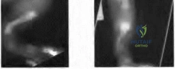
A 6-week-old female infant is undergoing screening for developmental dysplasia of the hip (DDH). An ultrasound of the hip is provided:
In the Graf classification system, the alpha angle is measured to assess the bony roof. Which anatomic structure determines the primary baseline used to establish this angle?
In the Graf classification system, the alpha angle is measured to assess the bony roof. Which anatomic structure determines the primary baseline used to establish this angle?
Explanation
In the Graf ultrasound method for DDH, three lines are drawn. The primary baseline is drawn parallel to the straight portion of the ilium. The second line is drawn along the bony roof of the acetabulum to measure the alpha angle, while the third line is drawn along the cartilaginous roof to measure the beta angle.
Question 2
A 12-year-old obese boy presents with left hip pain and an obligatory external rotation with hip flexion. The radiograph is shown:
He undergoes in situ pinning for a Slipped Capital Femoral Epiphysis (SCFE). Which of the following is the most reliable predictor for a future contralateral slip?
He undergoes in situ pinning for a Slipped Capital Femoral Epiphysis (SCFE). Which of the following is the most reliable predictor for a future contralateral slip?
Explanation
The modified Oxford bone age is the most accurate predictor of a contralateral slip in patients with a unilateral SCFE. A score derived from pelvic radiographs helps determine skeletal maturity; a score of 16 indicates a high risk for a subsequent contralateral slip, justifying prophylactic pinning.
Question 3
An 8-year-old boy presents with a painless limp of 6 months duration. Radiographs are provided:
According to the Herring classification for Legg-Calve-Perthes disease, which specific anatomic region is evaluated to determine the prognosis?
According to the Herring classification for Legg-Calve-Perthes disease, which specific anatomic region is evaluated to determine the prognosis?

Explanation
The Herring classification focuses on the height of the lateral pillar (the lateral one-third of the epiphysis) on an AP radiograph during the fragmentation stage of Perthes disease. Type A has no involvement, Type B maintains >50% lateral pillar height, and Type C has <50% height, which carries the worst prognosis.
Question 4
A 3-year-old girl presents with progressive bowing of her left leg. The standing radiograph is shown:
Which of the following radiographic parameters best differentiates infantile Blount disease from physiologic bowing?
Which of the following radiographic parameters best differentiates infantile Blount disease from physiologic bowing?
Explanation
The Levine-Drennan metaphyseal-diaphyseal angle (MDA) is used to differentiate physiologic bowing from infantile tibia vara (Blount disease). An MDA > 16 degrees indicates a high likelihood of progression to true Blount disease, whereas < 10 degrees is typical for physiologic bowing.
Question 5
A 6-year-old child sustains the elbow injury shown in the radiograph:
Following closed reduction and percutaneous pinning, the hand is pink, well-perfused, and warm with capillary refill < 2 seconds, but the radial pulse remains non-palpable. What is the most appropriate next step in management?
Following closed reduction and percutaneous pinning, the hand is pink, well-perfused, and warm with capillary refill < 2 seconds, but the radial pulse remains non-palpable. What is the most appropriate next step in management?
Explanation
A "pulseless, pink" hand after anatomic reduction and pinning of a supracondylar humerus fracture indicates adequate collateral circulation. Current guidelines recommend observation and close hospital monitoring, as the pulse often returns within days as vasospasm resolves. Vascular exploration is reserved for a pulseless, white (ischemic) hand.
Question 6
A 2-week-old infant is diagnosed with the condition shown:
The Ponseti method is initiated. What is the correct initial manipulative step to correct the cavus deformity prior to cast application?
The Ponseti method is initiated. What is the correct initial manipulative step to correct the cavus deformity prior to cast application?
Explanation
The first step in the Ponseti casting technique for idiopathic clubfoot is correcting the cavus. This is achieved by elevating the first metatarsal (first ray), which effectively supinates the forefoot so that it aligns with the supinated hindfoot. Subsequent casts will abduct the forefoot around the head of the talus.
Question 7
When evaluating a non-ambulatory 14-year-old patient with spastic quadriplegic cerebral palsy who has developed a progressive spinal deformity, what is the most common scoliotic curve pattern observed?
Explanation
Neuromuscular scoliosis, particularly in severe forms of cerebral palsy (spastic quadriplegia), classically presents as a long, sweeping C-shaped curve that often extends into the pelvis, causing pelvic obliquity and difficulty with seating. This contrasts with the classic right thoracic S-shaped curve seen in adolescent idiopathic scoliosis.
Question 8
A 10-year-old boy with spastic diplegic cerebral palsy develops a new-onset "crouch gait" one year after undergoing multi-level orthopedic lower extremity surgery. What is the most likely iatrogenic cause of this gait abnormality?
Explanation
Iatrogenic crouch gait is frequently caused by over-lengthening of the Achilles tendon. This weakens the plantarflexor-knee extension couple during the stance phase, causing the tibia to translate forward and the knee to subsequently collapse into flexion.
Question 9
Intravenous bisphosphonates are a mainstay of medical management for children with moderate to severe Osteogenesis Imperfecta (OI). What is the primary molecular target of nitrogen-containing bisphosphonates?
Explanation
Nitrogen-containing bisphosphonates (e.g., pamidronate, zoledronic acid) inhibit farnesyl pyrophosphate (FPP) synthase in the mevalonate pathway. This prevents the prenylation of small GTPases essential for osteoclast function and survival, leading to osteoclast apoptosis and decreased bone resorption.
Question 10
A 14-year-old boy presents with frequent ankle sprains and a rigid, painful flatfoot. Radiographs show a "C-sign" on the lateral ankle view. Which type of tarsal coalition is most likely, and at what age does it typically ossify and become symptomatic?
Explanation
The radiographic "C-sign" is indicative of a talocalcaneal (subtalar) coalition, which typically involves the middle facet. These coalitions ossify and become rigid/symptomatic between 12-16 years of age. Calcaneonavicular coalitions usually present earlier (8-12 years) and demonstrate the "anteater" sign on oblique radiographs.
Question 11
A 13-year-old girl presents after an external rotation injury to her ankle. She is diagnosed with a juvenile Tillaux fracture. Which ligament is responsible for the avulsion of the anterolateral distal tibial epiphysis?
Explanation
The juvenile Tillaux fracture is a Salter-Harris III fracture of the anterolateral distal tibial epiphysis. It occurs because the medial and central portions of the distal tibial physis close before the lateral portion. External rotation causes the AITFL to avulse the unfused anterolateral epiphysis.
Question 12
A newborn presents with bilateral absent radii and absent thumbs (radial clubhands). Before considering any surgical intervention, which of the following is an absolute mandatory hematologic screening test?
Explanation
Radial clubhand can be associated with multiple syndromes, notably Fanconi anemia, Holt-Oram, TAR, and VACTERL. Fanconi anemia is a fatal aplastic anemia if unrecognized, and surgery is contraindicated until it is ruled out. Chromosomal breakage testing using diepoxybutane (DEB) or mitomycin C is the gold standard for diagnosis.
Question 13
A 3-year-old child sustains a closed, isolated midshaft femur fracture with 1.5 cm of shortening. What is the most appropriate definitive management?
Explanation
For children aged 6 months to 4-5 years with isolated femur fractures and acceptable shortening (< 2 cm), early spica casting is the standard of care. Flexible intramedullary nailing is typically indicated for school-aged children (5-11 years).
Question 14
In the original Kocher criteria for differentiating pediatric septic arthritis from transient synovitis of the hip, which of the following laboratory/clinical parameters was NOT one of the four factors used?
Explanation
The original four Kocher criteria are: non-weight bearing, temperature > 38.5 C, ESR > 40 mm/hr, and WBC > 12,000 cells/mm3. CRP > 2.0 mg/dL was later validated and added by Caird et al., creating a 5-factor model, but it was not part of the original 4 criteria.
Question 15
In a child with developmental coxa vara, which of the following radiographic parameters is the strongest indication for a valgus proximal femoral osteotomy?
Explanation
The Hilgenreiner Epiphyseal Angle (HEA) is the angle between Hilgenreiner's line and a line drawn through the proximal femoral physis. A normal HEA is < 25 degrees. An HEA > 60 degrees is associated with inevitable progression of the coxa vara and is a definitive indication for surgical intervention (valgus osteotomy).
Question 16
A 10-year-old girl sustained a distal femoral physeal fracture 2 years ago and now presents with a progressive valgus deformity. MRI demonstrates a focal central physeal bridge. What is the generally accepted upper limit of physeal cross-sectional area involvement that remains amenable to bridge resection and interposition grafting?
Explanation
Physeal bar resection with interposition grafting (using fat, cranioplast, or bone wax) is generally indicated if the child has at least 2 years of skeletal growth remaining and the physeal bar involves less than 50% of the total cross-sectional area of the physis.
Question 17
The Wrisberg variant of the discoid lateral meniscus typically presents with a "snapping knee" in young children. This specific variant is anatomically defined by the congenital absence of which of the following structures?
Explanation
The Wrisberg variant of the discoid lateral meniscus lacks the normal posterior meniscotibial (coronary) ligament attachments, making it hypermobile. Its only posterior attachment is the meniscofemoral ligament of Wrisberg, leading to extrusion into the joint during extension and the classic 'snapping knee' presentation.
Question 18
Madelung deformity results from a localized growth disturbance of the volar-ulnar aspect of the distal radius physis. This condition is characterized by a tethering anomalous ligament known as:
Explanation
Vickers ligament is an anomalous, thickened volar radiolunate ligament that tethers the volar-ulnar aspect of the distal radius. This tethering restricts normal physeal growth in that region, leading to the characteristic volar and ulnar tilt of the distal radius articular surface seen in Madelung deformity.
Question 19
According to the Wiltse classification of pediatric spondylolisthesis, which type has the highest intrinsic risk of progression and often requires in situ fusion even for relatively low-grade slips?
Explanation
Type I (Dysplastic) spondylolisthesis is caused by congenital abnormalities of the upper sacrum or the neural arch of L5. It carries the highest risk of progression. Because the neural arch often remains intact, high-grade slips can cause severe cauda equina compression, making early surgical stabilization critical.
Question 20
A 9-year-old boy sustains a pathologic fracture through a centrally located, completely radiolucent lesion in the proximal humeral metaphysis. A "fallen leaf" sign is noted. What is the most appropriate INITIAL management after the fracture has been allowed to heal?
Explanation
The diagnosis is a Unicameral (Simple) Bone Cyst, as evidenced by the central location, patient age, and pathognomonic "fallen leaf" sign. Once the fracture has healed (which occasionally obliterates the cyst, though not reliably), the first-line active treatment is minimally invasive, typically involving aspiration and injection of corticosteroids or bone marrow aspirate.
Question 21
A 3-year-old child presents with bilateral bowing of the legs, worse on the left. Radiographs reveal depression of the medial tibial plateau with beaking of the medial metaphysis.
What is the primary pathological process driving this infantile deformity?
What is the primary pathological process driving this infantile deformity?
Explanation
The clinical scenario and radiograph describe infantile Blount disease (tibia vara). The primary pathogenesis involves the Heuter-Volkmann principle, where excessive compressive forces on the medial aspect of the proximal tibial physis inhibit its growth, while the lateral physis continues to grow normally, exacerbating the varus deformity.
Question 22
Which of the following is true regarding the most common tarsal coalitions?
Explanation
Calcaneonavicular (CN) coalitions are best visualized on an internal oblique radiograph of the foot. Talocalcaneal (TC) coalitions typically become symptomatic later (12-16 years) than CN coalitions (8-12 years). The 'C sign' is a radiographic indicator of a TC coalition, which most commonly involves the middle facet. Excision of a CN coalition typically utilizes the extensor digitorum brevis (EDB) for interposition, not the tibialis anterior.
Question 23
A 6-year-old child falls from monkey bars and sustains a displaced supracondylar humerus fracture.
Upon presentation, the hand is pink but pulseless. After closed reduction and percutaneous pinning, the hand remains pink and well-perfused (capillary refill < 2 seconds), but the radial pulse remains absent. What is the most appropriate next step in management?
Upon presentation, the hand is pink but pulseless. After closed reduction and percutaneous pinning, the hand remains pink and well-perfused (capillary refill < 2 seconds), but the radial pulse remains absent. What is the most appropriate next step in management?
Explanation
In a 'pink, pulseless' hand following reduction and pinning of a supracondylar humerus fracture, the hand is adequately perfused through collateral circulation despite the lack of a palpable radial pulse. The standard of care is admission for 24-48 hours of observation with serial neurovascular exams. Vascular exploration is reserved for a 'white, pulseless' hand that does not improve with fracture reduction.
Question 24
A 14-year-old adolescent sustains an ankle injury while playing soccer. Imaging demonstrates a Salter-Harris III fracture of the anterolateral distal tibia.
What is the primary deforming force and typical pattern of physeal closure in the distal tibia that predisposes to this specific fracture pattern?
What is the primary deforming force and typical pattern of physeal closure in the distal tibia that predisposes to this specific fracture pattern?
Explanation
This describes a Tillaux fracture. The distal tibial physis closes in a specific predictable pattern over an 18-month period: central, then anteromedial, then posteromedial, and finally the anterolateral portion. During this transitional period, the anterolateral physis remains open and vulnerable. An external rotation force causes the anterior inferior tibiofibular ligament (AITFL) to avulse the anterolateral epiphysis.
Question 25
A 5-year-old sustains an injury to the elbow. Radiographs reveal a displaced lateral condyle fracture.
Which of the following is a classic long-term complication associated with an untreated nonunion of this fracture type?
Which of the following is a classic long-term complication associated with an untreated nonunion of this fracture type?
Explanation
Nonunion of a lateral condyle fracture leads to progressive cubitus valgus deformity. This increasing valgus carrying angle causes stretch and friction on the ulnar nerve in the cubital tunnel over years, eventually resulting in tardy ulnar nerve palsy.
Question 26
A 13-year-old boy with a BMI of 32 presents with acute-on-chronic groin pain. He is unable to bear weight. Radiographs show a displaced Slipped Capital Femoral Epiphysis (SCFE).
Based on the Loder classification, what is the primary clinical significance of his inability to bear weight?
Based on the Loder classification, what is the primary clinical significance of his inability to bear weight?
Explanation
The Loder classification divides SCFE into stable (able to bear weight with or without crutches) and unstable (unable to bear weight even with crutches). Unstable SCFE has a significantly higher rate of avascular necrosis (AVN), reported to be between 10% and 47%, compared to less than 10% for stable slips.
Question 27
You are evaluating a 6-year-old girl with spastic quadriplegic cerebral palsy (GMFCS Level V). Her recent AP pelvis radiograph demonstrates a Reimer's migration percentage of 55% in the right hip. She is currently asymptomatic. What is the most appropriate recommendation?
Explanation
In cerebral palsy hip surveillance, a Reimer's migration percentage >30% warrants close monitoring, but a migration percentage >50% generally warrants surgical reconstruction (VDRO +/- pelvic osteotomy), even in an asymptomatic patient. This is to prevent progression to a painful, irreducible, dislocated hip.
Question 28
A 4-year-old boy with a known diagnosis of Osteogenesis Imperfecta Type I presents to the clinic. He has a history of multiple low-energy fractures. Which of the following describes the most likely genetic defect and its phenotypic expression in this patient?
Explanation
Osteogenesis Imperfecta (OI) Type I is the most common and mildest form. It is characterized by a quantitative defect (decreased production) in normal Type I collagen, leading to bone fragility, blue sclerae, and near-normal stature. Types II, III, and IV involve qualitative defects (structurally abnormal collagen) and are typically more severe.
Question 29
An infant with bilateral idiopathic clubfeet is undergoing Ponseti casting. After three casts, the treating physician notices the foot has become severely plantarflexed, the midfoot is deeply creased with a transverse plantar crease, and the heel is slipping out of the cast. What is the most appropriate modification to the standard Ponseti technique for this 'atypical' clubfoot?
Explanation
An 'atypical' or complex clubfoot is characterized by a short, stubby foot, severe plantar flexion, a deep transverse plantar crease, and a tendency for the cast to slip. The Ponseti method must be modified: the knee is flexed to 110 degrees to prevent cast slippage, and equinus and varus are corrected simultaneously rather than sequentially.
Question 30
A 5-year-old girl is noted to have a highly elevated, hypoplastic left scapula. Physical examination reveals limited shoulder abduction. Radiographs demonstrate an osseous connection between the superomedial angle of the scapula and the cervical spine. Which of the following is most strongly associated with this condition?
Explanation
The clinical picture describes Sprengel deformity (congenital elevation of the scapula). The osseous connection is an omovertebral bone, present in approximately 30% of cases. Sprengel deformity is highly associated with Klippel-Feil syndrome (congenital fusion of the cervical spine), as well as congenital scoliosis and renal anomalies.
Question 31
A newborn is evaluated for an absent right thumb and a radially deviated right wrist. Radiographs confirm complete absence of the radius. Which of the following diagnostic tests is most critical to perform next to rule out a potentially fatal associated condition?
Explanation
Radial longitudinal deficiency with an absent thumb is strongly associated with Fanconi anemia, Holt-Oram syndrome, and VACTERL. Fanconi anemia involves fatal aplastic anemia if undiagnosed, and chromosomal breakage analysis (using diepoxybutane) is the definitive screening test. TAR syndrome typically presents with a preserved thumb.
Question 32
A 6-week-old female with a completely dislocated left hip (Graf Type IV) is placed in a Pavlik harness. After 4 weeks of compliant, full-time wear, ultrasound demonstrates the hip remains persistently dislocated. What is the most appropriate next step in management?
Explanation
If a Pavlik harness fails to reduce a dislocated hip after 3 to 4 weeks, it must be discontinued to prevent 'Pavlik harness disease' (erosion and damage to the posterior acetabular wall). The next step is transitioning to a rigid abduction orthosis (e.g., Rhino Cruiser or Ilfeld brace) or proceeding directly to a closed reduction and spica casting.
Question 33
A 2-year-old child presents with significant leg length discrepancy, anteromedial bowing of the right tibia, and an equinovalgus foot. Radiographs reveal partial absence of the right fibula. Which of the following anomalies is most commonly associated with this condition?
Explanation
Fibular hemimelia is the most common congenital long bone deficiency. It is highly associated with anteromedial tibial bowing, absent lateral rays of the foot, tarsal coalition, ball-and-socket ankle joint, and hypoplasia of the lateral femoral condyle resulting in ACL and/or PCL deficiency (ACL deficiency being extremely common).
Question 34
A 6-year-old child with short trunk dwarfism, corneal clouding, and normal intelligence is diagnosed with Morquio syndrome (MPS IV). Which orthopedic manifestation requires the most urgent screening and potential surgical intervention in this condition?
Explanation
Morquio syndrome (Mucopolysaccharidosis Type IV) is characterized by an accumulation of keratan sulfate. A hallmark orthopedic issue is odontoid hypoplasia, which leads to severe atlantoaxial instability. This can cause cervical myelopathy, quadriplegia, or sudden death; thus, early screening and prophylactic posterior cervical fusion are often required.
Question 35
A 2-year-old child presents with a limp, fever of 38.5°C, and refusal to bear weight on the left leg. Aspiration of the hip yields purulent fluid. Which of the following organisms is the most common cause of joint infections in this specific age group and often requires specialized culture techniques (e.g., BACTEC blood culture bottles) for detection?
Explanation
Kingella kingae is a fastidious Gram-negative coccobacillus that has surpassed S. aureus as the most common cause of septic arthritis and osteomyelitis in children under the age of 4. It is notoriously difficult to grow on standard solid media, and its detection rate increases significantly when synovial fluid is inoculated directly into blood culture vials.
Question 36
A 10-month-old infant is diagnosed with an isolated 25-degree left thoracic curve. A spine radiograph is obtained to evaluate the rib-vertebra angle difference (RVAD) of Mehta. Which of the following statements regarding the RVAD is correct?
Explanation
In infantile idiopathic scoliosis, Mehta's Rib-Vertebral Angle Difference (RVAD) is measured at the apical vertebra. An RVAD > 20 degrees is highly predictive of curve progression. Phase 2 rib-head relationship (where the rib head overlaps the vertebral body) is also a strong predictor of progression. Most curves with an RVAD < 20 degrees resolve spontaneously.
Question 37
An 18-month-old boy presents with anterolateral bowing of the tibia and a recent fracture through the apex of the bow that shows no signs of healing. He has multiple café-au-lait spots. What is the most definitive surgical strategy to achieve union in this condition?
Explanation
Congenital pseudarthrosis of the tibia (CPT) is strongly associated with Neurofibromatosis Type 1 (NF1). It is notoriously difficult to heal. Plating alone almost uniformly fails. The standard of care involves excision of the hamartomatous tissue, intramedullary rodding (often spanning the ankle), and robust bone grafting (often utilizing BMP or vascularized fibula grafts).
Question 38
A 4-year-old patient with Spinal Muscular Atrophy (SMA) Type II is receiving Nusinersen (Spinraza) therapy via repeated intrathecal injections. The patient has developed a progressive 65-degree neuromuscular scoliosis. What specific surgical consideration must be planned regarding their future spine deformity correction?
Explanation
Nusinersen (Spinraza) is an antisense oligonucleotide administered via lifelong intrathecal injections to treat SMA. Because standard posterior spinal fusion typically obscures thecal access, surgeons must intentionally leave a lumbar interlaminar window (or install a subarachnoid catheter/port) to ensure the patient can continue receiving this life-saving medication postoperatively.
Question 39
An 8-year-old boy is diagnosed with Legg-Calvé-Perthes disease. Radiographs obtained during the fragmentation phase reveal that the lateral pillar of the femoral head maintains 60% of its original height. According to the Herring Lateral Pillar Classification, what group is this, and what is the typical recommendation for a child of this age?
Explanation
According to the Herring Lateral Pillar classification, Group A has 100% lateral pillar height, Group B maintains >50% height, and Group C has <50% height. For children aged 8 or older with Group B (or B/C border) hips, surgical containment (femoral or pelvic osteotomy) has been shown to result in significantly better spherical outcomes compared to non-operative treatment.
Question 40
A 3-year-old girl presents with massive, disproportionate overgrowth of her index and middle fingers on the right hand. The fingers are stiff and deviated. Intraoperative findings reveal massive enlargement of the digital nerves with fibrofatty infiltration. A somatic mutation in which of the following genes is most strongly associated with this isolated condition?
Explanation
Macrodactyly (nerve territory overgrowth) is characterized by fibrofatty hyperplasia of the nerve and its surrounding tissues (most commonly the median nerve distribution). It is classified under the PIK3CA-related overgrowth spectrum (PROS) and is driven by an isolated post-zygotic somatic mutation in the PIK3CA gene.
Question 41
A 5-year-old boy sustains a fall on an outstretched hand and presents with a completely displaced, extension-type supracondylar humerus fracture with no cortical contact (Gartland Type III).
Which of the following peripheral nerve injuries is most commonly associated with this specific fracture pattern?
Which of the following peripheral nerve injuries is most commonly associated with this specific fracture pattern?
Explanation
The anterior interosseous nerve (AIN) is the most commonly injured nerve in extension-type supracondylar humerus fractures. It typically presents with weakness of the flexor pollicis longus and flexor digitorum profundus to the index finger, resulting in an inability to make an "OK" sign.
Question 42
An infant is diagnosed with idiopathic congenital talipes equinovarus (clubfoot) and is undergoing the Ponseti method of serial casting.
According to the Ponseti method, what is the correct sequence of deformity correction?
According to the Ponseti method, what is the correct sequence of deformity correction?
Explanation
The correct sequence of correction in the Ponseti method is C-A-V-E: Cavus (by elevating the first ray), Adductus, Varus, and finally Equinus. Equinus is often corrected at the end with a percutaneous Achilles tenotomy.
Question 43
A 3-year-old girl is evaluated for bilateral severe genu varum. Standing radiographs demonstrate medial beaking of the proximal tibial epiphyses.
Measurement of the metaphyseal-diaphyseal (MD) angle is obtained. Which of the following MD angle measurements most strongly supports the diagnosis of infantile Blount's disease rather than physiologic bowing?
Measurement of the metaphyseal-diaphyseal (MD) angle is obtained. Which of the following MD angle measurements most strongly supports the diagnosis of infantile Blount's disease rather than physiologic bowing?
Explanation
An MD angle of greater than 16 degrees on standing AP radiographs indicates a high likelihood of progression to true infantile Blount's disease. Angles less than 10 degrees are typically associated with physiologic bowing that will spontaneously resolve.
Question 44
A 13-year-old boy presents with a painful, rigid flatfoot and a history of recurrent ankle sprains. Lateral weight-bearing radiographs reveal an elongated anterior process of the calcaneus forming an "anteater nose" sign.
Which specific tarsal coalition is most consistent with these radiographic findings?
Which specific tarsal coalition is most consistent with these radiographic findings?
Explanation
The "anteater nose" sign on a lateral radiograph is highly characteristic of a calcaneonavicular coalition. It represents the elongated anterior process of the calcaneus extending towards the navicular.
Question 45
An 18-month-old non-ambulatory child is brought to the emergency department with a spiral fracture of the left femur.
The parents state the child fell off a low sofa. What is the most critical next step in the management of this patient?
The parents state the child fell off a low sofa. What is the most critical next step in the management of this patient?
Explanation
Femur fractures in non-ambulatory infants, especially spiral fractures with a low-energy mechanism history, are highly suspicious for non-accidental trauma (NAT). Admission, a full skeletal survey, and a CPS consult are mandatory.
Question 46
A 4-year-old boy presents with a history of multiple recurrent long bone fractures, blue sclerae, and early hearing loss. Radiographs show osteopenia and multiple healed fractures in various stages of healing.
This condition is primarily caused by a genetic mutation affecting the synthesis of which type of collagen?
This condition is primarily caused by a genetic mutation affecting the synthesis of which type of collagen?
Explanation
Osteogenesis imperfecta is most commonly caused by mutations in the COL1A1 or COL1A2 genes, leading to defective or deficient Type I collagen. This collagen is the major structural protein in bone, sclerae, and teeth.
Question 47
A 9-year-old girl with cerebral palsy (GMFCS Level IV) has an AP pelvic radiograph demonstrating a Reimers migration percentage of 55% in the right hip. There are early osteoarthritic changes. What is the most appropriate definitive management?
Explanation
In cerebral palsy patients, a Reimers migration index >40-50% indicates significant hip subluxation requiring bony reconstructive surgery. The standard treatment is a proximal femoral varus derotational osteotomy (VDRO) combined with a pelvic osteotomy (e.g., Dega or San Diego).
Question 48
A 13-year-old male is diagnosed with a unilateral slipped capital femoral epiphysis (SCFE). Prophylactic in situ pinning of the contralateral hip is being considered. Which of the following is the strongest clinical indication for prophylactic contralateral pinning?
Explanation
Endocrine disorders (such as hypothyroidism, growth hormone deficiency, or renal osteodystrophy) dramatically increase the risk of a contralateral slip in SCFE patients. Prophylactic pinning of the unaffected hip is highly recommended in these populations.
Question 49
A 5-year-old boy presents with acute right hip pain, inability to bear weight, a temperature of 38.8°C (101.8°F), a WBC count of 14,000/mm³, and an ESR of 55 mm/hr. According to the Kocher criteria, what is the most appropriate next step in management?
Explanation
This patient meets all four Kocher criteria (non-weight bearing, fever, ESR > 40, WBC > 12k), predicting a 99% probability of septic arthritis. The next best step is an ultrasound-guided aspiration of the hip to confirm the diagnosis and identify the organism before surgical washout.
Question 50
A 3-month-old female infant is being treated with a Pavlik harness for developmental dysplasia of the hip (DDH). During follow-up, the mother notes the child is no longer actively kicking or extending the affected knee. Sensation appears intact. Which nerve is most likely compressed due to improper harness positioning?
Explanation
Femoral nerve palsy in a Pavlik harness is typically caused by excessive hip hyperflexion. It presents as an inability to actively extend the knee. The harness should be loosened or temporarily removed until function returns.
Question 51
An 8-year-old boy is diagnosed with Legg-Calve-Perthes disease. Radiographs reveal more than 50% loss of lateral pillar height. Which of the following factors at the time of presentation is the most reliable predictor of long-term prognosis?
Explanation
Age at clinical onset is one of the most critical prognostic factors in Legg-Calve-Perthes disease. Children who develop the disease after age 8 generally have a worse prognosis due to having less remaining time for remodeling before skeletal maturity.
Question 52
A 14-year-old gymnast presents with severe lower back pain and radicular pain in the L5 distribution. Radiographs demonstrate a Grade III L5-S1 isthmic spondylolisthesis. The surgeon is planning reduction and fusion. Which nerve root is at highest risk of injury during the surgical reduction of this high-grade slip?
Explanation
During the reduction of a high-grade L5-S1 spondylolisthesis, the L5 nerve root is placed under significant tension and is at the highest risk of stretch injury. Many surgeons opt for in situ fusion or partial reduction to minimize this specific neurological risk.
Question 53
A 14-year-old boy twisted his ankle while skateboarding. Anteroposterior radiographs show a Salter-Harris type III fracture of the anterolateral aspect of the distal tibial epiphysis. Avulsion by which of the following ligaments causes this specific fracture pattern?
Explanation
A Tillaux fracture is a Salter-Harris III fracture of the anterolateral distal tibial epiphysis. It is caused by an avulsion force from the anterior inferior tibiofibular ligament (AITFL) as the medial physis closes earlier than the lateral side in adolescents.
Question 54
An adolescent sustains a triplane fracture of the distal tibia following an external rotation injury. Radiographically, the fracture appears as a Salter-Harris Type III on the AP view and a Salter-Harris Type II on the lateral view. What is the overall Salter-Harris classification of a standard triplane fracture?
Explanation
A triplane fracture crosses the epiphysis, physis, and metaphysis, making it a Salter-Harris Type IV fracture by definition. It occurs during the transitional phase of distal tibial physeal closure.
Question 55
A 12-year-old elite baseball pitcher presents with progressive medial elbow pain, exacerbated during the late cocking phase of throwing. Radiographs demonstrate widening and irregularity of the medial epicondyle apophysis. What is the most likely diagnosis?
Explanation
Medial epicondyle apophysitis (Little League Elbow) is an overuse injury caused by repetitive valgus stress and traction on the open medial epicondyle apophysis in skeletally immature throwers. UCL tears are more typical in older, skeletally mature athletes.
Question 56
A 6-year-old girl presents with a painless "clunking" sound in her lateral knee during flexion and extension. MRI reveals a discoid lateral meniscus. In the Wrisberg variant of a discoid meniscus, which stabilizing anatomical structure is characteristically absent?
Explanation
The Wrisberg variant of a discoid meniscus lacks the normal posterior meniscotibial (coronary) ligament attachments. Its only posterior tether is the meniscofemoral ligament (of Wrisberg), resulting in a hypermobile meniscus that snaps during knee motion.
Question 57
An 8-year-old boy presents with mild right arm pain after throwing a ball. Radiographs reveal a central, lytic, well-circumscribed lesion in the proximal humeral metaphysis with a "fallen leaf" sign. Assuming the fracture has healed and the lesion remains asymptomatic, what is the most appropriate natural history expectation and management?
Explanation
The "fallen leaf" (or fallen fragment) sign is pathognomonic for a Unicameral Bone Cyst (UBC). Asymptomatic UBCs typically migrate away from the physis into the diaphysis and spontaneously resolve after skeletal maturity.
Question 58
A 10-year-old girl undergoes a knee radiograph for a minor contusion. An incidental eccentric, bubbly, multiloculated lucent lesion with a sclerotic rim is noted in the posteromedial metaphysis of the distal femur. What is the most common natural history of this lesion?
Explanation
This presentation is classic for a Non-Ossifying Fibroma (NOF) or fibrous cortical defect. They are benign, asymptomatic, self-limiting lesions that typically fill in with bone and spontaneously resolve by skeletal maturity.
Question 59
A 9-month-old infant is brought in for an abnormal leg shape. Examination reveals anterolateral bowing of the left tibia. Radiographs demonstrate diaphyseal narrowing and sclerosis at the apex of the bow. This condition is most strongly associated with which underlying genetic disorder?
Explanation
Anterolateral bowing of the tibia is the hallmark precursor to Congenital Pseudarthrosis of the Tibia (CPT). It is strongly associated with Neurofibromatosis Type 1 (NF1), which is present in over 50% of these patients.
Question 60
A 1-year-old infant with confirmed achondroplasia presents with hyperreflexia, clonus, and documented episodes of central sleep apnea. What is the most appropriate next diagnostic imaging modality to determine the underlying musculoskeletal etiology of these symptoms?
Explanation
Infants with achondroplasia are at high risk for foramen magnum stenosis, leading to cervicomedullary compression. Symptoms like central apnea, myelopathy, and hyperreflexia mandate an urgent MRI of the cervicomedullary junction to evaluate for surgical decompression.
Question 61
A 14-year-old boy presents with an unstable slipped capital femoral epiphysis (SCFE) and undergoes open reduction and internal fixation via a surgical hip dislocation approach. To minimize the risk of osteonecrosis, preserving the blood supply to the epiphysis is critical. Which vessel provides the primary blood supply to the femoral head in this patient?
Explanation
The deep branch of the medial femoral circumflex artery (MFCA) provides the primary blood supply to the femoral head in adolescents. Protection of this retinacular vessel is critical during surgical hip dislocation for SCFE.
Question 62
A 2-week-old infant is undergoing serial casting for idiopathic clubfoot using the Ponseti method. What is the correct sequence of deformity correction in this technique?
Explanation
The Ponseti method corrects deformities in the CAVE sequence: Cavus, Adductus, Varus, and finally Equinus. The cavus is corrected first by supinating the forefoot to align it with the hindfoot.
Question 63
A 6-year-old boy sustains a Gartland type III extension-type supracondylar humerus fracture. Following prompt closed reduction and percutaneous pinning, the hand remains pink and warm, but the radial pulse is absent. Capillary refill is less than 2 seconds. What is the most appropriate next step in management?
Explanation
A 'pink, pulseless' hand following reduction of a supracondylar fracture indicates adequate collateral perfusion. Observation is the standard of care, as the pulse often returns within a few days.
Question 64
A 2-year-old child presents with severe bilateral leg bowing. Radiographs demonstrate a metaphyseal-diaphyseal angle (Drennan angle) of 18 degrees with medial metaphyseal beaking.
What is the initial treatment of choice?
What is the initial treatment of choice?
Explanation
A metaphyseal-diaphyseal angle greater than 16 degrees suggests infantile Blount disease rather than physiologic bowing. In a child under 3 years of age with early-stage disease, KAFO bracing during weight-bearing is the first-line treatment.
Question 65
A 4-month-old infant is being treated with a Pavlik harness for developmental dysplasia of the hip (DDH). During a follow-up visit, the parents note the child is no longer actively kicking the knee into extension on the affected side. What is the most likely cause of this complication?
Explanation
Femoral nerve palsy in a Pavlik harness is caused by hyperflexion of the hip. Management involves adjusting the anterior strap to reduce flexion or temporarily discontinuing the harness until function returns.
Question 66
A 13-year-old boy complains of recurrent ankle sprains and a rigid, painful flatfoot. Clinical examination reveals peroneal spasticity. An oblique radiograph of the foot reveals an 'anteater nose' sign. Which condition is most likely present?
Explanation
The 'anteater nose' sign on an oblique radiograph of the foot represents an elongated anterior process of the calcaneus, characteristic of a calcaneonavicular coalition. Talocalcaneal coalitions are typically identified by a 'C-sign' on a lateral radiograph.
Question 67
A 7-year-old girl with spastic quadriplegic cerebral palsy (GMFCS Level V) presents with a progressively painful left hip. Radiographs show a Reimers Migration Percentage of 65% with breaking of the Shenton line, but no degenerative changes. What is the recommended surgical management?
Explanation
For a subluxated hip (Migration Percentage >50%) in a CP patient with reconstructable articular cartilage, a proximal femoral VDRO combined with a volume-reducing pelvic osteotomy (e.g., Dega or San Diego) is the gold standard to achieve a stable, concentric hip.
Question 68
When evaluating a patient with Legg-Calve-Perthes (LCP) disease, several demographic and radiographic factors guide prognosis. Which of the following is associated with a POOR clinical outcome?
Explanation
Female patients with LCP disease generally have a worse prognosis than age-matched males because they reach skeletal maturity earlier, allowing less time for the femoral head to remodel.
Question 69
A 13-year-old girl sustains an ankle injury. Radiographs reveal a juvenile Tillaux fracture. This fracture pattern is primarily caused by which mechanism of injury and involves avulsion by which ligament?
Explanation
A juvenile Tillaux fracture is an avulsion of the anterolateral distal tibial epiphysis caused by an external rotation force. The fragment is pulled by the anterior inferior tibiofibular ligament (AITFL) because the anterolateral physis is the last to close.
Question 70
According to the Loder classification, which clinical finding strictly defines a slipped capital femoral epiphysis (SCFE) as 'unstable'?
Explanation
The Loder classification defines a SCFE as unstable if the patient is unable to ambulate even with crutches. Unstable slips carry a significantly higher risk of avascular necrosis (up to 47%).
Question 71
A 3-year-old boy presents with a history of recurrent low-energy fractures, blue sclerae, and dentinogenesis imperfecta. Genetic testing is most likely to reveal a mutation affecting the synthesis of which structural protein?
Explanation
The patient's presentation is classic for Osteogenesis Imperfecta (OI), which is primarily caused by autosomal dominant mutations in the COL1A1 or COL1A2 genes, leading to defective Type I collagen synthesis.
Question 72
A 10-year-old boy sustains a medial epicondyle fracture of the humerus following a fall. Which of the following represents an absolute indication for open reduction and internal fixation?
Explanation
Incarceration of the medial epicondyle fragment in the elbow joint is an absolute indication for operative intervention. Displacement parameters (e.g., >5-15 mm) and non-progressive ulnar nerve symptoms remain relative indications.
Question 73
A 3-year-old, otherwise healthy boy sustains an isolated closed transverse fracture of the femoral diaphysis with 1.5 cm of shortening. What is the most appropriate definitive management?
Explanation
Early spica casting is the standard of care for isolated pediatric femur fractures in children aged 6 months to 4 or 5 years, provided there is less than 2-3 cm of initial shortening.
Question 74
A 12-year-old girl with a high-grade isthmic spondylolisthesis (Meyerding Grade IV) at L5-S1 undergoes surgical reduction and instrumented fusion. Postoperatively, she develops new-onset weakness in ankle dorsiflexion and extensor hallucis longus function. Injury to which nerve root is most likely responsible?
Explanation
The L5 nerve root is at the greatest risk for traction injury during the reduction of a high-grade L5-S1 spondylolisthesis. This typically presents with weakness in ankle dorsiflexion and great toe extension.
Question 75
An 8-year-old boy presents with a painless, snapping sensation in his lateral knee during extension. MRI confirms a discoid lateral meniscus. According to the Watanabe classification, which type lacks posterior capsular attachments and is highly hypermobile?
Explanation
The Wrisberg variant of a discoid meniscus lacks normal posterior meniscotibial attachments (coronary ligaments). It is attached posteriorly only by the meniscofemoral ligament of Wrisberg, leading to hypermobility and a clinical snapping knee.
Question 76
A newborn is noted to have anterolateral bowing of the tibia. Over the first year of life, the tibia fractures and fails to heal, progressing to a congenital pseudarthrosis of the tibia (CPT). This condition is most strongly associated with which underlying disorder?
Explanation
Congenital pseudarthrosis of the tibia with anterolateral bowing is highly associated with Neurofibromatosis type 1 (NF1), which is present in approximately 50% of these patients.
Question 77
A 7-year-old child sustains a significantly displaced radial neck fracture (Judet Type III) requiring percutaneous pin fixation. Following fracture healing and rehabilitation, what is the most common long-term functional complication?
Explanation
The most common complication following both nonoperative and operative treatment of pediatric radial neck fractures is a loss of forearm rotation, specifically decreased pronation and supination.
Question 78
A 13-year-old elite baseball pitcher complains of insidious-onset shoulder pain during the cocking phase of throwing. Radiographs reveal widening and irregularity of the proximal humeral physis.
What is the most likely diagnosis?
What is the most likely diagnosis?
Explanation
Little League Shoulder is an overuse injury causing proximal humeral epiphysiolysis. Radiographs classically show widening, demineralization, or sclerosis of the proximal humeral physis in adolescent throwing athletes.
Question 79
An infant is brought to the clinic with bilateral idiopathic clubfoot. The treating orthopedist decides to initiate serial casting using the Ponseti method. What is the correct sequence of deformity correction?
Explanation
The Ponseti method follows the CAVE sequence: Cavus, Adductus, Varus, and Equinus. The cavus is corrected first by supinating the forefoot to align it with the hindfoot, followed by correcting adductus and varus simultaneously, and finally equinus is addressed, often requiring an Achilles tenotomy.
Question 80
A 6-year-old boy sustains a completely displaced extension-type supracondylar humerus fracture.
Following closed reduction and percutaneous pinning, he cannot flex his interphalangeal joint of the thumb and the distal interphalangeal joint of the index finger. Which nerve is most likely injured?
Following closed reduction and percutaneous pinning, he cannot flex his interphalangeal joint of the thumb and the distal interphalangeal joint of the index finger. Which nerve is most likely injured?
Explanation
The anterior interosseous nerve (AIN), a branch of the median nerve, is the most commonly injured nerve in extension-type supracondylar humerus fractures. AIN palsy presents with an inability to make an "OK" sign due to weakness in the flexor pollicis longus and the flexor digitorum profundus to the index finger.
Question 81
A 4-year-old child presents with an acute onset of an inability to bear weight on the right leg, a temperature of 38.8°C, an ESR of 50 mm/hr, and a WBC count of 13,000/mm³. According to the Kocher criteria, what is the probability that this child has septic arthritis of the hip?
Explanation
This patient meets all four Kocher criteria: non-weight bearing, temperature >38.5°C, ESR >40 mm/hr, and WBC >12,000/mm³. The presence of all four criteria correlates with a 99% predictive probability of septic arthritis.
Question 82
A 13-year-old boy undergoes an urgent in situ pinning for an unstable slipped capital femoral epiphysis (SCFE) of the right hip.
Which of the following complications is he at the highest risk for developing compared to a patient with a stable SCFE?
Which of the following complications is he at the highest risk for developing compared to a patient with a stable SCFE?
Explanation
Unstable SCFE is defined by the inability to bear weight and is associated with a significantly higher risk of avascular necrosis (AVN), up to 50%, compared to almost 0% in stable SCFE. Urgent decompression and pinning are required to mitigate this risk.
Question 83
A 10-week-old female is being treated with a Pavlik harness for developmental dysplasia of the hip (DDH). At her follow-up appointment, the mother notes that the child is no longer kicking her right leg actively. On examination, there is decreased spontaneous active knee extension on the right. What is the most likely cause?
Explanation
Decreased active knee extension indicates a femoral nerve palsy, a known complication of the Pavlik harness caused by excessive hip flexion. If this occurs, the harness is typically removed or adjusted to reduce flexion until nerve function recovers.
Question 84
In evaluating a child with Legg-Calve-Perthes disease (LCPD),
which of the following is considered the single most significant prognostic factor for the final outcome of the hip?
which of the following is considered the single most significant prognostic factor for the final outcome of the hip?
Explanation
Age at disease onset is the most critical prognostic factor in LCPD. Children diagnosed before the age of 6 to 8 years have significantly better remodeling potential and superior long-term clinical outcomes compared to older children.
Question 85
A 3-year-old obese girl presents with progressive bowing of her left leg and a lateral thrust during gait. Radiographs reveal an increased metaphyseal-diaphyseal angle.
Which of the following is true regarding the initial management of infantile Blount's disease?
Which of the following is true regarding the initial management of infantile Blount's disease?
Explanation
Infantile Blount's disease is an osteochondrosis of the medial proximal tibial physis. KAFO bracing is most effective when initiated before age 3, particularly in patients with unilateral disease.
Question 86
A 13-year-old boy presents with a painful, rigid flatfoot and frequent ankle sprains. Radiographs show a "C-sign" on the lateral view. Which of the following is the most appropriate diagnostic imaging to confirm the exact location and extent of the suspected pathology?
Explanation
The clinical presentation and radiographic "C-sign" are indicative of a talocalcaneal tarsal coalition. A CT scan is the gold standard imaging modality for identifying the coalition, determining its extent, and planning surgical resection.
Question 87
A 6-year-old boy with spastic quadriplegic cerebral palsy is undergoing routine hip surveillance. His Gross Motor Function Classification System (GMFCS) level is V. What is the most widely accepted radiographic measurement used to monitor for hip displacement in this patient?
Explanation
Reimers migration percentage is the standard radiographic measurement used in cerebral palsy hip surveillance to quantify lateral displacement of the femoral head. A migration index greater than 30% typically prompts closer observation or surgical intervention.
Question 88
A 9-year-old girl complains of a painful, snapping sensation in her lateral knee when walking. MRI confirms a Wrisberg variant discoid lateral meniscus. What is the defining anatomical characteristic of this variant?
Explanation
The Wrisberg variant of a discoid meniscus lacks the normal posterior meniscotibial (coronary ligament) attachment, being stabilized posteriorly only by the ligament of Wrisberg. This leads to hypermobility and the classic "snapping knee" syndrome.
Question 89
An 18-month-old child is diagnosed with congenital pseudarthrosis of the tibia (CPT) showing classic anterolateral bowing. This condition is most strongly associated with which of the following systemic disorders?
Explanation
Congenital pseudarthrosis of the tibia (CPT) presents with anterolateral bowing and has a very strong association with Neurofibromatosis type 1 (NF1). Over 50% of patients with CPT have a diagnosis of NF1.
Question 90
A neonate with macrocephaly, frontal bossing, and rhizomelic short stature is diagnosed with achondroplasia. Which of the following gene mutations is responsible for this condition?
Explanation
Achondroplasia is caused by an activating (gain-of-function) mutation in the Fibroblast Growth Factor Receptor 3 (FGFR3) gene. This mutation pathologically inhibits chondrocyte proliferation in the proliferative zone of the physis, leading to impaired endochondral ossification.
Question 91
A 3-year-old boy sustains an isolated midshaft femur fracture. He is placed in a one-and-a-half hip spica cast. What is the acceptable amount of shortening in this age group to account for expected overgrowth?
Explanation
In pediatric femur fractures for children aged 2 to 10 years, 10 to 20 mm of bayonet apposition (shortening) is ideal and generally well-tolerated. This compensates for the expected femoral overgrowth resulting from fracture hyperemia.
Question 92
A 5-year-old boy with a history of multiple low-energy fractures, blue sclerae, and dentinogenesis imperfecta is treated with intravenous bisphosphonates. What is the primary mechanism of action of this medication in treating his condition?
Explanation
Osteogenesis imperfecta is a genetic defect in type I collagen. Bisphosphonates do not correct the collagen defect but inhibit osteoclast-mediated bone resorption, increasing overall bone density and decreasing the incidence of fractures.
Question 93
A 14-year-old gymnast presents with persistent low back pain exacerbated by extension. Oblique radiographs demonstrate a "collar on the Scotty dog" at L5. If the patient has a grade 2 isthmic spondylolisthesis, what defines a grade 2 slip according to the Meyerding classification?
Explanation
The Meyerding classification categorizes spondylolisthesis based on the percentage of forward translation of the superior vertebral body over the inferior one. Grade 2 corresponds to 26% to 50% translation.
Question 94
An 8-week-old infant presents with a left-sided neck mass and the head tilted to the left with the chin rotated to the right. A diagnosis of congenital muscular torticollis is made. What other orthopedic screening is most critical for this infant?
Explanation
Congenital muscular torticollis (CMT) is strongly associated with developmental dysplasia of the hip (DDH), occurring in up to 20% of cases. An ultrasound of the hips is recommended for screening in all infants with CMT.
Question 95
A 12-year-old active boy complains of anterior knee pain exacerbated by jumping. Examination reveals a prominent, tender tibial tubercle. Radiographs show fragmentation of the tibial tubercle apophysis. What is the most appropriate initial management?
Explanation
Osgood-Schlatter disease is a traction apophysitis of the tibial tubercle. It is self-limiting and best managed conservatively with activity modification, stretching, and non-steroidal anti-inflammatory drugs (NSAIDs).
None
Detailed Chapters & Topics
Dive deeper into specialized chapters regarding orthopedic-mcqs-online-010-pediatric
01
Chapter 1
12 min
Crucial Positioning for General Anaesthesia in Pediatric Orthopaedics
02
Chapter 2
21 min
Duchenne Muscular Dystrophy: Orthopedic Management, Early Signs & Surgical Anatomy
03
Chapter 3
17 min
Mastering Hemivertebra Unilateral Bar Diagnosis: 12 Cases
04
Chapter 4
27 min
Osteogenesis Imperfecta (OI) in Pediatric Orthopedics: Diagnosis, Biomechanics, and Surgical Management
05
Chapter 5
14 min
Pediatric Ankle Pain: Diagnosis Involves the Subtalar Joint
06
Chapter 6
26 min
Pediatric Clubfoot Deformity: Pathoanatomy, Biomechanics, and the Role of the Anterior Tibialis Tendon
07
Chapter 7
18 min
Pediatric Neurofibromatosis Type 1 & Congenital Pseudarthrosis of the Tibia: A Case Study
08
Chapter 8
39 min
Fibular Deficiency (Hemimelia): Epidemiology, Classification, Anatomy & Biomechanics
09
Chapter 9
13 min
Pediatric Tibial Osteofibrous Dysplasia: A Detailed Case Presentation
10
Chapter 10
22 min
Congenital Vertical Talus (CVT): Advanced Pathoanatomy, Diagnosis & Biomechanics
11
Chapter 11
20 min
Pediatric Distal Femoral Osteosarcoma: A Detailed Orthopedic Case Study
12
Chapter 12
17 min
Achondroplasia & Thoracolumbar Kyphosis: Pediatric Spinal Stenosis & Myelopathy Case Study
13
Chapter 13
27 min
Nonaccidental Injury (NAI) in Pediatric Orthopedics: Identification & Management Guide
14
Chapter 14
25 min
Congenital Knee Dislocation: Early Diagnosis & Effective Treatment
15
Chapter 15
19 min