Fibular Deficiency (Hemimelia): Epidemiology, Classification, Anatomy & Biomechanics
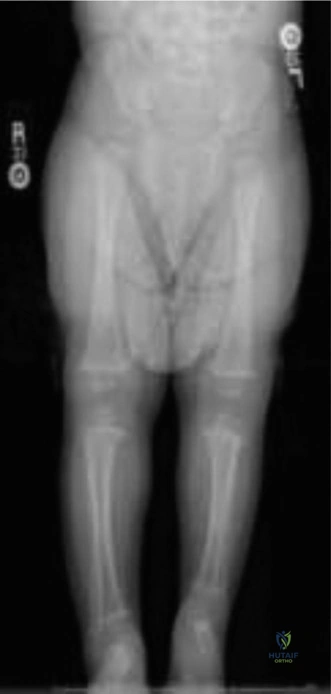
Key Takeaway
Fibular deficiency (fibular hemimelia) is the most common congenital lower limb deficiency, marked by partial or complete fibula absence. It causes limb length discrepancy, ankle valgus, and foot deformities. Classifications like Paley's (Type 1A, 1B, 2) are crucial for assessing severity and guiding treatment planning.
You are reviewing a 3-year-old child with a congenital deficiency of the lateral leg. Clinically, there is a significant limb length discrepancy and a valgus deformity of the ankle. Radiographic assessment is performed. Given the clinical features and the potential for treatment planning, what is the most important radiographic finding to categorize this condition according to the Paley classification, and what does this signify for the patient's prognosis?
Candidate: I would look for the presence of the distal fibular physis. If it is present, it is a Type 1A and has a better prognosis for lengthening. If the distal physis is absent, it is a Type 1B or 2, indicating more severe instability and likely a greater limb length discrepancy, which may steer the treatment towards either complex reconstruction or early consideration of amputation.
Candidates often focus solely on the "missing bone" (i.e., whether the fibula is partially or completely gone) without specifically identifying the distal physis. Failing to mention the distal physis misses the key prognostic indicator for growth potential and ankle joint stability.
The key finding is the status of the distal fibular epiphysis. Under Paley’s classification, Type 1A retains a distal physis, implying the fibula can grow proportionally with the tibia, allowing for successful lengthening. Type 1B (distal physis absent) and Type 2 (complete agenesis) signify a lack of lateral stabilization and progressive ankle valgus/talar instability. I would emphasize that the presence of the distal physis is the primary driver in deciding whether to attempt limb salvage vs. early conversion to Syme’s amputation in the presence of profound shortening.
The parents are concerned about the long-term functional outcome of the child's foot. The patient exhibits lateral ray deficiency and persistent equinovalgus. How do you assess the structural rigidity of this foot, and what are the implications of a tarsal coalition in this context?
Candidate: I would clinically assess the subtalar and midtarsal motion. If the foot is rigid, I would order a CT scan to look for tarsal coalitions, specifically calcaneonavicular or talocalcaneal. If present, these contribute to rigidity, making orthotic management difficult and often requiring surgical resection to restore enough flexibility for a plantigrade gait.
Neglecting the physical examination of joint flexibility or assuming all equinovalgus in fibular hemimelia is soft-tissue based. Failing to recognize that a tarsal coalition converts a flexible, braceable deformity into a rigid, non-accommodating one is a significant error in surgical planning.
Clinical assessment must include testing the passive range of motion of the subtalar joint. A rigid hindfoot in the setting of fibular deficiency is highly suggestive of a tarsal coalition. Radiographic plain films are often insufficient; a CT scan is mandatory for 3D characterization. The presence of a coalition changes the management strategy: a flexible deformity may be managed with an AFO, but a rigid coalition-related deformity often necessitates surgical resection or, in severe cases, complicates the decision for limb salvage versus primary Syme's amputation due to the poor functional outcomes of a rigid, non-plantigrade foot.
