Pediatric Neurofibromatosis Type 1 & Congenital Pseudarthrosis of the Tibia: A Case Study
Key Takeaway
Congenital pseudarthrosis of the tibia (CPT) is a rare dysplastic bone condition often associated with Neurofibromatosis Type 1 (NF1). Diagnosis involves characteristic clinical signs like café-au-lait macules and Lisch nodules, coupled with specific radiographic evidence of anterolateral tibial bowing and an 'hourglass' deformity, particularly in pediatric patients with a limp.
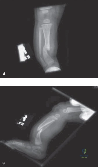
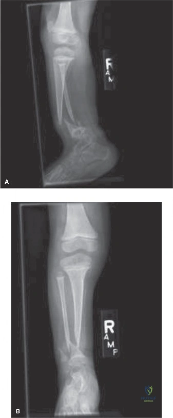
A 3-year-old child presents with a progressive right-sided limp and lower extremity deformity. Examination reveals multiple café-au-lait spots. You obtain the following radiographs. Describe your findings and provide your immediate clinical suspicion.
Candidate: The radiograph shows anterolateral bowing of the tibia and fibula. The medullary canal is sclerotic and the cortex appears thickened and narrowed at the apex. Given the age, the deformity, and the presence of café-au-lait spots, this is likely Neurofibromatosis Type 1 (NF1) associated with Congenital Pseudarthrosis of the Tibia (CPT).
Failing to classify the lesion using the Crawford system or omitting mention of the fibular involvement. Candidates often forget to explicitly state the "impending fracture" risk, treating it as a benign structural issue rather than a pre-fracture state requiring urgent planning.
Systematically address: 1. The radiographic pattern (Anterolateral bowing, medullary sclerosis, diaphyseal narrowing). 2. Classification (Crawford Type II, transitioning to III). 3. Etiology (NF1, identifying the need for multi-disciplinary review). 4. Prognosis (High risk of spontaneous fracture; surgery is mandated because the biological deficit prohibits normal healing).
You have decided to proceed with surgery. You perform the resection of the dysplastic hamartoma and prepare for fixation. Looking at the intraoperative image below, explain the rationale for this specific construct.
Candidate: The construct utilizes the "Charnley principle" of combined mechanical and biological fixation. The intramedullary rod provides internal splintage against bending, the circular frame provides axial compression and rotational stability, and the massive autograft creates a tibiofibular cross-union to provide a biological bridge across the pseudarthrosis site.
Focusing only on the hardware while ignoring the biological component (the autograft). Candidates who do not emphasize the necessity of radical resection of the hamartoma down to healthy, bleeding bone often fail, as they miss the underlying pathology of the condition.
Explain that CPT is a biological failure. Success requires: 1. Radical resection of the periosteal hamartoma. 2. Mechanical stability (Telescopic rod + Circular frame). 3. Biological enhancement (Autologous bone graft + potential use of BMP). 4. Acknowledging that the rod must be permanent and bracing is required until skeletal maturity to prevent recurrent deformity.
