Nonaccidental Injury (NAI) in Pediatric Orthopedics: Identification & Management Guide

Key Takeaway
Orthopedic surgeons identify Nonaccidental Injury (NAI) by recognizing specific fracture patterns inconsistent with reported mechanisms. Key indicators include metaphyseal corner fractures (bucket-handle), posterior/multiple rib fractures, long bone spiral fractures in non-ambulatory infants, and fractures at various healing stages. Careful evaluation of injury biomechanics and soft tissue signs is crucial for accurate diagnosis.
A 4-month-old infant is brought to the ED by their parents after they noticed the child was "favoring" their left leg. There is no clear history of trauma. On examination, the infant is irritable but has no fever. You obtain the following radiograph. What are the key features, what is your primary differential, and how do you proceed?
Candidate: The radiograph shows a metaphyseal corner fracture (bucket-handle fracture). This is highly suspicious for non-accidental injury (NAI). My immediate steps would be to stop the assessment, ensure the child is safe, document findings objectively, and initiate a full skeletal survey and multidisciplinary consultation with pediatrics and social services.
Jumping immediately into treatment options (e.g., "I would put the baby in a spica cast") before addressing the underlying diagnosis. Failure to state that the injury is "pathognomonic" for NAI in the absence of a high-energy accident, or failing to mention the mandatory multidisciplinary team (MDT) involvement.
The candidate identifies the "Metaphyseal Corner Fracture" (MCF) and states it is pathognomonic for NAI in a non-ambulatory infant. They categorize the management: 1) Immediate Safety: Admitting the child for protection. 2) Diagnostic Investigation: Formal skeletal survey (AAP guidelines), ophthalmology consult (for retinal hemorrhages), and lab work to rule out metabolic mimics (e.g., Osteogenesis Imperfecta, Rickets, Scurvy). 3) Reporting: Mandatory reporting to authorities/CPS. 4) Documentation: Meticulous, objective forensic recording without accusing parents during the initial interaction.
You are asked to review a 3-year-old child who sustained a femur fracture. The parents report a "fall from a height," but the fracture morphology is atypical. Looking at this image, what specific fracture pattern and associated risk factors should you be evaluating?
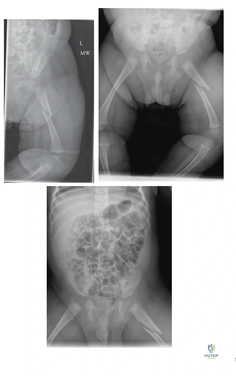
Candidate: I see a displaced femoral shaft fracture. I need to assess for cortical thinning or signs of rickets, and look for callus formation that might suggest a fracture at a different stage of healing. I must correlate the fracture type with the child's developmental age; a spiral fracture in a non-ambulatory infant would be much more suspicious than in a 3-year-old.
Ignoring the "medical mimics." Candidates often fail to mention checking for metabolic bone diseases like Osteogenesis Imperfecta or nutritional deficiencies like Vitamin D/Rickets, which can masquerade as NAI-related fractures.
The candidate should structure their response: "I would assess the fracture through three lenses: 1) Biological: Is the bone quality normal? (Rule out OI, Rickets). 2) Mechanical: Is the mechanism reported consistent with the fracture geometry? 3) Forensic: Are there occult fractures elsewhere? I would order a skeletal survey, check serum alkaline phosphatase, calcium, and phosphorus, and involve the Child Protection Team. Even if the child is 3, I would not assume this is accidental until the social history and physical exam confirm the mechanism."
