Pediatric Clubfoot Deformity: Pathoanatomy, Biomechanics, and the Role of the Anterior Tibialis Tendon

Key Takeaway
Pediatric clubfoot, or CTEV, is a complex foot and ankle deformity involving hindfoot equinus/varus, midfoot cavus, and forefoot adduction. It stems from contracted medial/posterior soft tissues and osseous abnormalities like talar displacement. The anterior tibialis tendon significantly contributes to forefoot adduction and cavus due to its altered pull, crucial for surgical planning and understanding persistent deformities.
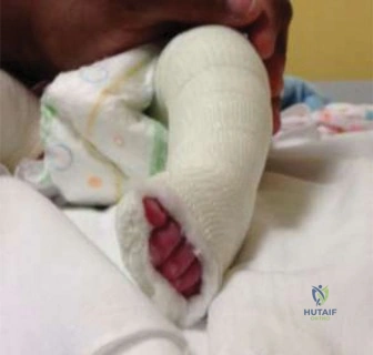
You are reviewing a 4-year-old child in the clinic who was treated for idiopathic clubfoot as an infant using the Ponseti method. The parents are concerned because the child has developed an inward-turning foot during the swing phase of gait, causing frequent tripping. On examination, there is dynamic forefoot supination upon active dorsiflexion. What is your diagnosis, and how would you manage this?
Candidate: The child has recurrent clubfoot deformity. Specifically, the dynamic supination suggests an overactive or pathologically positioned Anterior Tibialis Tendon (ATT). I would perform an Anterior Tibialis Tendon Transfer (ATTT) to the lateral cuneiform or cuboid to balance the foot.
Jumping directly to surgery without confirming brace compliance or failing to acknowledge the "dynamic" nature of the pathology. Candidates often forget to mention the diagnostic role of clinical assessment and misidentify the insertion site or the biomechanical goal of the transfer.
Start with a structured approach: Assessment, Diagnosis, and Management. 1. Assessment: Confirm the recurrence, check bracing history/compliance, and assess the dynamic deformity (the "tripod" gait). 2. Diagnosis: Recurrent clubfoot with dynamic forefoot supination secondary to anterior tibialis tendon imbalance. 3. Management: Confirm the need for ATTT. Explain the goal: detaching the ATT from the medial cuneiform/first metatarsal and re-routing it to the lateral cuneiform/cuboid. This converts the tendon's function from a supinator/invertor to an evertor/dorsiflexor, balancing the foot during the swing phase of gait.
Look at these radiographs of a child presenting with a resistant clubfoot. What are the key radiographic markers of uncorrected deformity, and how do they influence your surgical planning?
Candidate: I am looking at the AP and lateral views. On the AP view, I look for the talonavicular coverage angle, which should be reduced in clubfoot. On the lateral view, I assess the talocalcaneal angle and the talus-first metatarsal angle to evaluate equinus and cavus.
Listing angles without explaining what they represent pathologically. Candidates often fail to mention the "uncovering of the talar head" by the navicular, which is the hallmark of the medial column deformity.
Use a structured anatomical approach: 1. AP View: Look for the Talonavicular Coverage Angle. In a normal foot, the navicular is centered on the talar head. Medial subluxation indicates persistent forefoot adduction. 2. Lateral View: Look for the Talocalcaneal (Kite’s) Angle (reduced in hindfoot varus/equinus) and the Talo-first Metatarsal Angle. A plantarflexed first ray relative to the talus indicates persistent cavus. 3. Planning: These findings dictate whether I need a simple soft-tissue release, or if the deformity is so rigid that I must consider an osseous procedure like a dorsal closing wedge osteotomy or cuboid/calcaneal osteotomy.
Regarding the Cincinnati incision used in posteromedial releases, what are the primary neurovascular structures at risk, and how do you protect them?
Candidate: The Cincinnati incision is a transverse, curvilinear incision. Structures at risk are the posterior tibial neurovascular bundle, the saphenous nerve and vein, and the sural nerve.
Naming the structures but failing to describe the displacement of these structures in a clubfoot. In clubfoot, the bundle is often medial and deep, and its location is distorted by the surrounding fibrosis.
Structure by location: 1. Posterior to the Medial Malleolus: The Posterior Tibial Neurovascular Bundle. It must be identified proximal to the flexor retinaculum, where the anatomy is more reliable, and traced distally. 2. Anterior/Medial: The Saphenous Nerve and Vein. These are often encountered early in the skin incision and must be protected. 3. Surgical Strategy: Meticulous, blunt dissection and constant identification of the bundle before any soft-tissue release. I emphasize that in clubfoot, these structures are often contracted or scarred; therefore, gentle, systematic mobilization is key to preventing ischemic injury.
