Congenital Vertical Talus (CVT): Advanced Pathoanatomy, Diagnosis & Biomechanics
Key Takeaway
Congenital Vertical Talus (CVT) is a rare, rigid congenital foot deformity characterized by a vertically oriented talus, irreducible dorsal navicular dislocation, and hindfoot valgus. Clinically, it presents as a 'rocker-bottom' foot with a prominent plantar talar head. Diagnosis relies on clinical examination and specific radiographic findings, differentiating it from flexible flatfoot.
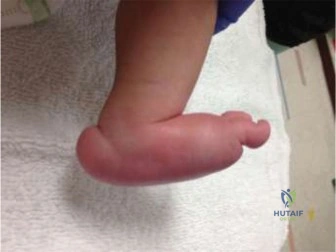
You are seeing a 9-month-old infant in the clinic. The parents are concerned about the appearance of the child's feet. On examination, you note a rigid, "rocker-bottom" deformity. The forefoot is abducted and dorsiflexed, and there is a prominent plantar bump. You order radiographs.
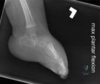
What is your diagnosis, and what are the three key radiographic features you are looking for to confirm this?
Candidate: The diagnosis is Congenital Vertical Talus (CVT). The key radiographic findings are a vertical orientation of the talus (equinus), a dorsal dislocation of the navicular on the talar head, and a fixed hindfoot valgus.
Failing to emphasize that radiographs must be "true lateral weight-bearing" or "simulated weight-bearing" to demonstrate the rigidity. Simply saying "vertical talus" without mentioning the talonavicular dislocation misses the pathognomonic feature.
The diagnosis is Congenital Vertical Talus. The three pathognomonic radiographic features, best seen on a lateral view in forced dorsiflexion, are: 1) A vertically oriented talus (fixed equinus), 2) Irreducible dorsal dislocation of the navicular on the talar head/neck, and 3) An increased talocalcaneal angle indicating hindfoot valgus. Meary’s angle will also be severely abnormal (apex plantar).
The child is scheduled for surgical correction. During the procedure, after performing the necessary releases, you achieve the reduction. You are currently confirming the final position with the C-arm.
What are the critical structures that must be released to allow this reduction, and what is your fixation strategy to maintain it?
Candidate: You need to release the contracted posterior and dorsal soft tissues. This includes the Achilles tendon, the posterior ankle capsule, and the dorsal talonavicular capsule. I would then use K-wires to hold the talonavicular and subtalar joints.
Omitting the peroneal tendons and the spring ligament complex. The peroneal tendons are often severely contracted and act as a tether; failing to address them will lead to an incomplete correction or early recurrence.
The release is comprehensive: 1) Posterior: Achilles lengthening and posterior ankle/subtalar capsulotomy. 2) Lateral: Peroneal tendon release/lengthening and calcaneocuboid capsule. 3) Medial/Dorsal: Deep deltoid ligament release and dorsal talonavicular capsulotomy. The reduction sequence involves dorsiflexing the talus, reducing the navicular onto the talar head, and correcting the hindfoot valgus. Fixation involves percutaneous K-wires: one across the talonavicular joint and one across the subtalar joint.
This condition is often associated with other syndromes. Name three associated conditions and explain why this is relevant to the surgical management of the patient.
Candidate: CVT is associated with arthrogryposis, spina bifida, and genetic syndromes like trisomy 18. It is relevant because these children may have complex systemic needs, and the risk of recurrence is higher in syndromic cases.
Ignoring the neurological implications. In patients with spina bifida, the potential for altered sensation, pressure sores, and the need for long-term orthotic management is significantly different compared to an isolated idiopathic case.
Associations include Arthrogryposis, Spina Bifida (myelomeningocele), and Trisomies (13, 15, 18). Relevance: 1) Systemic safety: Assessment for cardiac/pulmonary comorbidities is mandatory. 2) Prognosis: Syndromic feet are stiffer and have a much higher rate of recurrence. 3) Multidisciplinary care: These children require long-term bracing and potential repeat procedures, requiring a coordinated input from neurologists, geneticists, and orthotists.
