Clavicle Fracture: Comprehensive Orthopedic Guide to Epidemiology, Anatomy & Management
Key Takeaway
Surgical fixation for clavicle fractures is indicated for open fractures, neurovascular compromise, or severe displacement threatening skin. Relative indications include significant shortening (>15-20mm), extensive comminution, or for high-demand patients seeking early return to function. Lateral third fractures with coracoclavicular ligament disruption often also warrant operative management.
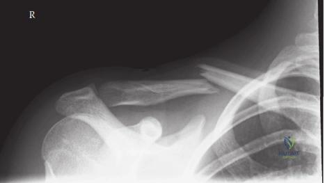
A 28-year-old male cyclist presents to the emergency department following a high-energy fall onto his right shoulder. He is in significant pain, and the skin over the mid-clavicle is tented but intact. Radiographs are provided below. Describe your clinical assessment and immediate management priorities.
Candidate: I would start with an ATLS assessment, specifically looking for associated thoracic injuries like a pneumothorax or scapular fracture. I would perform a thorough neurovascular exam of the upper limb—checking pulses and brachial plexus function. Given the significant displacement and skin tenting, I would consider urgent operative fixation after stabilizing the patient, likely using a precontoured locking plate.
Focusing purely on the fracture anatomy while ignoring the "floating shoulder" or life-threatening associated chest injuries. Failing to specifically mention the supraclavicular nerve at-risk during surgery or the specific risk of skin necrosis from pressure.
Systematically address: 1) Clinical status (triage for polytrauma); 2) Neurovascular status (specific mention of brachial plexus and subclavian vessels); 3) Skin integrity (impending rupture is an urgency); 4) Biomechanical rationale for surgery (shortening >2cm, displacement). Mention the need for a CT scan if there is comminution or suspicion of scapular/medial third involvement. Finally, state the surgical goal: anatomic restoration of length and rotation to avoid scapular dyskinesis.
You decide on operative fixation. What are the biomechanical advantages and potential drawbacks of choosing a superior plate vs. an anteroinferior plate for a midshaft fracture?
Candidate: Superior plating is biomechanically stronger because the superior surface is the tension side of the bone. However, it is more prominent, often leading to hardware irritation. Anteroinferior plating is less prominent and thus more comfortable, but it is biomechanically less robust and risks damaging the subclavian vessels if screws are too long.
Omitting the risk of injury to the neurovascular structures during anteroinferior screw placement or failing to acknowledge that superior plating is the gold standard for biomechanical stability.
Structure by biomechanics and clinical outcomes: 1) Superior plating: Acts as a tension band, higher moment of inertia, better resistance to bending; drawback is hardware prominence. 2) Anteroinferior plating: Smoother profile, less soft tissue irritation, but requires precise screw length to avoid injury to the subclavian artery/vein and brachial plexus in the infraclavicular fossa. Conclude with the importance of the "safe zone" for screw placement.
The patient returns 6 months post-operatively. He has no hardware pain, but serial radiographs show a persistent fracture gap with sclerotic margins. What is your management plan?
Candidate: This is a symptomatic nonunion. I would investigate metabolic causes (smoking, vitamin D) and then offer revision surgery: debridement of the fibrous tissue, freshening the bone ends, and repeat rigid internal fixation, likely with the addition of a bone graft such as autologous iliac crest.
Suggesting immediate revision surgery without optimizing the biological environment (e.g., smoking cessation) or ignoring the need for bone grafting in an atrophic nonunion.
Acknowledge this as an atrophic nonunion. Frame the response using the 'Diamond Concept' of bone healing: 1) Mechanical stability (rigid revision fixation); 2) Biological stimulation (debridement of fibrous tissue, autologous bone graft/BMP); 3) Osteoconductivity/Osteoinductivity. Mention checking for infection, smoking status, and systemic factors before revision.
