Pediatric Pain & Swelling: 16 Cases Solved & Diagnoses Revealed
Key Takeaway
Here are the crucial details you must know about Pediatric Pain & Swelling: 16 Cases Solved & Diagnoses Revealed. For an adolescent experiencing acute knee pain and swelling after an injury, particularly with an effusion and a challenging physical exam, an MRI is the next best diagnostic step. It accurately identifies soft tissue injuries, such as an ACL rupture or patellar dislocation, which are common causes of these acute knee symptoms in this age group.
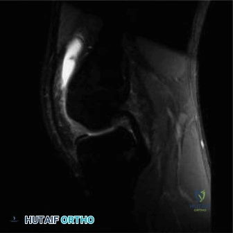
An 11-year-old male presents with persistent right distal thigh pain for six weeks following a minor soccer injury. He reports nocturnal pain and recent weight loss. On examination, he has a firm, fixed mass over the distal femoral metaphysis and restricted knee flexion. Based on the radiographs provided, what is your immediate clinical concern and your structured approach to further investigation?
Candidate: My main concern is a high-grade primary bone malignancy, most likely osteosarcoma. I would keep the patient non-weight bearing, order urgent bloods including inflammatory markers, and obtain an MRI of the entire femur to assess local extent. I would then perform a staging CT of the chest and refer to a specialist oncology center for a planned biopsy.
Failing to mention the biopsy technique or the risk of contaminating the operative field. Candidates often forget to mention that the biopsy should be performed at the institution that will provide definitive treatment, or they neglect to discuss the specific biopsy path (longitudinal, avoiding neurovascular bundles).
Start by describing the radiographic features: "Aggressive, permeative lesion with a sunburst periosteal reaction and Codman triangle, consistent with a primary malignant bone tumor." Structure the approach into: 1. Staging: MRI of the femur (for local extent/skip mets), CT chest (for pulmonary mets), and technetium bone scan. 2. Biopsy: Emphasize that the biopsy must be performed by the treating surgeon, via a longitudinal incision within the future excision zone, ensuring no compromise of major neurovascular structures. 3. Multidisciplinary: Early referral to an MDT including pediatric oncology, orthopedic oncology, and pathology. Mentioning the biopsy as a high-risk step for local recurrence is critical.
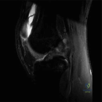
The MRI has been performed and confirms a 11.5cm distal femoral lesion. You are planning the surgical resection. What are the key factors that determine whether you proceed with limb salvage versus amputation, and how does this patient's age influence your reconstruction choice?
Candidate: Limb salvage is feasible if the neurovascular bundle is not encased and if functional reconstruction is possible. In this 11-year-old, we have to consider the remaining growth. Using a static prosthesis would cause a massive limb-length discrepancy, so I would look at an expandable endoprosthesis.
Forgetting to mention the "absolute contraindications" for limb salvage, such as encasement of the popliteal artery/sciatic nerve that cannot be salvaged, or massive soft tissue destruction precluding coverage.
Structure the answer into Oncologic and Functional determinants. 1. Oncologic: Ability to achieve wide surgical margins. Mention the importance of the neurovascular bundle and the muscle envelope. 2. Functional: The ability to preserve a functional extensor mechanism. 3. Growth-related: Acknowledge that in a child (Tanner stage II), a static prosthesis is unacceptable due to longitudinal growth potential. Recommend a non-invasive expandable endoprosthesis to allow for serial lengthening, minimizing the need for repeated revision surgeries.
During your definitive resection, you are preparing to reconstruct the soft tissues over the metallic endoprosthesis. Why is the choice of soft tissue coverage critical here, and what specific technique would you employ?
Candidate: Soft tissue coverage is vital because endoprostheses have poor soft tissue coverage, increasing the risk of infection and wound breakdown. I would use a gastrocnemius rotational flap to provide a vascularized layer between the skin and the implant.
Simply stating "use a flap" without explaining the "why" (vascularity, dead space, protection of the extensor mechanism) or the specific anatomy (medial vs. lateral gastrocnemius).
Connect the answer to the biology of oncology patients: 1. Rationale: These patients are often immunocompromised due to chemotherapy; thin, scarred skin over metal leads to high rates of periprosthetic infection and wound dehiscence. 2. Technique: Use of a rotational medial gastrocnemius flap. Explain that it provides robust, well-vascularized tissue that brings a new blood supply to the surgical site, fills dead space, and effectively creates a barrier between the metal implant and the skin surface. This is a critical nuance for achieving a durable limb salvage result.
