Anterior Shoulder Dislocation: Epidemiology, Pathoanatomy, Diagnosis & Management
Key Takeaway
Anterior shoulder dislocation is the anterior translation of the humeral head, typically from abduction-external rotation. Common in young, active individuals, it has high recurrence rates and frequently involves Bankart lesions, Hill-Sachs defects, and glenoid bone loss. Effective management requires understanding epidemiology, surgical anatomy, and tailored treatment strategies.
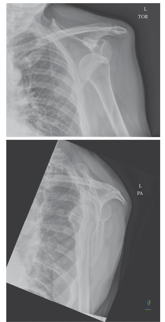
A 22-year-old rugby player presents to the clinic following his third anterior shoulder dislocation in 18 months. He is keen to return to competitive play. You are reviewing his imaging. Explain the anatomical structures involved in anterior shoulder stability and identify the key pathology depicted in the figure below.
Candidate: The shoulder is stabilized by the labrum, the glenohumeral ligaments, and the rotator cuff. The figure shows the labrum and ligaments. My patient has recurrent instability. I would look for a Bankart lesion where the labrum is torn off the glenoid, and a Hill-Sachs lesion on the humeral head.
Failure to categorize stabilizers into static and dynamic components. Candidates often list structures randomly without mentioning the Inferior Glenohumeral Ligament (IGHL) complex as the primary static stabilizer in abduction/external rotation—the position of dislocation.
Systematically structure the response: 1) Static Stabilizers: Emphasize the glenoid labrum (increasing surface area) and the IGHL complex, specifically the anterior band, as the primary restraint in abduction/external rotation. 2) Dynamic Stabilizers: Highlight the "concavity-compression" effect provided by the rotator cuff, specifically the subscapularis anteriorly. 3) Pathology Identification: Identify the Bankart lesion as the avulsion of the anteroinferior labrum and AIGHL. Mention the impact of bone loss (bony Bankart/glenoid erosion) and the Hill-Sachs lesion, noting the significance of an "engaging" lesion vs. "on-track" lesion.
The patient has recurrent instability. How do you decide between an arthroscopic Bankart repair and an open Latarjet procedure for this patient?
Candidate: I would choose based on whether he has bone loss. If he has a large glenoid bone defect, I would perform a Latarjet. If his bone stock is good, I would do an arthroscopic Bankart repair.
Providing a binary answer. A high-scoring candidate must discuss the "Goldilocks" zone of bone loss and integrate patient-specific factors like collision sports status and hyperlaxity.
Structured decision-making: 1) Bone Loss quantification: Use 3D-CT to assess glenoid bone loss (>15-20% warrants Latarjet). 2) Hill-Sachs characterization: Determine if the defect is "engaging" using the on-track/off-track concept. 3) Patient Profile: Collision athletes (e.g., rugby) have a higher recurrence rate; Latarjet is often preferred in these high-demand individuals even with borderline bone loss to ensure a definitive, stable result. 4) Tissue Quality: If the labral tissue is poor or scarred from previous failed repairs, Latarjet provides superior stability via the "triple effect" (bone block, conjoined tendon sling, and capsular repair).
During a Latarjet procedure, what are the critical risks you must warn the patient about in the consent process, specifically regarding long-term function?
Candidate: I would warn him about infection, stiffness, and the possibility of recurrence. I should also mention nerve damage like the axillary or musculocutaneous nerve, and potential hardware issues.
Listing complications without explaining their clinical impact. Failing to specifically mention the risk of degenerative joint disease (arthritis) which is a known long-term concern with Latarjet due to the altered glenoid surface.
Provide a structured "Specific, Serious, and Frequent" risk analysis: 1) Neurological: Musculocutaneous nerve is at greatest risk during dissection; axillary nerve is at risk with retraction. 2) Hardware: Screw prominence or failure requiring secondary removal. 3) Joint Health: Risk of accelerated osteoarthritis (OA) due to the non-anatomic reconstruction. 4) Structural: Non-union or migration of the coracoid graft. 5) Recurrence: Lower than arthroscopic, but still ~2-5%. 6) Functional: Permanent loss of some external rotation/abduction range.
