Complex Pediatric Distal Tibia Physeal Fracture: A Detailed Case Study
Key Takeaway
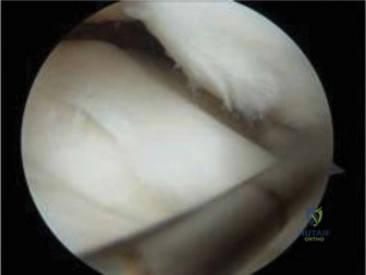
Complex pediatric distal tibia physeal fractures, often from high-energy rotational trauma, present with severe pain and gross deformity. Diagnosis mandates meticulous clinical examination, assessing neurovascular status and skin integrity. Comprehensive X-rays (AP, lateral, mortise views) are crucial to identify displaced intra-articular fractures and syndesmotic disruption, informing treatment focused on physeal preservation.
A 10-year-old male presents with a high-energy rotational injury to the ankle. After initial stabilization, these radiographs and the subsequent CT scan were obtained. Describe the injury pattern and explain why this specific classification is important for your management strategy.
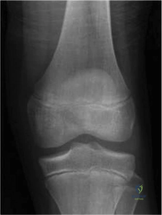
Candidate: This is a Triplane fracture of the distal tibia. It involves three planes: sagittal through the epiphysis, transverse through the physis, and coronal through the metaphysis. It is a transitional fracture seen in adolescents, and this case is complex due to the multi-fragmentary nature (four-part), requiring CT for surgical planning to ensure anatomical articular reduction.
Failing to mention the "transitional" nature of the injury or ignoring the clinical significance of the articular surface displacement. Candidates often forget to mention the syndesmotic injury frequently associated with these, or they fail to emphasize why the CT is mandatory (i.e., plain films underestimate the fragment size and articular step-off).
Identify this as a transitional injury occurring during asymmetrical physeal closure (central, then medial, then lateral). Highlight the "four-part" nature identified on CT: anteromedial epiphyseal, posterolateral epiphyseal, posterior metaphyseal, and the remaining tibial shaft. Emphasize that the goal is anatomical reduction of the articular surface to prevent post-traumatic arthritis and that the syndesmotic integrity must be assessed, as internal fixation of the fragments often stabilizes the syndesmosis, but if not, secondary stabilization is required.
You decide that closed reduction is insufficient. Describe your surgical approach and how you will manage the fixation to avoid long-term complications in this skeletally immature patient.
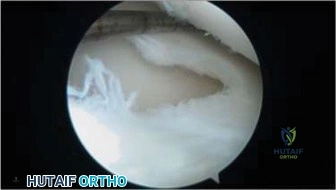
Candidate: I would utilize a dual approach: an anteromedial approach to address the medial malleolar/epiphyseal component and an anterolateral approach to visualize the syndesmosis and lateral epiphyseal fragment. Fixation would involve cannulated screws. I would avoid crossing the physis if possible, or if necessary, use fully threaded screws placed parallel to the physis.
Recommending heavy-duty plates or non-cannulated hardware without justifying the risk to the physis. Failing to address the "why" regarding the dual approach (the need to visualize the joint surface directly to ensure <2mm step-off).
Detail a structured approach: (1) Dual Incision: Anteromedial and anterolateral to allow joint-line visualization. (2) Reduction: Use K-wire joysticks for anatomical reduction of the articular fragments. (3) Physeal Sparing: Utilize 3.5mm fully threaded cannulated screws. Aim to stay within the epiphysis or metaphysis. If crossing the physis is unavoidable, emphasize the use of hardware that minimizes growth disturbance and note that these must be removed if they are felt to tether growth. Mention the importance of syndesmotic assessment post-reduction.
Post-operatively, the patient has achieved anatomical reduction. What are the long-term concerns you must discuss with the parents during the consent and follow-up process?
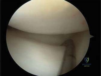
Candidate: The main concerns are physeal arrest leading to angular deformity or limb-length discrepancy, and post-traumatic arthritis due to the articular surface injury. I would counsel the parents that long-term follow-up is required until skeletal maturity.
Only mentioning "arthritis." Failing to discuss the specific risk of physeal bar formation and the need for serial clinical and radiographic monitoring to detect growth disturbances early, when intervention (e.g., bar resection or epiphysiodesis) might still be possible.
Categorize the risks: (1) Physeal complications: Growth arrest, angular deformity (varus/valgus), and limb length discrepancy due to physeal bar formation. (2) Articular surface: Post-traumatic arthritis secondary to chondral injury. (3) Functional: Chronic stiffness or syndesmotic pain. State the follow-up plan: periodic radiographs to monitor the physis until maturity and clinical assessment of gait and ROM.
