Acromioclavicular (AC) Joint Injuries: Epidemiology, Anatomy, Rockwood Classification & Management
Key Takeaway
Acromioclavicular (AC) joint injuries are common shoulder pathologies, classified by the Rockwood system into six types (I-VI). This system categorizes injuries based on AC and coracoclavicular ligament disruption and clavicle displacement, from mild sprains (Type I-II) to severe instability (Type IV-VI), guiding management decisions.
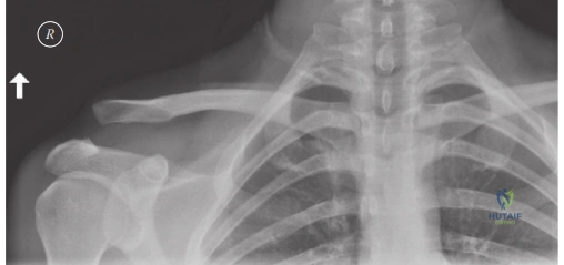
A 28-year-old rugby player presents after sustaining a direct blow to the superior aspect of the right shoulder. He is holding his arm in adduction. Clinical examination reveals a prominent distal clavicle and significant tenderness over the AC joint. You obtain a series of radiographs. Describe the biomechanical structures highlighted in this image and their primary functions.
Candidate: The image shows the coracoclavicular (CC) ligaments: the conoid and the trapezoid. The conoid is medial and resists superior translation, while the trapezoid is lateral and resists compression. They are essential for vertical stability of the AC joint.
Failing to distinguish between the primary functions of the two bundles. Candidates often confuse their insertions relative to the clavicle or miss the crucial role of the AC ligaments in horizontal stability. Also, failing to mention the normal anatomical CC distance (11-13mm) as a baseline for measuring displacement is a missed opportunity for higher marks.
Systematically categorize the stabilizers: The CC ligaments consist of the medial conoid (resists superior translation and anterior rotation) and the lateral trapezoid (resists axial compression). I would also highlight that the superior AC ligament is the primary restraint to horizontal (posterior) translation. Finally, I would mention that in a standard Zanca view, the normal CC distance is 11–13 mm, and this anatomy is critical for planning anatomical reconstruction to avoid over-tensioning.
The patient has a Rockwood Type III ACJ injury. He is a high-level manual laborer. How do you decide between non-operative and operative management, and what is your rationale?
Candidate: I would consider the patient's demand. Most Type III injuries are treated non-operatively with a sling and physiotherapy. However, because he is a manual laborer, I would assess for horizontal instability—Type IIIB—as these patients have poor outcomes with non-operative care. If he has persistent pain or significant horizontal translation, I would discuss surgical stabilization.
Suggesting that all Type III injuries require surgery. A high-scoring candidate must acknowledge the consensus that outcomes between operative and non-operative management are often equivalent in the general population, emphasizing the specific subsets (overhead athletes, laborers, Type IIIB) who deviate from the standard non-operative path.
Start by categorizing: Type IIIA (vertically unstable only) vs IIIB (horizontally and vertically unstable). For this manual laborer, I would evaluate his horizontal instability. I would offer non-operative management initially for all Type III cases, but if the patient demonstrates persistent pain, weakness, or dynamic overriding of the clavicle on cross-body adduction (Type IIIB), I would discuss anatomic CC ligament reconstruction, noting that while surgery improves contour, it does not guarantee a return to heavy-duty work without risk of hardware failure.
During a reconstructive procedure for a chronic Rockwood Type V injury, you are concerned about iatrogenic complications. What are the major risks during the dissection around the coracoid and how do you mitigate them?
Candidate: The main risks are coracoid fracture and injury to neurovascular structures, particularly the musculocutaneous nerve and the brachial plexus. I would use careful subperiosteal dissection and ensure my drill holes are centered on the coracoid base.
Failing to mention the "danger zone." Candidates often forget that the musculocutaneous nerve enters the conjoint tendon 3–5 cm distal to the coracoid. Also, ignoring the risk of coracoid fracture due to multiple or eccentric drill holes shows a lack of surgical planning maturity.
I would structure my response by anatomical danger: 1) Neurovascular: The musculocutaneous nerve is at risk 3–5 cm distal to the coracoid; I would avoid aggressive medial retraction. The brachial plexus is posterior and medial. 2) Osseous: Coracoid fracture is the most common technical error. I mitigate this by using a single, central drill hole or passing the graft around the coracoid rather than through it if the anatomy is small. 3) Vascular: Meticulous control of the acromial branch of the thoracoacromial artery is required to maintain a clear visual field.
