Acromioclavicular (AC) Joint Separation: Epidemiology, Classification & Biomechanics
Key Takeaway
The Rockwood classification categorizes acromioclavicular (AC) joint separations into six types, based on clavicular displacement and ligament disruption. Type I is a sprain; Type II, AC ligament tear; Type III, complete AC and CC ligament tears. Types IV-VI indicate severe displacement, often requiring surgery due to instability and functional impairment.
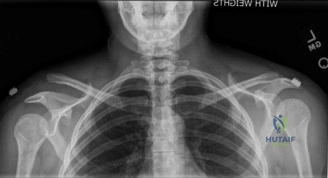
A 28-year-old rugby player presents after a direct impact to the superior aspect of his adducted shoulder. He complains of pain and a deformity at the shoulder. Please describe the radiographic evaluation required to diagnose an AC joint injury and interpret the following finding.

Candidate: I would order an AP view of the AC joint, a Zanca view—which is 10-15 degrees cephalic tilt—and an axillary view to assess posterior displacement. The provided image shows a Rockwood Type III or V injury with significant superior displacement of the distal clavicle relative to the acromion.
Candidates often forget the axillary view. The axillary view is critical to rule out a Type IV injury (posterior displacement), which significantly changes the surgical plan. Furthermore, failing to mention the CC distance compared to the contralateral side for quantification is a common error.
Structure the answer: 1. Imaging Suite (AP AC-joint, Zanca, Axillary). 2. Interpretation (Measure CC distance vs. contralateral side; check axillary view for posterior displacement). 3. Clinical Correlation (Type III vs. Type V). Mention that the Zanca view is specific for the AC joint as it avoids overlap with the scapular spine.
We are in the operating theater for a high-grade AC joint reconstruction using a suture-button construct. Describe the anatomical landmarks for drill placement and the specific safety considerations regarding the coracoid process.

Candidate: I target the footprint of the conoid and trapezoid ligaments on the clavicle, approximately 2-3 cm medial to the joint. For the coracoid, I drill from superior to inferior at the base of the process. I must avoid the medial aspect of the coracoid to protect the neurovascular structures.
Failing to emphasize the "medial and inferior" danger zone. Examiners want to hear explicit awareness of the brachial plexus and subclavian vessels located medial/inferior to the coracoid. Also, ignoring the risk of coracoid fracture due to improper drill path or tunnel spacing is a red flag.
Highlight the importance of the anatomical CC footprint (conoid and trapezoid). Detail the "Safe Zone": drilling the coracoid base 1–1.5 cm from the tip, directing the drill slightly obliquely to avoid medial neurovascular structures, and using a drill guide to prevent "plunging" which is the primary cause of iatrogenic neurovascular injury.
A patient returns at 6 months post-reconstruction with a recurrence of the distal clavicle prominence, but they are functionally asymptomatic. How do you manage this?
Candidate: If the patient is asymptomatic, I would recommend conservative management and reassurance. I would not operate based on the radiograph alone. If they develop pain, I would investigate further for hardware failure or AC joint arthritis.
Jumping to "revision surgery" because of the radiographic deformity. The FRCS examiner is testing clinical judgment—we treat patients, not X-rays. A "successful" reconstruction often involves a degree of residual superior prominence.
State clearly: "I treat the patient, not the X-ray." If asymptomatic, avoid re-operation. Explain that residual superior displacement is common and doesn't equate to failure. Outline a criteria-based decision if pain arises: evaluate for hardware breakage, AC joint degeneration (Mumford indication), or persistent instability impacting activities of daily living.
