Part of the Master Guide

Comprehensive Guide to AC Joint Injuries: Epidemiology, Surgical Anatomy & Biomechanics

Acromioclavicular (AC) Joint Injuries: Epidemiology, Anatomy, Rockwood Classification & Management

17 Apr 2026 15 min read 138 Views
Illustration of joint injury acromioclavicular - Dr. Mohammed Hutaif

Key Takeaway

Acromioclavicular (AC) joint injuries are common shoulder pathologies, classified by the Rockwood system into six types (I-VI). This system categorizes injuries based on AC and coracoclavicular ligament disruption and clavicle displacement, from mild sprains (Type I-II) to severe instability (Type IV-VI), guiding management decisions.
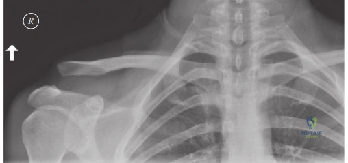
Introduction and Epidemiology

Acromioclavicular joint injuries are common pathologies encountered in orthopedic practice, comprising approximately 9 percent of all shoulder girdle injuries and up to 40 percent of all shoulder injuries in athletic populations. These injuries typically result from direct trauma to the superior aspect of the acromion, often with the arm adducted, driving the scapula inferiorly relative to the clavicle. This mechanism is frequently observed in contact sports such as football, rugby, and hockey, as well as high-velocity mechanisms including cycling accidents and falls from a height. Less commonly, an indirect mechanism involving a fall onto an outstretched hand can transmit force proximally through the humeral head into the acromion, resulting in isolated acromioclavicular ligament failure.

The incidence of acromioclavicular joint injuries demonstrates a bimodal distribution, with a primary peak in young, active males in their second and third decades of life, and a secondary peak in older individuals sustaining low-energy falls. Male patients are affected approximately five times more frequently than female patients. Concomitant intra-articular glenohumeral pathology is highly prevalent in high-grade acromioclavicular separations, with studies demonstrating that up to 15 to 30 percent of patients possess associated superior labral anterior-posterior tears, partial articular-sided supraspinatus tendon avulsions, or capsular injuries.

Rockwood Classification System

The severity of acromioclavicular joint injuries is universally classified using the Rockwood classification system, which categorizes injuries into six types based on the degree of ligamentous disruption, directional displacement of the clavicle relative to the acromion, and involvement of the deltotrapezial fascia.

  • Type I Sprain of the acromioclavicular ligaments without complete tear. The joint remains stable and radiographs appear normal.
  • Type II Complete tear of the acromioclavicular ligaments with intact coracoclavicular ligaments. Radiographs demonstrate mild superior subluxation of the clavicle, representing less than 25 percent increase in the coracoclavicular distance compared to the contralateral side.
  • Type III Complete tears of both the acromioclavicular and coracoclavicular ligaments. The clavicle is significantly displaced superiorly, typically demonstrating a 25 to 100 percent increased coracoclavicular distance. The deltotrapezial fascia remains intact.
  • Type IV Complete tears of the acromioclavicular and coracoclavicular ligaments with the distal clavicle displaced posteriorly into or through the trapezius muscle. This requires an axillary lateral radiograph for definitive diagnosis.
  • Type V Complete tears of the acromioclavicular and coracoclavicular ligaments with severe superior displacement of the clavicle, exceeding 100 percent of the normal coracoclavicular distance. This injury includes extensive stripping and detachment of the deltoid and trapezius fascia from the distal clavicle.
  • Type VI An extremely rare injury pattern characterized by complete tears of the acromioclavicular and coracoclavicular ligaments with inferior displacement of the clavicle beneath the coracoid process or acromion. This is typically the result of severe hyperabduction and external rotation trauma.

Surgical Anatomy and Biomechanics

A thorough understanding of the intricate anatomy and biomechanics of the acromioclavicular joint is paramount for effective diagnosis and surgical reconstruction. The acromioclavicular joint is a diarthrodial joint stabilized by a complex interplay of static ligamentous restraints and dynamic muscular forces.

Image

Static Stabilizers

The static stabilizers are divided into the acromioclavicular ligaments, the coracoclavicular ligaments, and the articular capsule.

The acromioclavicular ligaments include the superior, inferior, anterior, and posterior bundles. The superior acromioclavicular ligament is the thickest and most robust, blending intimately with the fibers of the deltoid and trapezius muscles. Biomechanically, the acromioclavicular ligaments provide the primary restraint to anterior-posterior horizontal translation of the clavicle on the acromion. Sectioning the superior acromioclavicular ligament results in profound horizontal instability.

The coracoclavicular ligaments are robust extra-articular structures crucial for vertical stability, connecting the base of the coracoid process to the inferior aspect of the clavicle. They consist of two distinct fascicles. The conoid ligament is the medial, conical structure originating from the posteromedial base of the coracoid and inserting onto the conoid tubercle of the clavicle, located approximately 45 millimeters medial to the distal articular surface of the clavicle. The conoid primarily resists superior translation and anterior rotation of the clavicle. The trapezoid ligament is the lateral, quadrilateral structure originating from the superior surface of the coracoid and inserting onto the trapezoid line, located approximately 30 millimeters medial to the distal clavicle. The trapezoid provides secondary resistance to superior translation and is the primary restraint against axial compression of the acromioclavicular joint. The normal radiographic distance from the superior aspect of the coracoid to the inferior aspect of the clavicle is typically 11 to 13 millimeters.

The articular disc is a fibrocartilaginous meniscus located within the joint space. It is typically complete in the first decade of life but undergoes rapid, predictable degeneration, often becoming a meniscoid remnant by the fourth decade. This degeneration is a normal physiological process and should not be inherently viewed as pathological, though it may predispose the joint to primary osteoarthritis.

Dynamic Stabilizers

The dynamic stabilizers consist primarily of the anterior deltoid and the superior trapezius musculature, which envelop the distal clavicle and acromion via the deltotrapezial fascia. This fascial sleeve provides significant superior stability to the distal clavicle. In high-grade separations, particularly Rockwood Type V injuries, this fascial envelope is violently stripped from the clavicle, allowing the clavicle to herniate subcutaneously. Meticulous surgical repair of this fascial layer is a critical component of any reconstructive procedure.

Neurovascular Anatomy

The primary vascular supply to the acromioclavicular joint arises from branches of the suprascapular artery and the acromial branch of the thoracoacromial trunk. Innervation is supplied by the lateral pectoral nerve, the axillary nerve, and the suprascapular nerve. When performing coracoid dissection, surgeons must remain acutely aware of the musculocutaneous nerve, which enters the conjoint tendon approximately 3 to 5 centimeters distal to the coracoid tip, and the underlying brachial plexus, which sits posteromedial to the coracoid base.

Indications and Contraindications

The management of acromioclavicular joint injuries is dictated by the Rockwood classification, patient activity level, chronicity of the injury, and associated comorbidities. While consensus exists for the extremes of the classification system, the management of Type III injuries remains highly debated within the orthopedic literature.

Indication Category Operative Management Non Operative Management
Rockwood Type I and II Rare. Considered only in chronic, refractory cases with persistent pain after >6 months of conservative therapy. Standard of care. Brief sling immobilization followed by early functional rehabilitation.
Rockwood Type III Overhead athletes, manual laborers, thin patients with prominent cosmetic deformity, and patients with significant horizontal instability (Type IIIB). Standard of care for the majority of patients. Functional outcomes often equal operative management without surgical risks.
Rockwood Type IV Standard of care. The posterior displacement into the trapezius prevents closed reduction and causes severe pain and dysfunction. Contraindicated unless the patient is medically unfit for surgery.
Rockwood Type V Standard of care. The severe superior displacement and deltotrapezial fascial stripping result in profound weakness and deformity. Contraindicated unless the patient is medically unfit for surgery.
Rockwood Type VI Standard of care. Severe displacement causes significant morbidity and potential neurovascular compromise. Contraindicated.
Chronic Symptomatic Persistent pain, fatigue, and weakness >3-6 months post-injury despite aggressive physical therapy. Initial management for all chronic presentations before considering surgical intervention.

Contraindications to surgical intervention include active local or systemic infection, severe medical comorbidities precluding anesthesia, highly non-compliant patients unable to adhere to strict postoperative rehabilitation protocols, and poor local soft tissue envelopes that would compromise wound healing.

Pre Operative Planning and Patient Positioning

Thorough preoperative planning relies heavily on high-quality radiographic evaluation to accurately classify the injury and assess for concomitant osseous pathology.

Radiographic Evaluation

Standard trauma series of the shoulder should be obtained, including a true anteroposterior view, a scapular Y view, and an axillary lateral view. The axillary lateral is absolutely critical for identifying the posterior clavicular displacement characteristic of a Rockwood Type IV injury.

The most sensitive imaging modality for evaluating the acromioclavicular joint is the Zanca view. This is obtained by tilting the X-ray beam 10 to 15 degrees cephalad and reducing the penetrance by 50 percent compared to a standard shoulder anteroposterior radiograph. This projects the acromioclavicular joint clear of the underlying scapular spine. Bilateral Zanca views on a single cassette can be obtained to compare the injured coracoclavicular distance to the contralateral normal side.

Cross-body adduction views can be utilized to assess dynamic horizontal instability, though their clinical utility is debated. Magnetic resonance imaging is not routinely indicated for isolated acromioclavicular joint injuries but should be strongly considered if concomitant intra-articular glenohumeral pathology is suspected, such as superior labral anterior-posterior tears or rotator cuff tears.

Patient Positioning and Setup

Surgical intervention is typically performed with the patient in the beach chair position. The head is secured, and the operative arm is draped free to allow for full manipulation during the procedure. A mechanical arm holder can be utilized to assist with reduction and maintain positioning. The surgical field must be prepared to allow access from the sternoclavicular joint to the lateral border of the acromion. Fluoroscopy is positioned either parallel to the patient or coming in from the contralateral side to ensure unobstructed orthogonal views of the clavicle and coracoid base during drilling and implant placement.

Detailed Surgical Approach and Technique

Surgical management of acromioclavicular joint instability is broadly divided into acute and chronic reconstructions. Acute injuries are generally defined as those treated within three to six weeks of the initial trauma. During this acute window, the native ligaments possess intrinsic healing potential, and surgical techniques primarily focus on reducing the joint and providing rigid or semi-rigid mechanical support to allow the native biology to heal. Chronic injuries, presenting after six weeks, lack this healing potential and require biologic augmentation, typically in the form of autograft or allograft tendon reconstruction.

Surgical Approaches

The most common approach is a superior longitudinal or saber incision. The incision is centered over the distal clavicle and extends anteriorly toward the tip of the coracoid process. Dissection is carried down through the subcutaneous tissue to the deltotrapezial fascia.

There is no true internervous plane for this approach. The surgeon must sharply incise the deltotrapezial fascia in line with the clavicle. Full-thickness subperiosteal flaps of the anterior deltoid and superior trapezius are elevated off the distal clavicle to expose the acromioclavicular joint and the superior surface of the clavicle. Retraction of the anterior deltoid distally and laterally allows visualization of the coracoid process. The clavipectoral fascia is incised lateral to the conjoint tendon to expose the base of the coracoid. Meticulous hemostasis is required, particularly regarding the acromial branch of the thoracoacromial artery.

Acute Injury Management

For acute injuries, modern surgical techniques favor arthroscopically assisted or open suspensory cortical button fixation. This technique restores the native biomechanics of the coracoclavicular ligaments without the need for rigid metallic hardware that requires subsequent removal.

  1. Coracoid Preparation The base of the coracoid is exposed. Using fluoroscopic guidance or arthroscopic visualization, a guide pin is placed centrally at the base of the coracoid. It is critical to place this pin centrally to avoid eccentric drilling, which drastically increases the risk of iatrogenic coracoid fracture.
  2. Clavicular Preparation Two guide pins are placed in the distal clavicle corresponding to the anatomic insertions of the conoid and trapezoid ligaments. The conoid pin is placed approximately 45 millimeters medial to the distal articular surface and slightly posterior. The trapezoid pin is placed approximately 30 millimeters medial and slightly anterior.
  3. Drilling and Passing Over-drilling of the guide pins is performed to accommodate the suspensory devices. Suture tapes and the cortical buttons are passed through the clavicle and shuttled beneath the coracoid.
  4. Reduction and Fixation The acromioclavicular joint is anatomically reduced. This is achieved by applying a superiorly directed force to the elbow to elevate the scapula while simultaneously applying an inferiorly directed force to the distal clavicle. Once anatomic reduction is confirmed clinically and fluoroscopically, the suspensory buttons are deployed and the suture tapes are tied securely over the clavicular cortex.

Chronic Injury Management

In chronic settings, the native ligaments are incompetent and scarred, necessitating biologic reconstruction. The anatomic coracoclavicular ligament reconstruction using a free tendon graft is the gold standard.

  1. Graft Preparation A semitendinosus autograft or allograft is prepared. The ends are whipstitched with high-strength non-absorbable suture.
  2. Tunnel Preparation Similar to the acute technique, anatomic bone tunnels are drilled in the clavicle at the conoid and trapezoid footprints. The coracoid is exposed, and the graft is looped beneath the base of the coracoid.
  3. Graft Passage and Fixation The limbs of the graft are passed through the respective clavicular tunnels (medial limb through the conoid tunnel, lateral limb through the trapezoid tunnel).
  4. Augmentation Because biologic grafts require months to incorporate, they must be protected with concurrent mechanical fixation. This is typically achieved by incorporating a suspensory cortical button system or high-strength suture tapes alongside the biologic graft.
  5. Tensioning The joint is reduced, the mechanical suspensory device is secured, and the biologic graft limbs are subsequently tensioned and fixed within the clavicular tunnels using interference screws or by tying the whipstitched sutures over a bone bridge.

Deltotrapezial Fascial Closure

Regardless of the technique utilized, meticulous closure of the deltotrapezial fascia is arguably the most critical step of the procedure. The robust fascial flaps must be imbricated and repaired securely over the superior aspect of the clavicle using heavy non-absorbable sutures. Failure to achieve a watertight, robust fascial closure will result in dynamic instability, hardware prominence, and high rates of clinical failure. If distal clavicle excision is performed concomitantly for pre-existing arthrosis, the remaining clavicle must be stabilized to prevent posterior translation.

Complications and Management

Surgical intervention for acromioclavicular joint injuries carries a unique set of potential complications. Surgeons must be prepared to identify and manage these effectively to optimize patient outcomes.

Complication Estimated Incidence Etiology and Salvage Strategy
Loss of Reduction 10 - 20% Etiology: Hardware failure, button pull-through, or biologic graft stretching. Salvage: If asymptomatic, observation is warranted. If symptomatic, revision to a robust biologic reconstruction with secondary mechanical augmentation is required.
Coracoid Fracture 1 - 5% Etiology: Eccentric drill hole placement, multiple drill holes, or drill holes exceeding 4 millimeters in diameter. Salvage: Fixation of the coracoid fracture if displaced, and revision of the acromioclavicular stabilization using alternative fixation points (e.g., passing grafts around the coracoid rather than through it).
Clavicular Fracture 2 - 6% Etiology: Stress risers from bone tunnels, particularly if placed too close together or too near the anterior/posterior cortices. Salvage: Open reduction and internal fixation of the clavicle with a superior or anterior-inferior plating system.
Distal Clavicle Osteolysis 5 - 15% Etiology: Micro-motion, rigid fixation (hook plates), or unrecognized intra-articular damage. Salvage: Arthroscopic or open distal clavicle excision (Mumford procedure) once the coracoclavicular ligaments have fully healed.
Infection 1 - 3% Etiology: Poor soft tissue envelope, prominent hardware leading to skin breakdown. Salvage: Aggressive irrigation and debridement, targeted intravenous antibiotics. Hardware retention is attempted until ligamentous healing occurs, but removal may be necessary in recalcitrant cases.
Adhesive Capsulitis 5 - 10% Etiology: Prolonged postoperative immobilization. Salvage: Aggressive physical therapy, intra-articular corticosteroid injections, and rarely arthroscopic capsular release.

Post Operative Rehabilitation Protocols

Postoperative rehabilitation must balance the protection of the surgical reconstruction with the prevention of glenohumeral stiffness. Protocols vary based on the specific surgical technique utilized and the quality of the patient's tissue, but generally follow a structured, multiphase approach.

Phase One Immobilization

From postoperative day zero to four weeks, the primary goal is strict protection of the healing ligaments and biologic grafts. The patient is placed in a shoulder sling with an abduction pillow. The sling must support the weight of the arm to prevent inferior traction on the scapula, which places direct stress on the coracoclavicular reconstruction. Active range of motion of the elbow, wrist, and hand is encouraged immediately to prevent distal stiffness. Pendulum exercises may be initiated at two weeks, but active shoulder elevation and reaching across the body are strictly prohibited.

Phase Two Early Motion

From four to eight weeks, the sling is gradually discontinued. The focus shifts to restoring passive and active-assisted range of motion. Forward elevation and external rotation in the scapular plane are progressed as tolerated. Cross-body adduction and internal rotation up the back are delayed until six to eight weeks to minimize stress on the posterior acromioclavicular capsule and the healing deltotrapezial fascia. Isometrics for the rotator cuff and deltoid can be initiated in a neutral position.

Phase Three Strengthening

From eight to twelve weeks, patients transition to active range of motion and progressive resistance exercises. Scapular dyskinesia is common following these injuries and must be addressed with targeted periscapular strengthening, focusing on the serratus anterior, rhomboids, and lower trapezius. Closed kinetic chain exercises are introduced.

Return to full, unrestricted activity and contact sports is generally delayed until four to six months postoperatively. Clearance for sport requires symmetric painless range of motion, normal scapulothoracic kinematics, and isokinetic strength testing demonstrating at least 90 percent strength compared to the uninjured contralateral extremity.

Summary of Key Literature and Guidelines

The management of acromioclavicular joint injuries continues to evolve, guided by biomechanical studies and clinical consensus guidelines.

Biomechanical literature, heavily championed by Mazzocca and colleagues, has established the superiority of anatomic coracoclavicular ligament reconstructions over non-anatomic historical procedures such as the Weaver-Dunn. The Weaver-Dunn procedure, which transfers the coracoacromial ligament to the distal clavicle, provides only 20 to 30 percent of the native ultimate load to failure. In contrast, dual-tunnel anatomic reconstructions utilizing free tendon grafts closely replicate the native kinematics and load-to-failure strength of the intact conoid and trapezoid ligaments.

The International Society of Arthroscopy, Knee Surgery and Orthopaedic Sports Medicine (ISAKOS) Upper Extremity Committee has provided critical consensus guidelines, particularly regarding the controversial Type III injuries. The committee subclassified Type III injuries into Type IIIA (vertically unstable but horizontally stable) and Type IIIB (vertically and horizontally unstable). The current consensus suggests that Type IIIA injuries are best managed non-operatively, while Type IIIB injuries, characterized by dynamic overriding of the clavicle on the acromion during cross-body adduction, often experience poor functional outcomes with conservative care and may benefit from early surgical stabilization.

Furthermore, recent systematic reviews comparing arthroscopic-assisted suspensory fixation to open techniques for acute injuries demonstrate equivalent functional outcomes and lower rates of superficial wound complications with the arthroscopic approach, though arthroscopy carries a steeper learning curve and longer initial operative times. The necessity of biologic augmentation in chronic cases remains an undisputed principle in the modern orthopedic literature, as isolated mechanical fixation in a chronic setting inevitably leads to hardware failure or loss of reduction.


Dr. Mohammed Hutaif
Medically Verified Content by
Prof. Dr. Mohammed Hutaif
Consultant Orthopedic & Spine Surgeon
Chapter Index