Solving Elbow Cases & Acromioclavicular Arthritis Pain
Key Takeaway
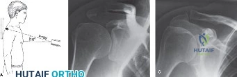
Your ultimate guide to Solving Elbow Cases & Acromioclavicular Arthritis Pain starts here. For elbow cases acromioclavicular pathology, initial diagnosis often involves a Zanca view radiograph, most accurate for AC joint detail. AC joint capsular ligaments predominantly ensure AP stability. When conservative treatments like PT fail for AC joint arthritis, an intra-articular injection is the recommended next step, providing diagnostic and therapeutic benefits.
You are in the ED. A 48-year-old male presents with a painful, deformed elbow following a fall from 3 meters. You are presented with the following radiograph. How do you describe this injury and what is your immediate management priority?
Candidate: The radiograph shows a posterolateral dislocation of the elbow. I can see a comminuted radial head fracture and a coronoid fracture. This is a "Terrible Triad" injury. My priority is to perform a neurovascular examination, provide analgesia, and then seek a CT scan to map the fractures before surgical planning.
Candidates often jump straight to "I will book this for surgery." They fail to mention a dedicated neurovascular exam, the importance of characterizing the coronoid fracture (O'Driscoll classification) for surgical approach planning, or the need for a CT scan to assess fragment size/comminution before committing to a specific implant strategy.
Start with a structured description: "This is a complex elbow dislocation with associated radial head and coronoid fractures, consistent with a Terrible Triad injury." Move to management: "1. Immediate neurovascular assessment and documentation. 2. Post-reduction radiographs and CT with 3D reconstruction to classify the coronoid (O'Driscoll) and radial head (Mason) fractures. 3. Stabilize in an above-elbow backslab. 4. Discuss with a consultant trauma lead, as this necessitates early surgical stabilization to address the LUCL, radial head, and coronoid to restore the elbow's structural integrity."
You have decided to proceed to surgery. You are planning to fix the coronoid fracture. What are the specific indications for a medial approach versus a lateral approach in a Terrible Triad injury, and how does your choice impact your ability to address the rest of the injury?
Candidate: A lateral approach is sufficient for the radial head and LUCL repair. However, if the coronoid fracture is large (O'Driscoll Type III) or involves the sublime tubercle, a medial approach is preferred because it allows direct visualization and stable fixation. A medial approach requires caution regarding the ulnar nerve.
Forgetting to mention the ulnar nerve is a fatal error. Also, failing to explain that the "holy trinity" of fixing the coronoid *first* is what dictates the secondary stability of the reconstruction.
Acknowledge the surgical sequence: Coronoid first, then Radial Head, then LUCL. Medial Approach is indicated for significant coronoid body fractures (O'Driscoll III) or medial facet involvement (Type II) to allow secure placement of suture anchors or a buttress plate. Crucial points: The ulnar nerve must be identified and protected, potentially transposed. This approach provides the best exposure for the anterior coronoid without over-stressing the lateral repair. If using a purely posterolateral approach, coronoid fixation is technically more demanding and often limited to smaller fragments.
During the procedure, you have fixed the coronoid and replaced the radial head. You are now testing stability. The elbow remains unstable in varus/valgus and shows a tendency to subluxate posterolaterally. What are your next steps?
Candidate: I would check the LUCL repair again. If that is stable and the elbow is still unstable, I would evaluate the MCL. If the joint is still not congruent, I would consider a hinged external fixator.
Suggesting a hinged external fixator before confirming the radial head prosthesis hasn't "overstuffed" the joint. Overstuffing is a common iatrogenic cause of stiffness and instability.
1. Exclude overstuffing: Ensure the radial head implant is not too large, which can push the joint into subluxation. 2. Address the LUCL: Ensure the LUCL is repaired to the isometric point on the lateral epicondyle. 3. Check MCL: If medial instability persists (valgus), address the MCL. 4. Last resort: Only after addressing all soft tissues and hardware, if instability remains, a hinged external fixator is used to bridge the joint through a protected range of motion while capsular healing occurs.
