ABOS Part I & OITE Orthopaedic Review: HPII, Hand & Pediatric Fractures | Part 22216
Key Takeaway
This ABOS Part I & AAOS OITE review module provides 22 advanced multiple-choice questions covering critical orthopaedic topics. It focuses on the diagnosis, surgical management, and rehabilitation of high-pressure injection injuries (HPII) to the hand, alongside pediatric femur, tibial tubercle, Tillaux, and triplane ankle fractures, offering essential preparation for board examinations.
ABOS Part I & OITE Orthopaedic Review: HPII, Hand & Pediatric Fractures | Part 22216
Comprehensive 100-Question Exam
00:00
Start Quiz
Question 1
A 45-year-old male industrial worker presents to the emergency department with a high-pressure injection injury to his non-dominant index finger. He states the injury occurred approximately 9 hours ago with paint thinner, but he initially felt minimal pain and only noticed a small puncture wound. Now, he reports increasing swelling and mild throbbing. Based on the case, which factor is most directly correlated with the severity of his injury and the high risk of long-term complications, including DIP joint contracture?
Explanation
Correct Answer: C
Explanation:
The case explicitly states, 'The severity of injury is directly related to the type of injectate, the volume injected, the pressure, the anatomical location, and, critically, the time elapsed between injury and surgical intervention.' It further emphasizes that 'Delayed presentation, often due to the initial innocuous appearance of the injury, is common and significantly correlates with poorer outcomes, including higher rates of infection, necrosis, amputation, and long-term joint stiffness and contracture.' Additionally, 'paint and paint thinner mixtures being among the most destructive due to their inflammatory and necrotic properties.' Therefore, the combination of a highly toxic injectate (paint thinner) and a significant delay to presentation (9 hours, exceeding the critical 6-hour window for optimal outcomes) is the most critical factor influencing the severity and risk of complications in this scenario.
- A. The patient's age and gender: While the case mentions the typical patient is a male industrial worker aged 30-50 years, this is an epidemiological observation, not a direct determinant of injury severity or outcome.
- B. The small initial appearance of the puncture wound: The case highlights that HPIIs 'often presenting with a deceptively small puncture wound,' which leads to delayed presentation. While deceptive, it's the delay it causes, not the wound size itself, that directly correlates with severity.
- D. The anatomical location (non-dominant index finger): The case notes the non-dominant index finger is the most frequently affected digit, making it a common site, but not the primary determinant of severity compared to injectate type and delay.
- E. The pressure at the nozzle (typically 2,000 to 10,000 psi): High pressure is a prerequisite for the injury mechanism, enabling deep penetration. However, the type of substance injected and the time it remains in the tissue are more critical for the severity of the subsequent chemical and inflammatory damage.
Question 2
A 30-year-old painter sustains a high-pressure paint injection injury to the volar aspect of his middle finger, specifically involving the DIP joint region. Despite immediate surgical debridement and aggressive post-operative therapy, he develops a severe, refractory DIP joint flexion contracture. Based on the surgical anatomy and biomechanics described in the case, which structure's involvement is most likely the primary cause of this specific contracture?
Explanation
Correct Answer: D
Explanation:
The case explicitly states under 'Biomechanics of Contracture Development' that 'The most common DIP joint contracture after HPII is flexion contracture, largely due to FDP tethering and volar plate/collateral ligament scarring.' It further details that 'Injected material can track along the tendon sheath, causing chemical tenosynovitis, adhesions, and eventually FDP tendon tethering and severe flexion contracture.' The FDP tendon is the primary flexor of the DIP joint, and its adherence within the fibrous flexor sheath (specifically the A5 pulley at the DIP joint level) is a direct cause of limited extension and fixed flexion.
- A. The oblique retinacular ligament (ORL): The ORL couples DIP and PIP motion. While its scarring can affect DIP motion, it's not described as the primary cause of severe flexion contracture in the same way FDP tethering is.
- B. The terminal extensor tendon: Damage or scarring to the terminal extensor tendon would primarily result in an extension lag or, if severely scarred dorsally, potentially an extension contracture, not a flexion contracture.
- C. The true collateral ligaments of the DIP joint: While collateral ligaments can become infiltrated, scarred, and shortened, contributing to restricted motion, the case specifically highlights FDP tethering as the primary driver for flexion contracture. Collateral ligament shortening primarily restricts the full arc of motion, making it difficult to achieve both full flexion and extension.
- E. The dorsal joint capsule: Scarring or thickening of the dorsal joint capsule would primarily lead to an extension contracture, not a flexion contracture.
Question 3
A 50-year-old male presents to the emergency department with a high-pressure grease injection injury to his thumb, sustained 4 hours prior. He reports mild pain and swelling, but his neurovascular status appears intact, and he has full active range of motion. Based on the indications and contraindications outlined in the case, what is the most appropriate next step in management?
Explanation
Correct Answer: B
Explanation:
The case unequivocally states under 'Indications for Surgical Intervention' that 'All suspected or confirmed high-pressure injection injuries' are a 'universal indication' for 'Primary Surgical Debridement (Urgent/Emergent).' It further emphasizes, 'Even in asymptomatic cases with a clear history of HPII, surgical exploration is indicated due to the deceptive nature of the injury and the high risk of delayed necrosis.' The time elapsed (4 hours) is well within the critical window for urgent intervention (ideally within 6 hours).
- A. Initiate broad-spectrum oral antibiotics and observe for 24 hours, given the mild symptoms: This is incorrect. The case stresses that HPIIs are deceptive and require immediate surgical intervention, not observation, even with mild symptoms. IV antibiotics are indicated, but not as a substitute for surgery.
- C. Obtain an MRI to delineate the extent of soft tissue involvement before deciding on surgery: While MRI can be helpful, the case states it 'should not delay urgent surgery.' The decision for surgery is based on the diagnosis of HPII, not on the extent shown by MRI.
- D. Apply a static splint in a position of safety and refer for outpatient hand therapy: This is incorrect. HPIIs are surgical emergencies. Non-operative management is not indicated for the acute phase.
- E. Administer corticosteroids to reduce inflammation and prevent fibrosis: The case notes that the use of corticosteroids is 'controversial' and 'Current guidelines generally do not recommend routine prophylactic corticosteroids.' They are not the primary or immediate management step.
Question 4
During the initial surgical debridement of a high-pressure injection injury to the ring finger, the surgeon needs to achieve wide exposure of the flexor tendon sheath, neurovascular bundles, and joint capsule while minimizing damage to critical structures and reducing the risk of secondary contractures. Based on the detailed surgical approach described in the case, which incision type is the most appropriate choice for this procedure?
Explanation
Correct Answer: C
Explanation:
The case explicitly states under 'Incisions for Initial Debridement': 'Mid-axial Incisions: The preferred incision for digital HPII. These incisions are made along the non-weight-bearing surfaces of the digits... Advantages: Provide excellent exposure of the neurovascular bundles, flexor tendon sheath, extensor mechanism, and joint capsules. They minimize damage to the volar skin and preserve dorsal lymphatic drainage.'
- A. A dorsal longitudinal incision along the midline of the digit: This would not provide adequate exposure to the volar structures (flexor sheath, neurovascular bundles) which are most commonly involved in HPII.
- B. A fish-mouth (racquet) incision extending from the fingertip to the palm: The case states, 'Fish-mouth (racquet) or Zigzag (Brunner) Incisions: While useful for wide exposure in certain hand injuries, they are generally avoided in acute HPII unless extensive skin necrosis dictates their use, due to the increased risk of skin edge necrosis, scarring, and secondary contractures.'
- D. A zigzag (Brunner) incision across the volar aspect of the digit: Similar to the fish-mouth incision, Brunner incisions are generally avoided in acute HPII due to the risk of skin edge necrosis and secondary contractures, unless extensive skin necrosis is already present.
- E. A transverse incision at the level of the DIP joint: A transverse incision would provide very limited exposure and would not allow for adequate debridement of the entire flexor sheath or tracking of injectate proximally or distally.
Question 5
A 60-year-old male presents with a high-pressure paint injection injury to his dominant index finger. He underwent immediate, aggressive surgical debridement and has been compliant with post-operative rehabilitation. Despite these efforts, he is concerned about potential long-term complications. Based on the 'Complications & Management' section of the case, which specific DIP joint-related complication has the highest reported incidence following significant HPIIs?
Explanation
Correct Answer: D
Explanation:
The 'Complications & Management' table in the case lists 'DIP Joint Contracture' with an incidence of 'Very high, 50-90% for significant injuries.' This is the highest incidence among the options provided for a specific DIP joint-related complication.
- A. Osteomyelitis: Incidence is listed as '<5-10%'.
- B. Complex Regional Pain Syndrome (CRPS): Incidence is listed as '5-20%'.
- C. Tendon rupture: While tendon adhesion is high, rupture is listed as 'less common, but severe.'
- E. Neuropathy / Sensory Loss: Incidence is listed as 'Up to 30%'.
Therefore, DIP joint contracture is the most common specific complication listed for significant HPIIs.
Question 6
A patient is 3 days post-operative following extensive debridement for a high-pressure hydraulic fluid injection injury to the middle finger, involving the DIP joint. The wounds are stable, and there are no signs of active infection. Based on the 'Post-Operative Rehabilitation Protocols' outlined in the case, what is the most crucial principle to initiate during this immediate post-operative phase to prevent DIP joint contracture?
Explanation
Correct Answer: C
Explanation:
Under 'Immediate Post-Operative Phase (Days 0-2 weeks)' and 'Initial Mobilization,' the case states: 'Affected DIP Joint: If wounds permit and there is no active infection or unstable fixation, very gentle, protected active DIP joint flexion and extension may be initiated within the first few days. This is crucial for preventing tendon adhesions and maintaining joint lubrication. The key is active, not passive, to promote tendon gliding.'
- A. Aggressive passive range of motion (PROM) exercises to stretch contracted tissues: The case advises 'Gentle, pain-free PROM may be introduced cautiously' in the intermediate phase, and warns 'Avoid forceful manipulation which can cause re-injury or trigger CRPS.' Aggressive PROM is contraindicated in the immediate phase.
- B. Prolonged static splinting of the DIP joint in full extension to prevent flexion: The case states, 'prolonged immobilization must be avoided.' While static splinting in a position of safety may be used for night wear, it is not the primary method for preventing contracture; early motion is.
- D. Immediate initiation of progressive resistive exercises for strengthening: Strengthening exercises are introduced in the 'Advanced Phase (Weeks 6-beyond),' not immediately post-op.
- E. Serial casting of the digit to achieve maximal correction of any developing contracture: Serial casting is a technique for 'significant, refractory contractures' in the 'Intermediate Phase,' not an immediate post-operative intervention for prevention.
Question 7
A 40-year-old mechanic sustained a high-pressure diesel fuel injection injury to his ring finger 8 months ago. He underwent initial debridement and has since completed an intensive 6-month hand therapy program, including dynamic splinting and scar management. Despite these efforts, he presents with a persistent 45-degree DIP joint flexion contracture that significantly impairs his ability to perform fine motor tasks. Radiographs show no significant joint destruction. Based on the case, what is the most appropriate next step in his management?
Explanation
Correct Answer: C
Explanation:
The case, under 'Secondary Surgical Management (for Established Contracture)' and 'Flexion Contracture,' states: 'If the FDP tendon is adherent, an extensive tenolysis is performed... If the volar plate and joint capsule are scarred and shortened, a volar plate release (often requiring resection of a portion) and anterior capsulotomy are performed to restore extension.' This patient has a significant, refractory flexion contracture (45 degrees) after extensive conservative management, with no joint destruction, making soft tissue release the indicated procedure.
- A. Continue with another 3 months of intensive hand therapy and splinting: The case indicates that surgical intervention for established contracture is considered after 'Failure of intensive non-operative therapy (typically >3-6 months).' This patient has already completed 6 months of intensive therapy, suggesting further conservative management is unlikely to be effective.
- B. Consider DIP joint arthrodesis in a functional position: Arthrodesis is described as a salvage procedure for 'severe, intractable contractures with significant joint destruction or pain.' The patient's radiographs show no significant joint destruction, making soft tissue release a more appropriate initial surgical option to preserve motion.
- D. Initiate a course of oral corticosteroids to reduce residual inflammation: The use of corticosteroids is controversial and generally not recommended for established contractures, especially after the acute inflammatory phase has subsided.
- E. Recommend hyperbaric oxygen therapy to improve tissue remodeling: HBO is considered an adjunct therapy, primarily for acute tissue salvage, and its role in established contracture remodeling is not a primary recommendation.
Question 8
A 28-year-old factory worker presents with a suspected high-pressure injection injury to his ring finger, sustained 2 hours ago. The entry wound is small, and he reports only mild discomfort. To aid in pre-operative planning and identify the extent of injected material and potential bony involvement, which diagnostic imaging modality is considered essential for initial evaluation, according to the case?
Explanation
Correct Answer: D
Explanation:
Under 'Pre-Operative Planning & Patient Positioning' and 'Imaging,' the case states: 'X-rays: Essential to identify injected material (e.g., paint, grease often appear radiopaque), bony involvement (fractures, osteomyelitis), and the presence of gas. Baseline X-rays are crucial for future comparisons.'
- A. Magnetic Resonance Imaging (MRI): While MRI 'Can delineate soft tissue involvement, tendon sheath tracking, and extent of chemical synovitis or necrosis,' the case notes it 'May be considered if diagnosis is uncertain or to guide extent of debridement in complex cases, but should not delay urgent surgery.' It is not listed as 'essential' for initial evaluation in the same way X-rays are.
- B. Computed Tomography (CT) scan: CT is not specifically mentioned as an essential initial imaging modality in the case for HPII.
- C. Ultrasound: Ultrasound is not mentioned in the case for diagnostic workup of HPII.
- E. Arteriogram: An arteriogram is an invasive procedure used to visualize blood vessels and is not part of the routine initial diagnostic workup for HPII unless specific vascular compromise requiring revascularization is suspected, which is rare in the acute phase.
Question 9
A 38-year-old male presents with a high-pressure injection injury to his middle finger. The emergency physician is attempting to ascertain the likely prognosis and potential for severe tissue damage. Based on the 'Introduction & Epidemiology' and 'Summary of Key Literature / Guidelines' sections of the case, which injected substance is consistently associated with the most destructive inflammatory response, highest rates of necrosis, and subsequent amputation?
Explanation
Correct Answer: E
Explanation:
The case explicitly states in the 'Introduction & Epidemiology' section: 'Non-toxic substances generally cause less severe reactions than toxic ones, with paint and paint thinner mixtures being among the most destructive due to their inflammatory and necrotic properties.' This is reinforced in the 'Summary of Key Literature / Guidelines' section: 'Literature consistently demonstrates that paint and paint thinner mixtures are the most damaging, leading to the highest rates of amputation and contracture.'
- A. Water: The case notes 'Water, air, and some solvents cause less severe reactions but still require prompt intervention.'
- B. Air: Similar to water, air causes less severe reactions.
- C. Hydraulic fluid: While hydraulic fluid is a common injectate and causes significant injury, the case specifically highlights paint and paint thinner as the most destructive.
- D. Diesel fuel: Diesel fuel is also a toxic injectate, but the case consistently identifies paint and paint thinner as having the highest destructive potential.
Question 10
A 40-year-old male presents with a high-pressure grease injection injury to his ring finger. He has a history of uncontrolled diabetes, is currently febrile (101.5°F), and has a white blood cell count of 18,000/µL, but is hemodynamically stable. Based on the 'Indications & Contraindications' section of the case, which statement regarding surgical intervention for his acute HPII is most accurate?
Explanation
Correct Answer: C
Explanation:
The case explicitly states under 'Contraindications' for 'Primary HPII Debridement': 'No absolute contraindications. Relative contraindications relate to patient stability.' It further clarifies: 'Patient instability: Severe systemic medical comorbidities precluding anesthesia may necessitate temporizing measures until stabilization, though this is rare given the limb-threatening nature of HPII.' While the patient has comorbidities and signs of infection, he is described as 'hemodynamically stable,' meaning he can tolerate surgery. The limb-threatening nature of HPII mandates immediate intervention.
- A. Surgical debridement should be delayed until his fever and elevated WBC count are normalized with antibiotics: This is incorrect. Delaying debridement for HPII significantly worsens outcomes. While antibiotics should be started immediately, they are an adjunct to, not a replacement for, urgent surgery.
- B. His uncontrolled diabetes is an absolute contraindication to immediate surgical debridement: This is incorrect. The case states there are 'No absolute contraindications' for primary HPII debridement. Diabetes is a comorbidity that increases risk but does not preclude urgent, limb-saving surgery.
- D. An MRI should be performed first to rule out osteomyelitis before proceeding with surgery: This is incorrect. MRI should not delay urgent surgery. While osteomyelitis is a potential complication, the immediate priority is debridement of the injected material and necrotic tissue.
- E. Only local wound care and broad-spectrum antibiotics are necessary, given his systemic instability: This is incorrect. The patient is described as hemodynamically stable, and HPII is a surgical emergency requiring debridement, not just conservative management.
Question 11
Following surgical debridement for a high-pressure injection injury to the DIP joint, a patient is in the intermediate phase of rehabilitation (Week 4). The hand therapist notes a persistent flexion contracture of 25 degrees at the DIP joint. Based on the 'Post-Operative Rehabilitation Protocols' and 'Key Principles for DIP Joint Contracture Prevention' in the case, which intervention is most appropriate to address this developing contracture?
Explanation
Correct Answer: C
Explanation:
Under 'Intermediate Phase (Weeks 2-6)' and 'Progressive Splinting,' the case states: 'Dynamic Splinting: Continue dynamic extension splinting for flexion contractures, with adjustable outriggers to apply gentle, constant stretch.' It also mentions 'Static Progressive Splinting: Utilize splints that allow for gradual, incremental adjustments to increase the stretch on the contracted structures.' These methods apply 'Sustained Low-Load Stretch,' which is a key principle for contracture prevention and management.
- A. Initiate forceful passive manipulation of the DIP joint to break adhesions: The case explicitly warns against this: 'Avoid forceful manipulation which can cause re-injury or trigger CRPS.'
- B. Discontinue all splinting to encourage natural movement: This is incorrect. Splinting, particularly dynamic or static progressive splinting, is crucial in the intermediate phase to address developing contractures.
- D. Refer for immediate surgical tenolysis and capsulotomy: Surgical intervention for established contracture is typically considered after 'Failure of intensive non-operative therapy (typically >3-6 months).' At Week 4, non-operative measures like splinting are the primary approach.
- E. Prescribe a period of complete immobilization to allow tissues to heal fully: The case emphasizes 'Early Mobilization' as the 'single most important factor' and states that 'Prolonged immobilization is cited as a major contributor to stiffness and contracture development.'
Question 12
A 7-year-old girl is transferred to the ER after suffering a right thigh injury playing tackle football. She has notable deformity and significant pain. Initial radiographs are shown below.
Which of the following is the most appropriate initial surgical treatment for this patient, considering her age and weight (35 kg)?
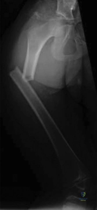
Explanation
Correct Answer: Flexible intramedullary nails
The patient is a 7-year-old girl weighing 35 kg with a diaphyseal femur fracture. According to current pediatric orthopedic guidelines, flexible intramedullary nails (FINs) are the optimal choice for femur fractures in children aged 6-8 years, and also for children 8 years to adolescent who are under 50 kg, especially for length-stable fractures. FINs offer the advantages of early mobilization and improved patient and family convenience compared to spica casting.
- Reduction and immediate spica cast: While spica casting is an option for this age group, flexible nailing is generally preferred due to better patient and family outcomes, allowing for earlier weight-bearing and easier care.
- Lateral trochanteric intramedullary nail fixation: This method is typically reserved for older children (at least 9 years of age or older) who weigh over 49 kg, or those with length-unstable fractures. The patient's age and weight do not meet these criteria, and there are risks of greater trochanteric apophyseal arrest and osteonecrosis of the femoral head with this approach.
- Traction followed by delayed spica casting: Traction is often used as a temporary measure or for very young children, but for a 7-year-old, definitive fixation with flexible nails is generally preferred over prolonged traction and casting.
- External fixation: External fixation is a viable option for pediatric femur fractures but is usually reserved for specific situations such as severely comminuted or open fractures, or in the context of damage control orthopaedics, none of which are indicated in this case.
Question 13
Following the decision to proceed with flexible intramedullary nailing for the 7-year-old girl's femur fracture, you measure the preoperative x-ray at the isthmus, finding it to be 7.5 mm. The goal for flexible intramedullary nail fixation is to achieve approximately 80% canal fill. Given this information, which combination of nails would be most appropriate?
Explanation
Correct Answer: Two 3-mm nails, 80% canal fill
The goal for flexible intramedullary nail fixation of a pediatric femur fracture is to achieve approximately 80% canal fill at the narrowest point (isthmus) to provide adequate stability without overstuffing the canal. The patient's isthmus measures 7.5 mm.
- Two 2.5-mm nails: The combined diameter would be 5.0 mm. (5.0 / 7.5) * 100% = 66.7% canal fill. This is less than the ideal 80% and could lead to inadequate fixation and risk of failure.
- Two 3-mm nails: The combined diameter would be 6.0 mm. (6.0 / 7.5) * 100% = 80% canal fill. This perfectly meets the target canal fill and provides optimal stability.
- One 3.0-mm nail and one 3.5-mm nail: The combined diameter would be 6.5 mm. (6.5 / 7.5) * 100% = 86.7% canal fill. While close to 80%, using two nails of different sizes can contribute to loss of reduction and malalignment. Additionally, exceeding 80% fill can increase the risk of complications.
- Two 3.5-mm nails: The combined diameter would be 7.0 mm. (7.0 / 7.5) * 100% = 93.3% canal fill. This significantly exceeds the 80% target, increasing the risk of iatrogenic fracture, malalignment, and other complications due to overstuffing the canal.
- One 2.5-mm nail and one 3.0-mm nail: The combined diameter would be 5.5 mm. (5.5 / 7.5) * 100% = 73.3% canal fill. This is below the ideal 80% and also involves nails of different sizes, which is generally less desirable.
Question 14
When discussing the potential complications of flexible intramedullary nailing for a pediatric femur fracture with the parents, which of the following is the most common complication you should inform them about?
Explanation
Correct Answer: Pain at the knee (insertion sites)
Flexible intramedullary nailing is a generally safe and effective procedure for pediatric femur fractures. While all surgical procedures carry risks, studies have shown relatively low rates of serious complications such as infection, significant bleeding, and loss of reduction (especially with length-stable fractures). The most common complication associated with flexible intramedullary nails is irritation of the soft tissues at the nail insertion sites, typically around the knee. This irritation can cause pain and discomfort, often necessitating hardware removal once the fracture has healed. To mitigate this, it is suggested that the nail ends be left no more than 25 mm out of the bone.
- Infection: While a risk with any surgery, infection rates for flexible nailing are low.
- Bleeding: Significant bleeding requiring transfusion is uncommon with this procedure.
- Loss of reduction: With proper nail sizing and technique for length-stable fractures, loss of reduction is infrequent.
- Nonunion: Nonunion is a rare complication in pediatric femur fractures, especially with appropriate fixation.
Question 15
When discussing the general complications of femur fractures in children with the family preoperatively, what information should you provide regarding the amount of overgrowth that may occur in a 7-year-old?
Explanation
Correct Answer: Ipsilateral overgrowth does occur, usually around 9 mm in 2 to 10-year-olds.
Femur fractures in children, particularly in the 2 to 10-year age range, are known to stimulate growth, leading to ipsilateral limb overgrowth. This phenomenon is thought to be related to hyperemia following the injury. While the range of overgrowth can vary (approximately 4 to 25 mm), the average amount of overgrowth observed in this age group is around 9 mm.
- There is no risk of overgrowth at this age; overgrowth only happens in children under 2: This is incorrect. Children under 2 years and those over 10 years are less likely to experience significant overgrowth compared to the 2-10 year age group.
- Ipsilateral overgrowth does occur, with an average of less than 5 mm: This underestimates the typical average overgrowth in this age group.
- Ipsilateral overgrowth does occur, usually between 15 and 20 mm in 2 to 10-year-olds: This overestimates the typical average overgrowth, although overgrowth within this range can occur in some cases.
- Overgrowth is a risk in children over 10, not in those younger than 10: This is incorrect. Children over 10 are less prone to significant overgrowth, while the 2-10 year age group is most susceptible.
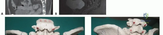
Question 16
A 14-year-old boy presents to the ER with acute right knee pain after a basketball injury. Radiographs and a CT scan are performed, with the CT scan shown below.
What condition is thought to be a significant risk factor for this type of fracture?
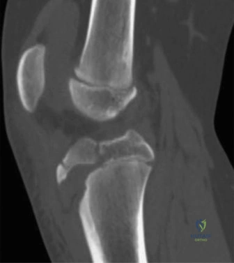
Explanation
Correct Answer: Osgood–Schlatter disease
Tibial tubercle fractures, as depicted in the image, occur more commonly in adolescents who have a history of Osgood–Schlatter disease. Osgood–Schlatter disease is an overuse injury characterized by repetitive strain across the tibial tubercle apophysis, leading to inflammation and microtrauma. While a direct causal relationship is not always demonstrated, the weakened apophysis in Osgood–Schlatter patients makes them more susceptible to avulsion fractures of the tibial tubercle.
- Patellofemoral syndrome: This condition involves anterior knee pain associated with overuse but is not specifically linked to an increased risk of tibial tubercle fractures.
- Sinding-Larsen–Johansson syndrome: Similar to Osgood–Schlatter, this is an overuse injury, but it affects the inferior pole of the patella, not the tibial tubercle, and therefore is not associated with tibial tubercle fractures.
- Patellar tendonitis: This is inflammation of the patellar tendon, which can cause pain but is not a known risk factor for avulsion fractures of the tibial tubercle itself.
- Chondromalacia patellae: This refers to softening and breakdown of the cartilage on the underside of the patella and is not a risk factor for tibial tubercle fractures.
Question 17
A 14-year-old boy sustains a tibial tubercle fracture. You are concerned about the risk of compartment syndrome. Which compartment and associated vessel are most commonly at risk with this injury?
Explanation
Correct Answer: Anterior compartment—recurrent anterior tibial artery
Tibial tubercle fractures, particularly displaced ones, carry a significant risk of developing compartment syndrome. Anatomical studies and clinical experience have shown that the anterior compartment is most commonly affected. The recurrent anterior tibial artery, which courses near the tibial tubercle, is particularly vulnerable to injury or compression in the setting of this fracture, contributing to the risk of anterior compartment syndrome. Therefore, close monitoring of the neurovascular status of the anterior compartment is crucial in the perioperative period, and many surgeons will perform a prophylactic anterior compartment fasciotomy during surgery.
- Anterior compartment—medial inferior geniculate artery: The medial inferior geniculate artery supplies the knee joint and surrounding structures but is not the primary vessel at risk for compartment syndrome in the anterior compartment with this specific fracture.
- Anterior compartment—anterior tibial artery: While the anterior tibial artery is the main artery of the anterior compartment, the recurrent anterior tibial artery is more directly implicated in the immediate vicinity of the tibial tubercle fracture.
- Lateral compartment—recurrent anterior tibial artery: The recurrent anterior tibial artery is associated with the anterior compartment, not the lateral compartment.
- Lateral compartment—fibular artery: The fibular artery (also known as the peroneal artery) supplies the lateral and posterior compartments but is not typically at risk with an isolated tibial tubercle fracture.
Question 18
A 13-year-old male presents with acute right ankle pain after a soccer match. Radiographs, including the AP view shown below, reveal a transitional ankle fracture.
Which of the following answers correctly pairs the eponym commonly used to describe this injury with the affected anatomic structure?

Explanation
Correct Answer: Tillaux fracture; AITFL (anterior inferior tibiofibular ligament)
The radiograph displays a Tillaux fracture, which is a specific type of transitional ankle fracture seen in adolescents. It involves an avulsion fracture of the anterolateral distal tibial epiphysis. The anterior inferior tibiofibular ligament (AITFL), a key syndesmotic ligament, inserts onto this fragment. The injury typically occurs due to external rotation forces on the foot, causing the AITFL to avulse a piece of the epiphysis.
- Tillaux fracture; ATFL (anterior talofibular ligament): The ATFL is the most commonly injured ligament in lateral ankle sprains but is not directly involved in a Tillaux fracture.
- Chopart fracture; ATFL (anterior talofibular ligament): Chopart injuries involve the midtarsal joint (talonavicular and calcaneocuboid joints) and are distinct from distal tibial physeal fractures.
- Chopart fracture; AITFL (anterior inferior tibiofibular ligament): This option incorrectly combines the Chopart injury with the AITFL.
- Chaput fracture; ATFL (anterior talofibular ligament): A Chaput fracture is another eponym for an avulsion fracture of the anterior inferior tibial tubercle, which is essentially the same anatomical injury as a Tillaux fracture but is more commonly used in adult ankle fracture classifications. However, it is associated with the AITFL, not the ATFL.
Question 19
A 13-year-old male presents with a left ankle injury sustained during a soccer match. Radiographs, including the AP and lateral views shown below, are obtained.
Based on the available imaging, which of the following best describes the classic radiographic appearance of this injury?
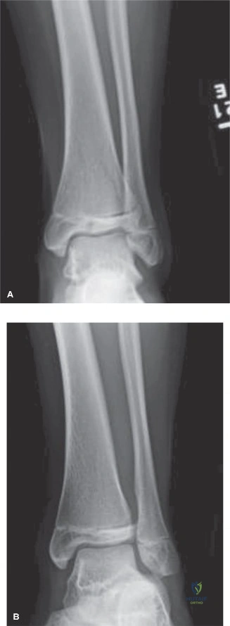
Explanation
Correct Answer: Triplane fracture; Salter–Harris II on sagittal view; Salter–Harris III on anteroposterior (AP) view
The images depict a triplane fracture of the distal tibia, a complex transitional ankle fracture occurring during physeal closure. The characteristic radiographic appearance of a triplane fracture is a Salter–Harris II fracture pattern on the lateral view (sagittal plane) and a Salter–Harris III fracture pattern on the anteroposterior (AP) or coronal view. This is because the fracture involves the epiphysis, physis, and metaphysis in multiple planes.
- Salter–Harris III on sagittal view; Salter–Harris II on anteroposterior (AP) view: This reverses the classic appearance.
- Salter–Harris III on sagittal view; Salter–Harris III on anteroposterior (AP) view: This would imply a Salter-Harris III in both planes, which is not the classic description of a triplane fracture.
- Tillaux fracture: A Tillaux fracture is a Salter-Harris III fracture of the anterolateral distal tibial epiphysis only, without a metaphyseal component, and is typically seen as a Salter-Harris III on AP/coronal views. It does not have the Salter-Harris II component on the lateral view.
Question 20
A 13-year-old male is diagnosed with a triplane ankle fracture. You decide to perform a closed reduction and long-leg splint application. Which of the following best describes the primary reduction maneuver for this injury?
Explanation
Correct Answer: Traction, internal rotation of the foot, and dorsiflexion of the ankle
The classic reduction maneuver for triplane ankle fractures involves sustained axial traction to disimpact the fragments, followed by maximal internal rotation and supination of the foot, combined with dorsiflexion of the ankle. This maneuver aims to reverse the typical external rotation and pronation mechanism of injury and to achieve anatomical reduction of the articular surface.
- Traction, internal rotation of the foot, and plantarflexion of the ankle: Plantarflexion would not help reduce the fracture and may even exacerbate displacement.
- Traction, external rotation of the foot, and dorsiflexion of the ankle: External rotation is typically the mechanism of injury, so applying external rotation would worsen the displacement.
- Traction, external rotation of the foot, and plantarflexion of the ankle: Both external rotation and plantarflexion are incorrect for reduction.
- Traction, abduction of the foot, and plantarflexion of the ankle: Abduction and plantarflexion are not the correct components of the reduction maneuver for this fracture.
Question 21
After attempting a closed reduction for a triplane ankle fracture in a 13-year-old male, you recognize the importance of re-evaluating the fracture alignment. Which of the following is the most appropriate next step in evaluating the reduction quality, especially concerning the articular surface?
Explanation
Correct Answer: Computed tomography scan (CT) without contrast of the left ankle
The primary goal after reducing a triplane ankle fracture is to assess the anatomical alignment of the articular surface and the physeal gap. While MRI is excellent for soft tissue evaluation (ligaments, tendons, cartilage), CT scanning is considered the gold standard for evaluating bony injury, fracture alignment, and articular congruity, especially in complex fractures like triplane fractures where displacement can be subtle on plain radiographs. A non-contrast CT scan provides sufficient detail for this purpose, and contrast enhancement would not add significant useful information for assessing bony alignment.
- Magnetic resonance imaging (MRI) without contrast: While it can show bony detail, CT is superior for precise assessment of fracture fragments and articular step-off. MRI is more for soft tissue.
- Magnetic resonance imaging (MRI) with contrast: Contrast is typically used for evaluating vascularity, tumors, or infection, not primarily for post-reduction bony alignment.
- Delayed gadolinium-enhanced MRI of cartilage (dGEMRIC): This specialized MRI technique is used to evaluate articular cartilage health and injury, but it is not the immediate post-reduction imaging of choice for assessing fracture alignment.
- Computed tomography scan (CT) with contrast: Contrast is not necessary for evaluating bony alignment and articular congruity in this setting.
Question 22
Nine months after his initial presentation and treatment for a triplane ankle fracture, a 13-year-old male returns for follow-up. You inform him about the most likely long-term sequela of his injury. Which of the following is the most probable outcome at this point?
Explanation
Correct Answer: Posttraumatic physeal closure of the distal tibia that does not require additional treatment
Transitional ankle fractures, such as Tillaux and triplane fractures, occur during the period of physiological physeal closure. By the time these injuries occur, a significant portion of the growth plate has already fused. Therefore, while some degree of posttraumatic physeal closure may occur, it is typically not clinically significant and does not lead to substantial limb length discrepancy or angular deformity requiring corrective osteotomy. The remaining growth potential is usually minimal.
- Posttraumatic physeal closure of the distal tibia that requires corrective valgus/varus/extension osteotomy: These are unlikely because significant growth arrest leading to angular deformity or limb length discrepancy is rare with transitional fractures due to the timing of the injury during natural physeal closure.
- Nonunion of the Thurston–Holland fragment: The Thurston–Holland fragment is associated with Salter-Harris II fractures (a component of triplane fractures). While nonunion is a theoretical complication of any fracture, it is exceedingly rare in pediatric physeal fractures, especially with appropriate treatment, and has not been reported as a common sequela for triplane fractures.
Question 23
A 28-year-old mechanic sustains a high-pressure injection injury to his long finger. Which of the following injected substances is associated with the highest rate of eventual amputation?
Explanation
Question 24
A 35-year-old worker sustains a high-pressure injection injury to the volar aspect of his left thumb. The injected fluid travels proximally. Into which deep space is the fluid most likely to directly track?
Explanation
Question 25
A 6-year-old boy crushes his finger in a door, presenting with a clinically flexed distal phalanx, a laceration at the nail fold, and the proximal nail plate resting superficial to the eponychium. Radiographs show a widening of the distal phalanx physis. What is the most appropriate management?
Explanation
Question 26
A 5-year-old girl falls onto an outstretched hand and sustains a minimally displaced (<2 mm) lateral condyle fracture of the humerus. Which of the following represents the most significant long-term complication if this fracture goes on to nonunion?
Explanation
Question 27
A 6-year-old boy sustains an extension-type Gartland III supracondylar humerus fracture. Post-reduction, he is unable to flex the interphalangeal joint of his thumb and the distal interphalangeal joint of his index finger. Which nerve is most likely injured?
Explanation
Question 28
During surgical exploration of a high-pressure paint injection injury to the index finger, what is the recommended approach to minimize the risk of secondary tissue ischemia and optimize clearance of the injected material?
Explanation
Question 29
A 22-year-old male sustains a fracture-dislocation of the thumb carpometacarpal joint. Radiographs show a small volar-ulnar base fragment of the first metacarpal remaining articulated with the trapezium. Which muscle is primarily responsible for the proximal and dorsal displacement of the main metacarpal shaft?
Explanation
Question 30
Which of the following is considered an acceptable radiographic parameter for nonoperative management of a diaphyseal both-bone forearm fracture in an 8-year-old child?
Explanation
Question 31
A 6-month-old infant is brought to the emergency department with swelling and decreased spontaneous movement of the right leg. Radiographs reveal a spiral fracture of the midshaft femur. What is the most critical next step in management?
Explanation
Question 32
A 25-year-old sustains a transverse fracture of the proximal phalanx shaft of the long finger. What is the characteristic deformity of this fracture, and which structures are responsible?
Explanation
Question 33
A worker inadvertently triggers a high-pressure grease gun into his palm. Despite minimal initial symptoms, the surgeon insists on urgent operative debridement. If neglected, what is a potential severe systemic consequence specific to certain industrial solvent injections, aside from local tissue necrosis?
Explanation
Question 34
A 12-year-old gymnast falls and sustains an acute posterior elbow dislocation. After closed reduction, a post-reduction radiograph shows the medial epicondyle is missing from its anatomic location and is incarcerated within the joint space. What is the absolute indication for operative intervention in this scenario?
Explanation
Question 35
A 5-year-old boy presents with a displaced Gartland III supracondylar humerus fracture. The hand is pink but the radial pulse is absent. After closed reduction and percutaneous pinning, the hand remains pink and well-perfused with a capillary refill of 2 seconds, but the radial pulse remains unpalpable. What is the most appropriate next step?
Explanation
Question 36
A 4-year-old child sustains a phalangeal neck fracture of the index finger with 90 degrees of dorsal angulation. Attempted closed reduction is unsuccessful due to soft tissue interposition. Which structure is most commonly interposed, blocking reduction in this specific pediatric fracture?
Explanation
Question 37
In cases of high-pressure injection injuries to the hand, a delay in definitive surgical debridement beyond what timeframe is most strongly associated with a significantly increased risk of amputation?
Explanation
Question 38
Which of the following injected materials is associated with the highest risk of eventual amputation in high-pressure injection injuries (HPII) to the hand?
Explanation
Question 39
A patient sustains a high-pressure grease injection to the volar aspect of the index finger. If the injected material ruptures proximally through the flexor tendon sheath, which deep palmar space is most immediately at risk for direct involvement?
Explanation
Question 40
A 6-year-old child falls on an outstretched hand and sustains an extension-type supracondylar humerus fracture. Radiographs demonstrate posteromedial displacement of the distal fragment. Which nerve is most at risk of injury in this specific displacement pattern?
Explanation
Question 41
A 5-year-old boy is diagnosed with a displaced lateral condyle fracture of the humerus. If this fracture is managed non-operatively and goes on to nonunion, what is the classic long-term complication?
Explanation
Question 42
A 25-year-old sustains a transverse fracture of the proximal phalanx shaft, which presents with apex-volar angulation. Which distinct deforming forces are responsible for this classic fracture pattern?
Explanation
Question 43
During closed reduction and pinning of a Bennett fracture, the surgeon must overcome specific deforming forces. Which anatomic structure maintains the small anteromedial articular fragment in its anatomic position?
Explanation
Question 44
A 13-year-old boy presents with a Salter-Harris III fracture of the anterolateral aspect of the distal tibial epiphysis (Tillaux fracture).
Which ligamentous structure is responsible for avulsing this fragment?
Explanation
Question 45
A 35-year-old mechanic presents 2 hours after accidentally discharging a high-pressure grease gun into his non-dominant index finger. The entry wound is only 2 mm with mild swelling, and capillary refill is intact. What is the most appropriate management?
Explanation
Question 46
A 10-year-old boy sustains a traumatic elbow dislocation. Following closed reduction, radiographs reveal an associated medial epicondyle fracture. Which of the following is an absolute indication for operative fixation of the medial epicondyle?
Explanation
Question 47
A 22-year-old man punches a wall and sustains a 5th metacarpal neck fracture (Boxer's fracture). To prevent clinically significant pseudoclawing and functional deficit, what is the generally accepted maximum volar angulation for this specific digit?
Explanation
Question 48
An 8-year-old boy sustains a midshaft both-bone forearm fracture. Non-operative management with cast immobilization is planned. What is the maximum acceptable angulation for this fracture in this age group to still allow for adequate spontaneous remodeling?
Explanation
Question 49
A 5-year-old presents with a severely displaced supracondylar humerus fracture. The radial pulse is absent, but the hand is warm, pink, and has a brisk capillary refill. Following closed reduction and percutaneous pinning, the hand remains 'pink and pulseless.' What is the next best step in management?
Explanation
Question 50
A 28-year-old man with chronic wrist pain is diagnosed with Scaphoid Nonunion Advanced Collapse (SNAC). Which of the following joints is characteristically spared from degenerative arthritic changes in a SNAC wrist, allowing for a proximal row carpectomy or four-corner fusion?
Explanation
Question 51
Which of the following factors independently dictates a 100% amputation rate in high-pressure injection injuries of the hand?
Explanation
Question 52
A 24-year-old mechanic sustains a high-pressure injection injury to the volar aspect of his right index finger. Which of the following injected materials carries the HIGHEST risk of eventual amputation?
Explanation
Question 53
A 10-year-old boy presents with a Seymour fracture of his right middle finger. Which of the following is the most appropriate definitive management?
Explanation
Question 54
In proximal phalanx shaft fractures of the hand, what is the typical apex deformity, and which intrinsic muscles are primarily responsible?
Explanation
Question 55
A 32-year-old male sustains a high-pressure injection injury to his palm. He presents to the ED 2 hours post-injury. Which of the following interventions is CONTRAINDICATED in the emergency department?
Explanation
Question 56
A 6-year-old boy falls on an outstretched hand and sustains an extension-type supracondylar humerus fracture. Upon examination, he cannot actively flex the interphalangeal joint of his thumb. Which nerve is most likely injured?
Explanation
Question 57
A 25-year-old professional boxer sustains a displaced fracture of the 2nd metacarpal neck. What is the maximum acceptable volar angulation for this digit to maintain optimal functional outcome?
Explanation
Question 58
Which of the following factors is considered the most significant prognostic indicator for the likelihood of eventual amputation following a high-pressure injection injury to the hand?
Explanation
Question 59
A 9-year-old girl is evaluated for a lateral condyle fracture of the humerus. Which of the following accurately describes a Milch Type II fracture?
Explanation
Question 60
A 30-year-old male sustains a Bennett fracture. Which muscle is primarily responsible for the proximal and dorsal displacement of the metacarpal shaft?
Explanation
Question 61
A 7-year-old child presents with a pink, pulseless hand following a closed reduction and percutaneous pinning of a displaced supracondylar humerus fracture. Capillary refill is brisk and the child is comfortable. What is the next most appropriate step in management?
Explanation
Question 62
Which injected material in a high-pressure injection injury to the finger carries the highest risk of subsequent amputation?
Explanation
Question 63
A 32-year-old sustains a high-pressure injection injury of the index finger volar pulp. What is the recommended surgical incision to adequately explore the flexor tendon sheath while minimizing complications?
Explanation
Question 64
An 8-year-old boy presents with a crush injury to his distal middle finger. Radiographs demonstrate a displaced Salter-Harris I fracture of the distal phalanx. The proximal nail plate sits outside the eponychial fold. What is the most appropriate management?
Explanation
Question 65
A 5-year-old sustains a displaced phalangeal neck fracture of the proximal phalanx. Which complication is most frequently associated with failure to anatomically reduce this fracture pattern?
Explanation
Question 66
A 25-year-old boxer sustains a closed, isolated, spiral fracture of the fifth metacarpal shaft. Which clinical finding is an absolute indication for operative intervention?
Explanation
Question 67
A 28-year-old mechanic sustains a high-pressure injection injury to his palm with diesel fuel. He presents to the ED 2 hours post-injury. Which factor is considered the most critical determinant of eventual amputation risk in this injury?
Explanation
Question 68
A 34-year-old basketball player sustains a dorsal fracture-dislocation of the PIP joint of his middle finger. Radiographs reveal a volar lip fragment involving 45% of the articular surface. Which surgical treatment is most appropriate?
Explanation
Question 69
A 6-year-old boy presents with a completely displaced extension-type supracondylar humerus fracture.
On examination, he is unable to actively flex the interphalangeal joint of his thumb. Which nerve is most likely injured?
Explanation
Question 70
A 5-year-old girl falls on an outstretched hand and sustains a Milch Type II lateral condyle fracture of the humerus with 3 mm of displacement. What is the standard management for this injury?
Explanation
None
