ABOS Part I Orthopaedic Review: Calcaneal Fractures & Spinal Osteoid Osteoma | Part 22206
Key Takeaway
This ABOS Part I Orthopaedic Review module offers 21 advanced MCQs on critical orthopaedic topics. It comprehensively covers displaced intra-articular calcaneal fractures, including diagnosis, surgical management, and complications. Additionally, it delves into spinal osteoid osteoma, focusing on its clinical presentation, diagnostic imaging, and treatment modalities like RFA and surgical excision.
ABOS Part I Orthopaedic Review: Calcaneal Fractures & Spinal Osteoid Osteoma | Part 22206
Comprehensive 100-Question Exam
00:00
Start Quiz
Question 1
A 32-year-old male presents to the emergency department after falling from a ladder, landing on his heels. Radiographs confirm a displaced intra-articular calcaneal fracture. Given the high-energy mechanism and the epidemiology of calcaneal fractures, which of the following associated injuries should the orthopedic surgeon prioritize screening for during the initial comprehensive evaluation?
Explanation
Correct Answer: C
Explanation:
The case explicitly states that calcaneal fractures are predominantly high-energy injuries, often resulting from axial loading mechanisms such as falls from height. It further highlights that the incidence of associated injuries is considerable, with reports of concurrent spine fractures (thoracolumbar region) in 10-15% of cases. This makes screening for thoracolumbar spine fractures a critical component of the initial comprehensive evaluation for a patient presenting with a calcaneal fracture from a fall from height.
- A. Distal radius fracture: While falls can cause distal radius fractures, the specific mechanism of landing on the heels from a height primarily transmits axial load through the lower extremities and spine, making a thoracolumbar spine fracture a more commonly associated injury with calcaneal fractures.
- B. Cervical spine fracture: While any high-energy trauma warrants a general spine assessment, the case specifically identifies the thoracolumbar region as the most common site for associated spine fractures with calcaneal injuries due to the axial loading mechanism.
- D. Patella fracture: Patella fractures are typically associated with direct trauma to the knee or forceful quadriceps contraction, not a primary axial load through the heel.
- E. Humeral shaft fracture: Humeral shaft fractures are generally not associated with calcaneal fractures from an axial loading mechanism to the heels.
Question 2
A 48-year-old male sustains a calcaneal fracture after a motor vehicle collision. Initial lateral radiographs of the foot reveal a Böhler's angle of 10 degrees and a Gissane's angle of 160 degrees. Based on these findings and the provided case information, what do these angular measurements primarily indicate?
Explanation
Correct Answer: C
Explanation:
The case describes Böhler's Angle (Tuber Joint Angle) as normally ranging from 20-40 degrees, and a decrease signifies loss of calcaneal height and subtalar joint depression. The Critical Angle of Gissane normally ranges from 120-145 degrees, and a decrease indicates depression of the lateral portion of the posterior facet. In this patient, a Böhler's angle of 10 degrees (significantly decreased from the normal 20-40 degrees) directly indicates a significant loss of calcaneal height and subtalar joint depression. A Gissane's angle of 160 degrees (increased from the normal 120-145 degrees) also suggests a disruption and likely depression of the posterior facet, as the angle typically widens when the lateral portion of the posterior facet is depressed and the lines diverge more acutely.
- A. Restoration of calcaneal height and subtalar joint congruity: This is incorrect. The measured angles are outside the normal range, indicating pathology, not restoration or congruity.
- B. Normal calcaneal morphology and stable posterior facet: This is incorrect. Both angles are abnormal, indicating significant disruption.
- D. Isolated anterior process fracture without articular involvement: While anterior process fractures exist, the changes in Böhler's and Gissane's angles are specific to posterior facet depression and loss of calcaneal height, indicating intra-articular involvement.
- E. Medial wall blowout fracture with tarsal tunnel impingement: While possible in calcaneal fractures, these angles do not directly assess medial wall integrity or tarsal tunnel impingement. They are primarily indicators of posterior facet depression and calcaneal height.
Question 3
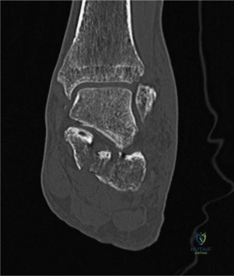
During surgical planning for a displaced intra-articular calcaneal fracture, the surgeon reviews the patient's CT scans. The axial CT image below demonstrates significant widening and displacement of the posterior facet. This finding is most consistent with which Sanders classification type?
Explanation
Correct Answer: D
Explanation:
The provided image and description indicate 'significant intra-articular calcaneal fracture with widening and displacement of the posterior facet.' The Sanders Classification categorizes intra-articular fractures based on the number and location of primary fracture lines through the posterior facet. Sanders Type IV is defined as a highly comminuted fracture of the posterior facet. Significant widening and displacement, especially with multiple fragments implied by 'highly comminuted,' are characteristic of a Type IV fracture. While the image itself doesn't explicitly show all fracture lines, the description of 'significant intra-articular fracture with widening and displacement' points towards a severe, comminuted pattern.
- A. Sanders Type I: This refers to non-displaced intra-articular fractures, which is inconsistent with 'significant widening and displacement.'
- B. Sanders Type II: This describes two-part fractures, which are less severe than 'significant widening and displacement' typically implies.
- C. Sanders Type III: This describes three-part fractures, also less severe than the highly comminuted nature suggested by 'significant widening and displacement' in the context of the most severe Sanders type.
- E. Extra-articular fracture: The description explicitly states 'intra-articular calcaneal fracture,' ruling out an extra-articular classification.
Question 4
A 28-year-old male presents with a displaced intra-articular calcaneal fracture (Sanders Type III) after a fall from a roof. He is a heavy smoker and has poorly controlled diabetes mellitus. The soft tissue envelope is severely compromised with extensive blistering and early signs of skin necrosis. Which of the following is the most appropriate initial management strategy?
Explanation
Correct Answer: E
Explanation:
The case lists 'critically compromised soft tissue envelope (e.g., extensive blistering, skin necrosis, severe open fractures with gross contamination)' as an absolute contraindication to definitive open reduction internal fixation. In such situations, the priority is soft tissue management and temporary stabilization. External fixation provides temporary stability, helps maintain length, and allows for soft tissue recovery, which is crucial before any definitive internal fixation can be considered. The patient's heavy smoking and poorly controlled diabetes are also relative contraindications that further complicate wound healing and increase the risk of complications with immediate ORIF.
- A. Immediate open reduction internal fixation (ORIF) via extensile lateral approach: This is contraindicated due to the severely compromised soft tissue envelope, which would lead to unacceptably high rates of wound complications and infection.
- B. Delayed ORIF after 7-14 days, once soft tissue swelling subsides: While delayed surgery is generally preferred, the presence of 'extensive blistering and early signs of skin necrosis' indicates a more severe soft tissue injury that requires immediate attention beyond just waiting for swelling to subside. Definitive ORIF is still contraindicated until the soft tissue is healthy.
- C. Non-operative management with cast immobilization and strict non-weight bearing: While non-operative management is an option for certain fractures or patients with severe comorbidities, a displaced intra-articular Sanders Type III fracture typically benefits from operative intervention if the soft tissues allow. However, the immediate priority is soft tissue management and temporary stabilization, not definitive non-operative care, especially if surgical reconstruction is still a goal.
- D. Urgent fasciotomy due to high risk of compartment syndrome: While compartment syndrome is a potential complication, the scenario describes 'extensive blistering and early signs of skin necrosis,' which are signs of severe soft tissue injury, not necessarily compartment syndrome. Fasciotomy is indicated for confirmed compartment syndrome, not as a primary response to skin necrosis.
Question 5
A 55-year-old male is undergoing open reduction internal fixation (ORIF) of a displaced intra-articular calcaneal fracture via an extensile lateral approach. During the approach, the surgeon must be particularly vigilant to protect which neurovascular structure that courses along the lateral aspect of the hindfoot and is highly vulnerable?
Explanation
Correct Answer: C
Explanation:
The case explicitly states under 'Surgical Anatomy & Biomechanics' that 'The sural nerve courses along the lateral aspect of the hindfoot and is particularly vulnerable during lateral surgical approaches.' Furthermore, under the 'Extensile Lateral Approach (ELA)' section, it emphasizes that 'Protection of the sural nerve, which lies subcutaneously, is paramount' during full-thickness flap elevation.
- A. Posterior tibial nerve: This nerve is part of the tarsal tunnel contents and lies on the medial aspect of the ankle, not the lateral, making it vulnerable during medial approaches or medial wall blowout fractures, but not typically the extensile lateral approach.
- B. Deep peroneal nerve: This nerve is located in the anterior compartment of the leg and innervates the dorsum of the foot. It is not typically at high risk during a lateral calcaneal approach.
- D. Superficial peroneal nerve: This nerve is also in the lateral compartment of the leg and innervates the lateral aspect of the lower leg and dorsum of the foot. While it can be at risk in more anterior lateral approaches to the ankle, the sural nerve is the primary concern for the extensile lateral approach to the calcaneus.
- E. Medial plantar nerve: This nerve is a terminal branch of the posterior tibial nerve, located on the plantar aspect of the foot, and is not at direct risk during a lateral calcaneal approach.
Question 6
During the reduction sequence of a displaced intra-articular calcaneal fracture via an extensile lateral approach, the surgeon aims to restore the posterior facet congruity. Which of the following calcaneal fragments serves as the most crucial and reliable reference point for achieving anatomical reduction of the posterior facet?
Explanation
Correct Answer: D
Explanation:
The case explicitly states under 'Biomechanics of Fracture' and reiterated under 'Reduction Sequence' that the 'Sustentacular Fragment: Often remains attached to the talus via the strong interosseous ligament and usually maintains its position relative to the talus. This makes it a crucial reference point for anatomical reduction.' It further emphasizes under 'Posterior Facet Disimpaction and Reduction' that 'The sustentaculum tali fragment, which usually remains anatomically aligned with the talus, serves as the primary reference point for restoring the posterior facet.'
- A. Lateral wall fragment: This fragment is often comminuted and displaced laterally, and its reduction is secondary to the posterior facet. It acts as a buttress after posterior facet reduction.
- B. Tuberosity fragment: While important for restoring calcaneal length and Achilles tendon attachment, it is often rotated and displaced and is not the primary reference for posterior facet congruity.
- C. Anterior process fragment: This fragment is anterior to the subtalar joint and not directly involved in the reduction of the posterior facet.
- E. Thalamic fragment: This is the main weight-bearing portion of the posterior facet, which is often depressed and comminuted. It is the fragment that needs to be reduced, not a reference point for its own reduction.
Question 7
A 40-year-old construction worker undergoes ORIF for a Sanders Type III calcaneal fracture. Post-operatively, he develops persistent pain, stiffness, and limited subtalar motion, despite appropriate rehabilitation. Radiographs show some residual articular incongruity. Based on the case, what is the most common long-term complication following calcaneal fracture fixation, even with anatomical reduction, that this patient is likely experiencing?
Explanation
Correct Answer: C
Explanation:
The case explicitly identifies 'Post-traumatic Subtalar Arthritis' as the 'Most common long-term sequela, even with anatomical reduction,' with an incidence of 30-70%. The patient's symptoms of persistent pain, stiffness, and limited subtalar motion, along with residual articular incongruity, are classic signs of developing subtalar arthritis.
- A. Nonunion: While a possible complication, nonunion is rare (1-2%) for calcaneal fractures and not the most common long-term issue.
- B. Peroneal tendon impingement: This is a common late complication (10-20%) due to residual calcaneal widening, but post-traumatic subtalar arthritis is more prevalent and directly related to articular incongruity.
- D. Tarsal tunnel syndrome: This is also a possible late complication (5-10%) due to medial wall impingement or fibrosis, but less common than subtalar arthritis.
- E. Complex Regional Pain Syndrome (CRPS): CRPS can occur (2-5%) and cause pain and stiffness, but post-traumatic subtalar arthritis is a more direct and common consequence of articular injury and residual incongruity.
Question 8
A 35-year-old male is 6 weeks post-ORIF of a displaced intra-articular calcaneal fracture. He is progressing well in rehabilitation. According to the typical post-operative rehabilitation protocol outlined in the case, what is the most appropriate weight-bearing status for this patient at this stage?
Explanation
Correct Answer: D
Explanation:
The case outlines a structured rehabilitation protocol. Phase 2 (Early Mobilization, 2-8 weeks) specifies 'Continue NWB' for weight bearing. It also states that the patient transitions to a 'removable walking boot or controlled ankle motion (CAM) walker' during this phase. Therefore, at 6 weeks, the patient should still be strictly non-weight bearing but typically in a removable boot for controlled motion and protection.
- A. Full weight bearing (FWB) in a supportive shoe: This is typically achieved much later, in Phase 4 (3-6+ months), after radiographic evidence of healing and progression through PWB.
- B. Partial weight bearing (PWB) with crutches in a CAM walker: Progression to PWB usually begins in Phase 3 (8-12+ weeks), not at 6 weeks.
- C. Strict non-weight bearing (NWB) in a posterior splint: While NWB is correct, the patient would typically have transitioned from a posterior splint (Phase 1, 0-2 weeks) to a removable boot by 6 weeks.
- E. Weight bearing as tolerated (WBAT) in a walking boot: WBAT is generally not recommended for calcaneal fractures until much later in the rehabilitation process, after a period of controlled PWB, due to the risk of disrupting fixation or malunion.
Question 9
A 42-year-old male undergoes ORIF of a displaced intra-articular calcaneal fracture. During the procedure, the surgeon utilizes intraoperative fluoroscopy to confirm anatomical reduction and stable fixation, as shown in the image below. Which specific fluoroscopic views are most crucial for assessing posterior facet congruity and restoration of calcaneal angles?

Explanation
Correct Answer: D
Explanation:
The case states under 'Imaging Modalities' that 'Broden's views (oblique views of the subtalar joint) are sometimes used to evaluate posterior facet congruity.' It also mentions that 'The lateral view allows assessment of Böhler's and Gissane's angles.' Under 'Reduction Sequence,' it reiterates that 'Frequent fluoroscopic imaging (lateral, axial, Broden's views) is used to confirm articular reduction and restore Böhler's and Gissane's angles.' Therefore, Broden's views are specifically designed for subtalar joint assessment, and the lateral view is essential for the calcaneal angles.
- A. Anteroposterior (AP) and oblique views of the foot: While standard foot views, they are not the primary views for detailed assessment of subtalar joint congruity or calcaneal angles.
- B. Lateral and axial (Harris) views of the calcaneus: The lateral view is crucial, but the axial (Harris) view primarily assesses calcaneal widening and varus/valgus alignment, not directly posterior facet congruity as effectively as Broden's views.
- C. Mortise and AP ankle views: These views are for the ankle joint and do not provide detailed information about the calcaneus or subtalar joint.
- E. Calcaneocuboid and talonavicular joint views: These views focus on the midfoot joints and are not the primary views for posterior facet reduction or calcaneal angles.
Question 10
A 50-year-old male with a history of heavy smoking and peripheral vascular disease undergoes ORIF for a displaced intra-articular calcaneal fracture via an extensile lateral approach. Two weeks post-operatively, he develops significant wound dehiscence and necrosis along the incision site. Based on the case, what is the most appropriate initial management strategy for this complication?
Explanation
Correct Answer: C
Explanation:
The case lists 'Wound Dehiscence/Necrosis' as an early complication (5-25% incidence) and specifies its management as 'Local wound care, strict elevation, debridement of necrotic tissue, negative pressure wound therapy (NPWT), skin grafting/flaps, delayed primary closure. May require hardware removal.' Given the patient's comorbidities (heavy smoking, peripheral vascular disease) which predispose to wound complications, a conservative but aggressive approach to wound management is indicated initially.
- A. Immediate hardware removal and wound closure: Hardware removal is typically considered if the wound infection is deep and persistent, or if stable union has occurred. It's not the immediate first step for dehiscence and necrosis, especially if the fracture is not yet healed.
- B. Oral antibiotics and continued observation: While antibiotics may be indicated if infection is present, observation alone is insufficient for necrosis. Debridement and active wound management are crucial.
- D. Urgent re-exploration and primary closure with a local flap: A local flap might be considered later if the wound cannot be closed by other means, but immediate re-exploration and primary closure are unlikely to succeed in the presence of necrosis and compromised tissues. Debridement and creating a healthy wound bed are prerequisites.
- E. Therapeutic anticoagulation to improve flap viability: Anticoagulation is not a standard treatment for wound dehiscence or necrosis in this context and could increase bleeding complications.
Question 11
The Canadian Orthopaedic Trauma Society (COTS) Randomized Controlled Trial (2001) significantly influenced the management of displaced intra-articular calcaneal fractures. What was the primary conclusion of this landmark study regarding operative versus non-operative management?
Explanation
Correct Answer: C
Explanation:
The case explicitly states under 'Summary of Key Literature / Guidelines' that the 'Canadian Orthopaedic Trauma Society (COTS) Randomized Controlled Trial (2001)... demonstrated that while ORIF improved functional outcomes for certain subgroups (e.g., younger, non-smoking females, patients not on Workers' Compensation), it did not show a universal benefit over non-operative care for all patients.' This highlights the importance of patient selection and comorbidities.
- A. Open reduction internal fixation (ORIF) universally provides superior functional outcomes for all patients: This contradicts the COTS trial's finding that there was no universal benefit.
- B. Non-operative management is always superior to ORIF due to lower complication rates: While non-operative management avoids surgical complications, the COTS trial did not conclude it was always superior, especially for specific patient subgroups who benefited from ORIF.
- D. The choice of management (operative vs. non-operative) has no impact on long-term functional outcomes: This is incorrect, as the study found improved outcomes for certain subgroups with ORIF.
- E. Minimally invasive approaches are superior to extensile lateral approaches for all fracture types: The COTS trial predates the widespread adoption and comparison of minimally invasive approaches and focused on operative versus non-operative management in general. The case mentions minimally invasive approaches as a growing area of literature, but not as a primary conclusion of COTS.
Question 12
A 16-year-old male presents with a 6-month history of insidious onset low back pain. The pain is consistently worse at night, often waking him from sleep, but is dramatically relieved by a single dose of ibuprofen. Physical examination reveals mild tenderness over the lumbar spine. Neurological examination is unremarkable. Based on the most characteristic clinical presentation described in the case, what is the primary pathophysiological mechanism responsible for the dramatic pain relief with NSAIDs?
Explanation
Correct Answer: C
Explanation:
The case explicitly states that the classic symptomology of osteoid osteoma, particularly the intense nocturnal pain and dramatic relief with NSAIDs, is attributed to the high concentration of prostaglandin E2 (PGE2) and prostacyclin within the nidus. These prostaglandins are produced by proliferating osteoblasts and associated inflammatory cells, sensitizing nociceptors and contributing to the pain. NSAIDs exert their analgesic effect by inhibiting cyclooxygenase (COX) enzymes, thereby reducing the synthesis of prostaglandins, including PGE2. This direct inhibition of the primary pain mediator explains the dramatic relief.
- Option A (Direct mechanical compression of nerve roots by the nidus): While osteoid osteomas can rarely cause neurological symptoms due to impingement, this is not the primary mechanism for the characteristic nocturnal pain or its dramatic relief by NSAIDs. Mechanical compression would likely cause more consistent radicular pain, less responsive to NSAIDs alone.
- Option B (Reduction of muscle spasm induced by the lesion): Muscle spasm can be a secondary effect of spinal osteoid osteoma, particularly in pediatric scoliosis. However, NSAIDs primarily target the inflammatory cascade rather than directly acting as muscle relaxants. The dramatic relief points to a more direct inhibition of the pain-generating substances.
- Option D (Decreased vascular congestion and edema surrounding the lesion): Prostaglandins do contribute to increased vascularity and edema, and NSAIDs can indirectly reduce these. However, the direct and most significant effect of NSAIDs in this context is the inhibition of prostaglandin synthesis, which is the upstream cause of both pain and some of the vascular changes.
- Option E (Stabilization of microfractures within the reactive sclerotic bone): Osteoid osteoma is a benign tumor, not primarily a condition of microfractures. While reactive sclerosis occurs, the pain is not typically due to structural instability or microfractures that would be 'stabilized' by NSAIDs.
Question 13
A 12-year-old girl presents with a painful, progressive right thoracic scoliosis. Imaging reveals an osteoid osteoma in the right posterior elements of the T8 vertebra. The curve is concave towards the right. Based on the biomechanical principles discussed in the case, what is the most likely underlying mechanism for the development of this scoliotic deformity?
Explanation
Correct Answer: C
Explanation:
The case explicitly states that pediatric spinal osteoid osteomas can induce a painful scoliotic deformity, with the curve concave towards the side of the lesion. The mechanism is believed to be persistent muscle spasm on the side of the lesion, which over time can lead to structural changes. The pain from the osteoid osteoma causes the paraspinal muscles on the affected side to contract defensively, leading to a sustained spasm that pulls the spine into a scoliotic curve. Excision of the nidus typically resolves the pain and allows for spontaneous correction of the scoliosis in many cases, especially if performed before significant structural changes occur.
- Option A (Direct structural weakening of the vertebral body leading to collapse): While osteoid osteomas involve bone, they are typically small and cause reactive sclerosis, not direct structural weakening leading to vertebral body collapse, especially in the posterior elements.
- Option B (Asymmetrical growth plate stimulation on the side of the lesion): While growth plate involvement can occur, the primary mechanism for scoliosis in this context is not described as asymmetrical growth stimulation but rather muscle spasm.
- Option D (Compensatory postural changes due to leg length discrepancy): Leg length discrepancy can cause compensatory scoliosis, but the case describes a lesion-specific cause for the scoliosis, directly linked to the osteoid osteoma.
- Option E (Neurological imbalance causing paraspinal muscle atrophy): Neurological symptoms are rare with osteoid osteomas, and the mechanism described is muscle spasm, not atrophy due to neurological imbalance.
Question 14
A 22-year-old male presents with intractable right L5 radiculopathy and severe low back pain, unresponsive to NSAIDs. CT scan reveals an osteoid osteoma nidus within the right L5 pedicle, with significant reactive sclerosis impinging on the neural foramen. The surgical team plans an open excision. During the approach, what is the most critical anatomical structure to protect, given the lesion's location and the patient's symptoms?
Explanation
Correct Answer: C
Explanation:
The case highlights that pedicles define the medial and lateral boundaries of the spinal canal and superior and inferior boundaries of the neural foramen. It explicitly states, 'Lesions within the pedicle can directly impinge upon traversing nerve roots or the spinal cord itself, necessitating precise localization and careful resection.' The patient's presentation with L5 radiculopathy directly implicates the L5 nerve root as the structure being compressed and therefore the most critical to protect during resection of a pedicle lesion. Damage to this nerve root could worsen the patient's neurological deficit.
- Option A (The superior articular process of L5): While important for facet joint integrity, it is not the primary structure causing radiculopathy from a pedicle lesion, nor is its protection as critical as the nerve root in this scenario.
- Option B (The L5 spinous process): The spinous process is a posterior midline structure, generally remote from a pedicle lesion causing neural foramen impingement.
- Option D (The L4-L5 interspinous ligament): This ligament is important for spinal stability but is not directly impinged by a pedicle lesion causing radiculopathy, nor is it the most critical structure to protect in this context.
- Option E (The L5 transverse process): The transverse process is a lateral projection. While a lesion here could be problematic, a pedicle lesion causing radiculopathy is more directly related to the neural foramen and the nerve root.
Question 15
A 30-year-old patient undergoes open surgical excision of a large osteoid osteoma involving the posterior elements of L4. During the procedure, the surgeon performs an extensive bilateral facetectomy at L4-L5 to achieve complete nidus removal. Based on the biomechanical principles outlined in the case, what is the most significant iatrogenic complication that must be anticipated and potentially addressed intraoperatively?
Explanation
Correct Answer: C
Explanation:
The case specifically warns about iatrogenic instability: 'Excessive removal of the posterior elements, particularly bilateral facetectomies or extensive unilateral facetectomy with pedicle involvement, can compromise the tension band effect of the posterior ligamentous complex and lead to segmental instability. Pre-operative assessment of the anticipated resection volume and consideration for prophylactic instrumentation and fusion are crucial, especially in the lumbar spine.' An extensive bilateral facetectomy at L4-L5 would significantly compromise the posterior column's stability, necessitating consideration for spinal instrumentation and fusion to prevent progressive deformity and pain.
- Option A (Increased risk of dural tear): While dural tears are a potential complication of spinal surgery, they are more directly associated with laminectomy or decompression near the dura, not primarily with facetectomy itself.
- Option B (Compromise of the anterior column stability): Facetectomy primarily affects the posterior column. The anterior column (vertebral body and disc) would not be directly compromised by this procedure.
- Option D (Postoperative hematoma formation): Hematoma is a general complication of any surgery, but not the most significant biomechanical consequence of extensive bilateral facetectomy.
- Option E (Damage to the erector spinae muscles): While muscle stripping occurs during exposure, the primary concern with extensive bilateral facetectomy is the loss of bony stability, not just muscle damage.
Question 16
A 19-year-old male is scheduled for open surgical excision of an osteoid osteoma located in the L3 lamina. The surgeon plans a posterior midline approach. Which of the following best describes the primary internervous plane utilized during the subperiosteal dissection to expose the posterior elements?
Explanation
Correct Answer: C
Explanation:
The case explicitly states: 'For posterior approaches to the spine, the primary internervous plane is typically between the erector spinae group (innervated by posterior rami of spinal nerves) and deeper muscles like the multifidus (also innervated by posterior rami). Subperiosteal dissection directly off the spinous processes and laminae minimizes muscle damage and preserves vascularity.' This describes the anatomical plane used to access the posterior elements of the spine while minimizing damage to muscle innervation.
- Option A (Between the psoas major and quadratus lumborum muscles): This plane is relevant for anterior or anterolateral approaches to the lumbar spine, not a posterior midline approach.
- Option B (Between the rectus abdominis and external oblique muscles): These are anterior abdominal wall muscles, irrelevant for a posterior spinal approach.
- Option D (Between the latissimus dorsi and trapezius muscles): These are superficial back muscles, but the primary internervous plane for deep spinal exposure is deeper, involving the erector spinae group.
- Option E (Between the gluteus maximus and gluteus medius muscles): These are gluteal muscles, relevant for hip or pelvic surgery, not a direct posterior spinal approach.
Question 17
A 25-year-old female presents with a 9-month history of severe, NSAID-refractory low back pain due to an osteoid osteoma in the L4 pedicle. She has no neurological deficits and no spinal deformity. She has failed a trial of maximal non-operative management. Which of the following is the most appropriate initial intervention for this patient?
Explanation
Correct Answer: C
Explanation:
The case, particularly the 'Summary of Key Literature / Guidelines' section, clearly states: 'Numerous studies... have established radiofrequency ablation (RFA) as the preferred first-line treatment for most accessible osteoid osteomas, including many in the spine. RFA offers high success rates (typically 80-95%), minimal invasiveness, lower morbidity, shorter recovery times, and reduced costs compared to open surgery.' The patient's intractable, NSAID-refractory pain is a clear indication for intervention, and without neurological deficits or spinal deformity, RFA is the preferred minimally invasive option before considering open surgery.
- Option A (Continue NSAIDs and observe for spontaneous resolution): The patient has already failed maximal non-operative management, including NSAIDs, making continued observation inappropriate. Spontaneous resolution is rare, especially in spinal lesions.
- Option B (Initiate a course of oral corticosteroids): Corticosteroids are not a standard treatment for osteoid osteoma. While they have anti-inflammatory properties, they do not address the underlying lesion and carry significant side effects with prolonged use.
- Option D (Proceed directly to open surgical excision and fusion): Open surgical excision is typically reserved for specific indications where percutaneous methods are less suitable or have failed, such as neurological deficits, progressive deformity, or failure of RFA. Fusion would only be considered if instability is anticipated or present, which is not indicated here.
- Option E (Recommend intensive physical therapy and chiropractic adjustments): While physical therapy can be part of conservative management, it is unlikely to resolve the pain from an osteoid osteoma that has failed NSAIDs. Chiropractic adjustments are not a recognized treatment for osteoid osteoma.
Question 18
A 10-year-old boy presents with a painful scoliosis and an osteoid osteoma in the T7 lamina. The lesion is located very close to the spinal cord, and there is concern about potential thermal injury to neural structures if percutaneous radiofrequency ablation (RFA) is attempted. Which of the following is the most appropriate management strategy in this scenario?
Explanation
Correct Answer: C
Explanation:
The case outlines specific operative indications, including 'Atypical Location/Deep Lesions: Lesions that are technically challenging or unsafe for percutaneous ablation due to proximity to critical neurovascular structures (e.g., spinal cord, major nerve roots, major vessels) or complex anatomy.' In this scenario, the proximity to the spinal cord makes RFA risky due to thermal injury. Therefore, open surgical excision, which allows for direct visualization and precise removal while protecting neural structures, becomes the most appropriate management strategy.
- Option A (Proceed with RFA, but use a lower temperature setting): Lowering the temperature may reduce efficacy and still pose a risk to the spinal cord. This does not negate the fundamental safety concern.
- Option B (Observe with NSAIDs, as spontaneous resolution is common in children): The patient has painful scoliosis, which is a progressive deformity and an indication for intervention. Spontaneous resolution is less common in spinal lesions, and observation would allow the deformity to worsen.
- Option D (Attempt cryoablation (CNA) as a safer percutaneous alternative): While cryoablation is an alternative to RFA, the case mentions it as 'an effective alternative with similar success rates' but does not explicitly state it is inherently safer near the spinal cord in all situations. The primary concern is proximity to critical neural structures, which often makes any percutaneous ablation risky, favoring open excision for direct visualization and protection.
- Option E (Administer systemic chemotherapy to shrink the lesion): Osteoid osteoma is a benign tumor and does not respond to chemotherapy.
Question 19
A 35-year-old male presents with chronic, severe low back pain. Initial plain radiographs are unremarkable. Given the high clinical suspicion for an osteoid osteoma, which imaging modality is considered the gold standard for definitively diagnosing and precisely localizing the nidus in the spine?
Explanation
Correct Answer: D
Explanation:
The case explicitly states under 'Pre-Operative Planning': 'Computed Tomography (CT) Scan: This is the gold standard for diagnosing and localizing osteoid osteomas. A high-resolution CT scan with thin axial and sagittal cuts is essential. It clearly delineates the central lucent nidus, the surrounding reactive sclerosis, and its precise relationship to cortical bone, medullary cavity, and vital adjacent structures (spinal canal, neural foramen, vessels).'
- Option A (Magnetic Resonance Imaging (MRI) with contrast): MRI is valuable for assessing soft tissue inflammation, peri-nidal edema, and neural impingement, and for differentiating from other entities. However, it is 'less effective than CT for visualizing the nidus directly' and is not the gold standard for nidus localization.
- Option B (Plain radiographs with oblique views): Plain radiographs are often normal or show subtle sclerosis and have 'Limited utility for precise localization in the spine.'
- Option C (Bone scintigraphy (Technetium-99m)): Bone scintigraphy is 'Highly sensitive for osteoid osteoma ('double-density sign' or 'hot spot') but lacks specificity and anatomical detail. Useful for identifying the general area of involvement when clinical suspicion is high but initial radiographs are equivocal.' It is not for precise localization.
- Option E (Ultrasound): Ultrasound has no role in the diagnosis or localization of osteoid osteoma within bone.
Question 20
A surgeon is preparing a patient for a posterior lumbar spine fusion. The patient is positioned prone on a specialized spinal surgical frame. What is the primary biomechanical advantage of ensuring the patient's abdomen hangs freely in this position?
Explanation
Correct Answer: B
Explanation:
Under 'Patient Positioning,' the case states: 'The patient is typically positioned prone on a specialized spinal surgical frame... This position offers several advantages: Minimizes Abdominal Compression: Allows the abdomen to hang freely, reducing intra-abdominal pressure. This decreases epidural venous bleeding, which significantly improves visualization in the surgical field.' Reduced epidural bleeding is crucial for clear visualization and safer dissection in the spinal canal.
- Option A (Facilitates easier access for anterior column instrumentation): Prone positioning is for posterior approaches. Anterior column instrumentation would typically require an anterior approach.
- Option C (Prevents pressure ulcers on the anterior abdominal wall): While padding is important to prevent pressure injuries, the primary biomechanical advantage of free abdominal hang is related to venous pressure, not just skin protection.
- Option D (Optimizes lumbar lordosis for fusion): While some frames can optimize spinal alignment, the specific advantage of free abdominal hang is about venous pressure, not primarily lordosis.
- Option E (Allows for better visualization of the iliac crest for bone graft harvest): While the iliac crest may be accessible in this position, the primary biomechanical advantage described is related to intra-abdominal and epidural venous pressure.
Question 21
A 28-year-old patient is undergoing open surgical excision of an osteoid osteoma located deep within the L5 pedicle, close to the traversing S1 nerve root. The surgeon has exposed the posterior elements. Which intraoperative localization technique offers the highest accuracy for guiding the resection to the nidus while minimizing bone removal and risk to neural structures?
Explanation
Correct Answer: D
Explanation:
The case emphasizes the criticality of intraoperative nidus localization, especially for deep or complex lesions. It states: 'Advanced Navigation (O-arm / Intraoperative CT): Increasingly utilized for complex spinal cases. Fusion of pre-operative CT data with intraoperative imaging allows for real-time, highly accurate 3D guidance to the nidus, minimizing dissection and improving resection accuracy.' For a deep pedicle lesion close to a nerve root, this 3D real-time guidance is superior for precision and safety.
- Option A (Intraoperative fluoroscopy (C-arm) alone): Fluoroscopy is essential for verifying the correct spinal level but provides only 2D images and is less precise for deep, 3D localization of a small nidus within a pedicle.
- Option B (Visual and tactile identification of the nidus): The case states this is 'unreliable for definitive localization,' especially for deep lesions.
- Option C (Pre-operative CT-guided wire localization): While useful for very small or deeply situated nidi, it is a pre-operative measure. The wire can migrate, and intraoperative confirmation with real-time 3D imaging (like O-arm) offers superior dynamic guidance during the actual resection.
- Option E (Gamma probe detection after Technetium-99m injection): The case mentions this as 'less common than CT-guided techniques in spinal surgery' and primarily identifies areas of increased metabolic activity, lacking the precise anatomical detail for guiding resection in a delicate area like the pedicle.
Question 22
What is the primary anatomic structure evaluated to determine the Sanders classification for intra-articular calcaneal fractures on a coronal CT scan?
Explanation
Question 23
A 16-year-old male presents with painful scoliosis. Radiographs demonstrate a levoscoliosis (convexity to the left). An osteoid osteoma is identified in the thoracic spine. Which of the following is the most likely location of the lesion?
Explanation
Question 24
During surgical fixation of a displaced intra-articular calcaneal fracture via an extensile lateral approach, which of the following fragments remains attached to the talus and serves as the "constant" reference point for reduction?
Explanation
Question 25
A 19-year-old female presents with axial low back pain, particularly bothersome at night and remarkably relieved by ibuprofen. Initial radiographs are equivocal. Which of the following imaging modalities is considered the gold standard for identifying the exact location of the nidus of a suspected spinal osteoid osteoma?
Explanation
Question 26
According to the Essex-Lopresti classification of calcaneal fractures, what distinguishes a joint-depression type fracture from a tongue-type fracture?
Explanation
Question 27
A 42-year-old male undergoes open reduction and internal fixation of a Sanders type III calcaneus fracture via a standard extensile lateral approach. Which of the following represents the most frequent complication associated with this specific surgical approach?
Explanation
Question 28
A 21-year-old male undergoes surgical excision of a suspected osteoid osteoma in the L4 lamina. Histopathological examination of the nidus is most likely to reveal which of the following?
Explanation
Question 29
A patient presents with a severe heel injury after a fall. Normal lateral radiographs of the calcaneus typically exhibit a Böhler's angle in which of the following ranges?
Explanation
Question 30
Both osteoid osteomas and osteoblastomas are benign bone-forming tumors that commonly affect the posterior elements of the spine. Which of the following clinical or radiographic features most reliably differentiates an osteoblastoma from an osteoid osteoma?
Explanation
Question 31
A 55-year-old male complains of severe lateral hindfoot pain and a feeling of instability when walking on uneven ground, two years after nonoperative management of a displaced intra-articular calcaneus fracture. Physical exam reveals focal tenderness just distal to the lateral malleolus. What is the most likely cause of his symptoms?
Explanation
Question 32
A 35-year-old smoker sustains a closed Sanders Type II calcaneal fracture with significant soft tissue swelling and fracture blisters. What is the most appropriate timing for open reduction and internal fixation via an extensile lateral approach?
Explanation
Question 33
While percutaneous radiofrequency ablation (RFA) is often the treatment of choice for osteoid osteoma, it is generally contraindicated for which of the following spinal lesions?
Explanation
Question 34
According to the Buckley et al. randomized controlled trial comparing operative versus nonoperative treatment of displaced intra-articular calcaneal fractures, which patient demographic demonstrated significantly better functional outcomes with operative management?
Explanation
Question 35
A 24-year-old male with a documented osteoid osteoma of the T11 pedicle asks why his pain is uniquely worse at night and responsive to aspirin. This symptom profile is primarily mediated by local tumor production of which of the following substances?
Explanation
Question 36
A 70-year-old diabetic female presents with a displaced tongue-type calcaneus fracture with posterior skin blanching over the Achilles insertion. What is the most urgent orthopaedic intervention required?
Explanation
Question 37
A 14-year-old girl is diagnosed with a spinal osteoid osteoma causing a secondary painful scoliosis. The lesion is successfully treated with percutaneous radiofrequency ablation. What is the expected outcome of her scoliosis following definitive treatment?
Explanation
Question 38
When applying a lateral locking plate for a calcaneus fracture via an extensile lateral approach, screw placement into the sustentaculum tali must be carefully directed to avoid injury to which of the following medial structures?
Explanation
Question 39
A 40-year-old roofer falls 20 feet, sustaining bilateral intra-articular calcaneal fractures. Due to the axial loading mechanism of injury, what is the approximate incidence of associated spinal fractures in this patient population?
Explanation
Question 40
A 28-year-old male presents with chronic back pain and stiffness. Imaging reveals a 2.5 cm radiolucent lesion in the L3 posterior elements with mild surrounding sclerosis. The patient reports his pain is dull, continuous, and only mildly relieved by ibuprofen. What is the most likely diagnosis?
Explanation
Question 41
A 45-year-old male sustains a severely comminuted, intra-articular calcaneal fracture. On the initial lateral radiograph, a pathognomonic "double density" sign is observed. Which of the following anatomical phenomena primarily accounts for this radiographic finding?
Explanation
Question 42
A 14-year-old male presents with persistent mid-back pain that is worse at night and relieved by ibuprofen. Standing radiographs demonstrate a structural scoliosis. If this patient has an osteoid osteoma of the spine, what is the expected relationship between the lesion and the scoliotic curve?
Explanation
Question 43
According to the Essex-Lopresti classification of calcaneal fractures, which of the following defines a "tongue-type" fracture pattern?
Explanation
Question 44
A 16-year-old male with a spinal osteoid osteoma reports dramatic relief of his severe night pain following the administration of nonsteroidal anti-inflammatory drugs (NSAIDs). Which molecular mediator is most directly responsible for this classic pain response?
Explanation
Question 45
When performing an extensile lateral approach for the open reduction and internal fixation of a calcaneal fracture, creating a full-thickness "no-touch" subperiosteal flap is critical to minimize wound necrosis. Which artery provides the primary blood supply to the apex of this surgical flap?
Explanation
Question 46
A 13-year-old girl is diagnosed with a spinal osteoid osteoma that has caused a painful secondary scoliosis. Her symptoms began 8 months ago. If she undergoes successful radiofrequency ablation (RFA) of the nidus, what is the most likely natural history of her spinal deformity?
Explanation
Question 47
Which of the following best describes the basis of the Sanders classification system for intra-articular calcaneal fractures?
Explanation
Question 48
A 22-year-old male presents with chronic axial back pain. Imaging reveals a radiolucent lesion in the posterior elements of L3 with surrounding sclerosis. The nidus measures 2.4 cm in diameter. Histologically, the lesion shows interlacing trabeculae of woven bone. What is the most likely diagnosis?
Explanation
Question 49
A 45-year-old heavy smoker sustains a displaced intra-articular calcaneal fracture. The surgeon plans an extensile lateral approach. The viability of the full-thickness lateral soft tissue flap is most heavily dependent on blood supply from which of the following arteries?
Explanation
Question 50
A 14-year-old boy presents with back pain that is worse at night and significantly relieved by NSAIDs. Physical exam reveals a painful scoliosis. Radiographs demonstrate a sclerotic lesion in the lumbar spine. Which of the following correctly describes the expected relationship between the lesion and the scoliotic curve?
Explanation
Question 51
In displaced intra-articular calcaneus fractures, the sustentaculum tali is often referred to as the 'constant fragment' because it remains anatomically aligned with the talus. Which of the following ligamentous structures is primarily responsible for maintaining this relationship?
Explanation
Question 52
An 18-year-old male is diagnosed with an osteoid osteoma of the L4 pedicle. Advanced imaging reveals the nidus is located 4 mm from the L4 exiting nerve root. What is the most appropriate definitive management?
Explanation
Question 53
A 55-year-old male presents to the emergency department with a severe heel injury after falling from a roof. Clinical examination reveals profound posterior skin blanching over the heel. Radiographs demonstrate a tongue-type calcaneus fracture. What is the most critical next step in management?
Explanation
Question 54
The primary fracture line in an intra-articular calcaneus fracture typically occurs due to axial loading of the talus into the calcaneus. This primary line divides the calcaneus into two major fragments and runs in which of the following directions?
Explanation
Question 55
According to the landmark Buckley et al. randomized controlled trial comparing non-operative versus operative treatment for displaced intra-articular calcaneus fractures, which patient subgroup demonstrated significantly better outcomes with surgical fixation?
Explanation
Question 56
The intense night pain classically associated with an osteoid osteoma is mediated by high local concentrations of which of the following substances?
Explanation
Question 57
A patient with a poorly reduced calcaneus fracture develops chronic lateral hindfoot pain and a noticeable limp. Clinical examination and imaging reveal lateral wall 'blowout.' This specific deformity most commonly leads to impingement of which anatomical structure?
Explanation
Question 58
What is the most reliable clinical indicator that the soft tissues are amenable to a safe extensile lateral approach for definitive open reduction and internal fixation of a closed calcaneus fracture?
Explanation
Question 59
A 15-year-old girl has had painful scoliosis for 10 months due to an osteoid osteoma located in the L3 lamina. If the patient undergoes successful surgical resection of the lesion today, what is the most likely natural history of her scoliotic curve?
Explanation
Question 60
The Sanders classification system is widely used for preoperative planning of calcaneal fractures. This classification is primarily based on the number and location of fracture lines through which of the following structures on a coronal CT scan?
Explanation
Question 61
During the extensile lateral approach to the calcaneus, the vertical limb of the incision is made just lateral to the Achilles tendon, and the horizontal limb runs just superior to the glabrous skin of the heel. Which nerve is at greatest risk during the elevation of this full-thickness flap?
Explanation
Question 62
A 22-year-old male presents with persistent back pain. Radiographs demonstrate a sclerotic lesion in the T12 pedicle. Which of the following imaging characteristics reliably differentiates an osteoid osteoma from an osteoblastoma?
Explanation
Question 63
A 40-year-old construction worker is evaluated 2 years after non-operative management of a highly comminuted, displaced intra-articular calcaneus fracture. He complains of unremitting lateral hindfoot pain and stiffness. Radiographs show severe subtalar joint space narrowing, subchondral sclerosis, and osteophyte formation. What is the most appropriate definitive surgical intervention?
Explanation
Question 64
In evaluating a lateral radiograph of a normal calcaneus, the angle formed by a line drawn from the highest point of the anterior process to the highest point of the posterior facet, intersecting with a line from the highest point of the posterior facet to the highest point of the posterior tuberosity, is known as Böhler's angle. What is the normal range for this angle?
Explanation
Question 65
A 35-year-old male falls from a height of 15 feet, sustaining a comminuted calcaneus fracture. Given the mechanism of axial loading, what is the most common associated skeletal injury that the treating surgeon must routinely screen for?
Explanation
Question 66
Which of the following describes the primary advantage of utilizing a sinus tarsi approach rather than an extensile lateral approach for the open reduction and internal fixation of a displaced calcaneal fracture?
Explanation
Question 67
A 28-year-old male sustains a Sanders Type III calcaneal fracture. Which of the following findings on an axial CT scan represents the most common direction of displacement for the primary posterolateral (tuberosity) fragment?
Explanation
Question 68
A 10-year-old child undergoes radiofrequency ablation for an osteoid osteoma of the L5 vertebral body. Compared to osteoid osteomas in the appendicular skeleton, those located in the spine have a higher association with which of the following?
Explanation
Question 69
A 45-year-old male undergoes open reduction and internal fixation of a calcaneal fracture via an extensile lateral approach. Which of the following arterial structures is most at risk during the creation of the full-thickness subperiosteal flap, and provides the primary blood supply to the corner of this flap?
Explanation
Question 70
A 14-year-old boy presents with a painful left thoracic scoliosis. He reports waking up at night with severe back pain that is completely relieved by ibuprofen. Radiographs and CT imaging reveal a lesion in the posterior elements of the thoracic spine. Which of the following best describes the typical relationship between this lesion and the spinal deformity?
Explanation
Question 71
In the setting of a displaced intra-articular calcaneal fracture, the primary fracture line courses obliquely through the posterior facet. Which of the following fragments is considered the "constant fragment", and what structures maintain its anatomic relationship to the talus?
Explanation
Question 72
The profound night pain associated with a spinal osteoid osteoma is primarily mediated by which of the following mechanisms?
Explanation
Question 73
A 60-year-old diabetic patient presents with a closed, displaced tongue-type calcaneal fracture with significant tension and blanching of the posterior heel skin. What is the most appropriate next step in management?
Explanation
Question 74
A 19-year-old female is diagnosed with an osteoid osteoma of the L4 pedicle. A CT scan
reveals the nidus is located 6 mm from the adjacent exiting nerve root. Which of the following is the most appropriate surgical treatment?
Explanation
Question 75
According to the Canadian Orthopaedic Trauma Society (Buckley et al.) multicenter randomized controlled trial on displaced intra-articular calcaneal fractures, which patient cohort demonstrated significantly better clinical outcomes with operative management compared to non-operative management?
Explanation
Question 76
A 16-year-old male complains of severe, progressively worsening low back pain at night. Initial radiographs are unremarkable. An MRI is ordered and shows extensive bone marrow edema in the L3 pedicle and pars interarticularis, raising suspicion for a malignancy or infection. What is the most appropriate next imaging step to confirm the suspected diagnosis of osteoid osteoma?
Explanation
Question 77
The Sanders classification for calcaneal fractures is based on the number and location of articular fracture lines through the posterior facet. On which specific imaging view is this classification system determined?
Explanation
None
