ABOS Orthopedic Board Review: Primary Bone Tumors, Chondromas, & MSK Pathology | Part 9
Key Takeaway
Orthopedic bone tumors are diverse lesions, with primary malignant tumors accounting for 0.2-0.5% of all malignancies. They frequently affect children, metastasize primarily to the lung, and present with nonspecific symptoms leading to delayed diagnosis. Benign variants like periosteal chondromas and enchondromas, including Ollier's disease, are also key topics in orthopedic pathology.
ABOS Orthopedic Board Review: Primary Bone Tumors, Chondromas, & MSK Pathology | Part 9
Comprehensive 100-Question Exam
00:00
Start Quiz
Question 1
A 35-year-old asymptomatic woman undergoes radiographs after a minor knee contusion. Imaging reveals a well-defined intramedullary lesion with "popcorn" calcifications in the distal femur.
What is the most appropriate next step in management?
Explanation
Question 2
Which of the following MRI findings is most indicative of a low-grade chondrosarcoma rather than an enchondroma in a long bone?
Explanation
Question 3
A 20-year-old male presents with a painless mass over his proximal humerus. Imaging shows a surface lesion < 3 cm with a "saucerized" appearance of the underlying cortex and an intact sclerotic margin.
What is the diagnosis?
Explanation
Question 4
A 28-year-old man presents with a pathologic fracture of the proximal phalanx of the ring finger after jamming his hand. Radiographs reveal an expansile, lucent lesion.
What is the most appropriate definitive management?
Explanation
Question 5
A 15-year-old boy presents with multiple enchondromas and multiple dark blue, compressible soft tissue nodules on his extremities. He is at highest risk for developing which of the following?
Explanation
Question 6
A 45-year-old male with a known distal femur osteochondroma reports recent onset of pain and growth of the mass. An MRI is obtained. Which cartilage cap thickness is considered the threshold most concerning for malignant transformation in an adult?
Explanation
Question 7
A 16-year-old boy presents with knee pain. Radiographs reveal a lucent lesion in the epiphysis of the proximal tibia with a thin sclerotic rim. Histology shows polygonal cells with grooved nuclei and "chicken-wire" calcifications. What is the diagnosis?
Explanation
Question 8
A 40-year-old man presents with chronic hip pain. Imaging demonstrates a lytic lesion in the femoral head epiphysis. Biopsy reveals malignant chondrocytes with abundant clear cytoplasm. This lesion is frequently misdiagnosed clinically and radiographically as which of the following?
Explanation
Question 9
A 65-year-old woman with a known low-grade chondrosarcoma presents with sudden, rapid enlargement of the lesion. Biopsy reveals a bimorphic pattern: a low-grade cartilage tumor adjacent to a high-grade non-cartilaginous sarcoma. What is the expected prognosis?
Explanation
Question 10
A biopsy of a bone tumor reveals a biphasic pattern consisting of islands of well-differentiated hyaline cartilage interspersed with highly cellular areas of small, round blue cells in a hemangiopericytoma-like vascular pattern. What is the diagnosis?
Explanation
Question 11
Mutations in the isocitrate dehydrogenase 1 and 2 (IDH1/IDH2) genes are pathognomonic and most frequently associated with which of the following skeletal conditions?
Explanation
Question 12
A 25-year-old male presents with a painful, eccentric, metaphyseal radiolucent lesion in the proximal tibia. Histology shows lobules of myxoid and chondroid tissue separated by fibrous septa containing multinucleated giant cells. What is the recommended treatment?
Explanation
Question 13
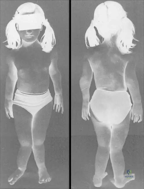
A 12-year-old girl is evaluated for multiple bony protuberances around her knees and ankles, leading to forearm bowing and short stature. The underlying genetic mutation for this condition primarily affects the synthesis of which of the following?
Explanation
Question 14
A 55-year-old male is diagnosed with a grade 2 conventional chondrosarcoma of the proximal femur. There is no evidence of metastasis. What is the mainstay of treatment?
Explanation
Question 15
A 32-year-old female presents with an incidental finding on a hand radiograph obtained after mild trauma. She has no pain at rest or night pain. Radiographs reveal a centrally located, lytic lesion in the proximal phalanx with stippled calcifications and no cortical breakthrough. What is the most appropriate next step in management?
Explanation
Question 16
A 25-year-old male presents with a painless lump on his proximal humerus. Radiographs demonstrate a surface lesion causing saucerization of the underlying cortex with a well-defined sclerotic margin and no medullary involvement.
What is the most likely diagnosis?
Explanation
Question 17
Which of the following imaging features is the most reliable indicator for differentiating a low-grade (Grade 1) chondrosarcoma from a benign enchondroma in a long bone?
Explanation
Question 18
Patients with Ollier disease and Maffucci syndrome frequently possess somatic mosaic mutations in which of the following genes?
Explanation
Question 19
A 32-year-old woman presents with a pathologic fracture of her proximal phalanx through a previously asymptomatic lytic lesion with stippled calcifications. What is the most appropriate initial management?
Explanation
Question 20
A 22-year-old male presents with knee pain. Radiographs reveal an eccentric, heavily scalloped lytic lesion in the proximal tibial metaphysis with a sclerotic rim. Histology shows lobules of myxoid and chondroid tissue with stellate cells and hypercellular peripheries. What is the diagnosis?
Explanation
Question 21
A 14-year-old boy presents with chronic right knee pain. Radiographs show a well-circumscribed lytic lesion in the distal femoral epiphysis. Histological examination reveals mononuclear cells with longitudinal nuclear grooves and areas of "chicken-wire" calcification. What is the most likely diagnosis?
Explanation
Question 22
In a skeletally mature patient with a solitary osteochondroma, malignant transformation to secondary chondrosarcoma is best indicated on MRI by a cartilage cap thickness greater than:
Explanation
Question 23
Which of the following is the histological hallmark of a dedifferentiated chondrosarcoma?
Explanation
Question 24
A 35-year-old male is noted to have an asymptomatic, 1.5 cm well-circumscribed lesion in the proximal phalanx with stippled calcifications found incidentally on X-ray for a sprained finger.
What is the most appropriate management?
Explanation
Question 25
Mutations in the EXT1 and EXT2 genes, seen in multiple hereditary exostoses (MHE), lead to the impaired synthesis of which of the following?
Explanation
Question 26
What is the recommended surgical management for a biopsy-proven atypical cartilaginous tumor (Grade 1 chondrosarcoma) contained entirely within the medullary canal of the distal femur?
Explanation
Question 27
A 55-year-old male presents with deep pelvic pain. Radiographs show a large destructive mass in the ilium with "popcorn" calcifications. Core biopsy confirms Grade II chondrosarcoma. What is the most appropriate treatment?
Explanation
Question 28
A 28-year-old female presents with multiple hard, bony swellings in her hands and several bluish soft-tissue nodules on her forearm that contain phleboliths on X-ray. What is her most likely diagnosis?
Explanation
Question 29
A 17-year-old boy presents with a firm mass on the surface of his proximal tibia. Imaging demonstrates a subperiosteal lesion causing saucerization of the outer cortex, bordered by solid reactive sclerosis.
There is no marrow invasion. What is the most likely diagnosis?
Explanation
Question 30
Which of the following distinct histological patterns is characteristic of mesenchymal chondrosarcoma?
Explanation
Question 31
The IDH1 and IDH2 mutations commonly found in central chondrosarcomas and enchondromatosis syndromes promote tumorigenesis by producing which of the following oncometabolites?
Explanation
Question 32
A 45-year-old male presents with hip pain. Radiographs reveal a lytic lesion in the proximal femoral epiphysis. Biopsy reveals cells with distinct borders, clear cytoplasm, and central round nuclei in a background of cartilaginous matrix with scattered giant cells. Diagnosis?
Explanation
Question 33
Patients with Multiple Hereditary Exostoses (MHE) have an increased lifetime risk of malignant transformation compared to the general population. What is the approximate rate of malignant transformation in MHE?
Explanation
Question 34
A 65-year-old male with a known history of Ollier disease presents with a rapidly enlarging, painful mass in his left hand over the past 3 months.
Given the sudden clinical change, what is the most likely complication?
Explanation
Question 35
When evaluating a cartilaginous lesion of the medullary canal, which of the following histological findings strongly favors a diagnosis of low-grade chondrosarcoma over an enchondroma?
Explanation
Question 36
A 35-year-old asymptomatic male incidentally discovers a 3 cm medullary lesion with stippled "rings and arcs" calcification in the proximal humerus.
What is the most appropriate next step in management?
Explanation
Question 37
A 28-year-old male presents with a painless palpable mass over the proximal humerus. Radiographs show a small surface lesion causing cortical saucerization with a well-defined sclerotic margin.
What is the most likely diagnosis?
Explanation
Question 38
A 12-year-old boy with multiple enchondromatosis presents with a rapid increase in the size of a thigh lesion. He is also noted to have multiple soft tissue hemangiomas. Which genetic mutation is most strongly associated with this specific syndrome?
Explanation
Question 39
A 15-year-old boy presents with knee pain. Radiographs reveal a 2 cm eccentric, purely lytic lesion in the epiphysis of the distal femur. Histology demonstrates mononuclear cells with grooved nuclei and "chicken-wire" calcifications. What is the most common complication following the standard surgical treatment of this lesion?
Explanation
Question 40
A 22-year-old female presents with a moderately painful, eccentric, radiolucent lesion in the metaphysis of the proximal tibia. Histology reveals a lobular architecture with hypercellular peripheries and hypocellular centers containing stellate cells in a myxoid background. What is the most appropriate treatment?
Explanation
Question 41
A 48-year-old male with a known history of an osteochondroma on his distal femur notices recent enlargement and increasing pain. MRI demonstrates a cartilaginous cap thickness of 2.5 cm. What is the most likely diagnosis?
Explanation
Question 42
A 30-year-old female sustains a closed, non-displaced fracture of her proximal phalanx after minor trauma. Radiographs reveal an underlying expansile, well-circumscribed radiolucent lesion with stippled calcifications.
What is the most appropriate management?
Explanation
Question 43
A 35-year-old presents with a painless, swollen ring finger. Radiographs show a centrally located, lytic lesion in the proximal phalanx with stippled calcifications.
What is the most appropriate initial management?
Explanation
Question 44
A 12-year-old boy presents with right knee pain. Radiographs reveal an eccentric, lytic surface lesion causing cortical scalloping on the proximal medial tibia without medullary extension.
What is the most likely diagnosis?
Explanation
Question 45
A 45-year-old woman is evaluated for a new painful mass in her proximal humerus. She has a known history of Ollier disease. Radiographs show a destructive lesion arising from a pre-existing calcified intramedullary tumor. Which of the following genetic mutations is most strongly associated with her underlying syndrome?
Explanation
Question 46
A 25-year-old male presents with chronic knee pain. Imaging reveals an epiphyseal lytic lesion in the distal femur. Histology demonstrates mononuclear cells with longitudinal nuclear grooves and areas of eosinophilic matrix with "chicken-wire" calcifications. What is the diagnosis?
Explanation
Question 47
Which of the following radiographic features best distinguishes a low-grade intramedullary chondrosarcoma from a benign enchondroma in a long bone?
Explanation
Question 48
A 22-year-old patient presents with a palpable mass on the posterior surface of the proximal humerus.
A biopsy confirms a periosteal chondroma. What is the recommended treatment for a symptomatic periosteal chondroma?
Explanation
Question 49
A 16-year-old male is diagnosed with an osteoid osteoma of the proximal femur. Which of the following best describes the molecular etiology of his severe nocturnal pain?
Explanation
Question 50
A 14-year-old girl complains of increasing right thigh pain. Radiographs demonstrate a diaphyseal permeative lytic lesion with an "onion skin" periosteal reaction. A biopsy is planned. Which cytogenetic abnormality is most likely to be found?
Explanation
Question 51
A patient with suspected Maffucci syndrome presents with a rapidly growing hand mass.
Besides a high risk for malignant transformation to chondrosarcoma, patients with Maffucci syndrome are at a significantly increased risk for which of the following?
Explanation
Question 52
A 30-year-old woman presents with knee pain. Radiographs show an eccentric, lytic epiphyseal lesion extending to the subchondral bone in the proximal tibia. A biopsy demonstrates multinucleated giant cells in a background of uniform mononuclear cells. What is the molecular target of the pharmacological agent used for unresectable cases of this tumor?
Explanation
Question 53
A 9-year-old boy presents with a pathologic fracture of the proximal humerus after a minor fall. Radiographs reveal a central, lytic, well-circumscribed diaphyseal-metaphyseal lesion with a "fallen leaf" sign. Which of the following is true regarding this condition?
Explanation
Question 54
A 24-year-old male presents with multiple asymmetric nodular deformities of the hands and feet, along with multiple dark-blue soft tissue masses. Genetic testing would most likely reveal a somatic mutation in which of the following genes?
Explanation
Question 55
A 30-year-old female presents with a closed, minimally displaced pathologic fracture of the proximal phalanx of the index finger. Radiographs show an expansile, centrally located lytic lesion with stippled calcifications.
What is the most appropriate initial management?
Explanation
Question 56
Which of the following histologic features is the most reliable discriminator for diagnosing a low-grade chondrosarcoma rather than a benign enchondroma?
Explanation
Question 57
A 15-year-old boy presents with a painless mass over the proximal humerus. Radiographs show a small (< 3 cm), cortically based lucent lesion with a sclerotic margin and cortical saucerization.
What is the most likely diagnosis?
Explanation
Question 58
Which of the following is an accurate characteristic regarding the inheritance and genetic profile of Ollier disease?
Explanation
Question 59
A 45-year-old man with a known asymptomatic distal femur enchondroma presents with new-onset, progressive night pain over the past 3 months. Imaging shows increased radiolucency and endosteal scalloping > 2/3 of the cortical thickness. Biopsy confirms secondary chondrosarcoma. What is the most appropriate definitive treatment?
Explanation
Question 60
In evaluating an intramedullary cartilaginous lesion of the distal femur, which MRI finding best distinguishes a bone infarct from an enchondroma?
Explanation
Question 61
A 32-year-old woman is diagnosed with an atypical cartilaginous tumor (Grade 1 chondrosarcoma) of the proximal humerus. What is the currently recommended surgical management?
Explanation
Question 62
A 22-year-old female presents with multiple hard bony protuberances and bluish soft-tissue nodules on her hands. Radiographs reveal multiple enchondromas. What is her lifetime risk of developing any malignancy?
Explanation
Question 63
A 34-year-old male presents with acute pain in his ring finger after a minor jamming injury. Radiographs show a pathologic fracture through a central lytic lesion with stippled calcifications in the proximal phalanx.
What is the most appropriate management?
Explanation
Question 64
A 60-year-old male presents with deep thigh pain. Imaging reveals a large distal femoral metaphyseal lesion. Biopsy demonstrates well-differentiated hyaline cartilage sharply demarcated from a high-grade pleomorphic spindle cell sarcoma. What is the diagnosis?
Explanation
Question 65
A 35-year-old male presents with chronic hip pain. Radiographs demonstrate a lytic lesion with a sclerotic margin localized specifically to the proximal femoral epiphysis. Histological analysis shows cells with abundant clear cytoplasm and distinct cell membranes, mixed with areas of conventional chondrosarcoma. What is the most likely diagnosis?
Explanation
Question 66
Which of the following histological features is the most reliable for distinguishing a low-grade conventional chondrosarcoma from a benign enchondroma?
Explanation
Question 67
A 12-year-old boy presents with multiple cartilaginous lesions predominantly affecting the right side of his body, causing limb-length discrepancy. Genetic testing of the lesional tissue is most likely to reveal a somatic mutation in which of the following genes?
Explanation
Question 68
A 25-year-old male presents with multiple enchondromas predominantly affecting the right hand and leg, along with multiple soft-tissue venous malformations. What genetic mutation is most strongly associated with this condition, and what is the approximate risk of malignant transformation of the bone lesions?
Explanation
Question 69
A 30-year-old right-hand-dominant carpenter presents with acute pain in his right ring finger after a minor twisting injury. Radiographs show a well-circumscribed, lucent lesion in the proximal phalanx with a pathologic fracture.
What is the most appropriate initial management for this patient?
Explanation
Question 70
A 15-year-old male presents with a painless mass on the proximal humerus. Radiographs demonstrate a 2.5 cm surface lesion with underlying cortical saucerization and a sclerotic rim.
Biopsy is performed. Which of the following histologic features is characteristic of this lesion and must not be overinterpreted as malignancy?
Explanation
Question 71
A 14-year-old boy presents with progressive knee pain. Radiographs reveal a 2 cm eccentric, purely lytic lesion in the distal femoral epiphysis with a thin sclerotic margin. MRI shows extensive surrounding bone marrow edema. What is the most likely histologic finding upon biopsy of this lesion?
Explanation
Question 72
A 22-year-old woman presents with chronic, dull pain in her proximal leg. Radiographs reveal an eccentric, heavily scalloped, lytic metaphyseal lesion in the proximal tibia with a distinct sclerotic rim. There is no matrix calcification. What is the most appropriate definitive treatment for this condition?
Explanation
Question 73
A 35-year-old man presents with a hard mass on the distal femur. Imaging shows a 6 cm lobulated surface mass with dense ring-and-arc calcifications. The lesion is elevating the periosteum but the underlying cortex is intact without medullary involvement.
What radiographic feature most strongly differentiates this from a periosteal chondroma?
Explanation
Question 74
A 40-year-old patient undergoes an MRI for a suspected meniscal tear. An incidental intramedullary lesion is found in the distal femur. It demonstrates a serpentine, well-demarcated border with a low-signal rim on both T1 and T2 sequences. How is this lesion radiographically distinct from an enchondroma?
Explanation
Question 75
A 32-year-old right-hand-dominant male presents with sudden pain in his right ring finger after a mild twisting injury. Radiographs demonstrate a pathologic fracture through a centrally located, lytic, expansile lesion with stippled calcifications in the proximal phalanx. What is the most appropriate management strategy?
Explanation
Question 76
Which of the following genetic mutations is primarily responsible for the development of multiple enchondromatosis (Ollier disease)?
Explanation
Question 77
A 55-year-old male presents with worsening thigh pain. Biopsy of a proximal femur lesion reveals a bimorphic histological pattern showing a low-grade hyaline cartilage tumor abruptly juxtaposed with a high-grade spindle cell sarcoma. Which of the following is the most likely diagnosis?
Explanation
Question 78
A 22-year-old male presents with a slow-growing, painless mass on his proximal humerus.
Imaging reveals a 2.5 cm surface lesion causing saucerization of the underlying cortex with a sclerotic margin. What is the definitive treatment to minimize local recurrence?
Explanation
Question 79
A 45-year-old male presents with chronic, mild hip pain. Radiographs reveal a lytic lesion in the epiphysis of the proximal femur. Histological examination shows large cells with distinct borders, abundant clear cytoplasm, and interspersed woven bone. What is the diagnosis?
Explanation
Question 80
A 16-year-old male undergoes curettage and bone grafting for an epiphyseal lesion in the proximal humerus. Histopathology reveals mononuclear cells with clefted, "coffee-bean" nuclei and areas of fine, pericellular "chicken-wire" calcification. Despite its benign nature, what complication must be monitored for?
Explanation
Question 81
A 24-year-old female presents with a painless, eccentric, expansile metaphyseal lesion in her proximal tibia. Histopathology demonstrates a lobular architecture with stellate and spindle cells embedded in a myxoid background, with multinucleated giant cells clustered at the lobular peripheries. What is the most likely diagnosis?
Explanation
Question 82
A 60-year-old female undergoes an MRI of her knee for a suspected meniscal tear. An incidental intramedullary distal femur lesion is identified. Which of the following MRI findings best distinguishes a bone infarct from an enchondroma?
Explanation
Question 83
A 30-year-old male presents with a destructive lesion in the mandible. Biopsy reveals a bimorphic pattern consisting of islands of well-differentiated hyaline cartilage surrounded by sheets of highly cellular, undifferentiated small round blue cells with hemangiopericytoma-like vessels. What is the diagnosis?
Explanation
Question 84
A 14-year-old boy with multiple hemangiomas and venous malformations is found to have multiple asymmetric cartilaginous lesions in his hands and long bones. He is diagnosed with Maffucci syndrome. Compared to Ollier disease, he is at an inherently higher risk for which of the following?
Explanation
Question 85
In evaluating an intramedullary cartilaginous lesion of the proximal humerus, which of the following clinical or radiographic features is the most reliable indicator of malignant transformation to a chondrosarcoma?
Explanation
Question 86
An asymptomatic 45-year-old male has an incidental finding on a hand radiograph.
The image shows a well-circumscribed, central lucent lesion in the proximal phalanx with focal stippled calcifications. No cortical destruction is noted. What is the recommended management?
Explanation
Question 87
A 50-year-old male with a known history of multiple hereditary exostoses (MHE) reports rapid growth of a previously stable osteochondroma on his pelvis. On MRI, what cartilage cap thickness threshold is most highly suspicious for secondary chondrosarcoma in an adult?
Explanation
Question 88
Which of the following inheritance patterns correctly describes the genetic transmission of multiple enchondromatosis (Ollier disease)?
Explanation
Question 89
Primary chondrosarcoma most frequently arises in which of the following anatomic locations, which also happens to present significant surgical challenges and carries a poorer prognosis?
Explanation
Question 90
A 50-year-old male is diagnosed with an Atypical Cartilaginous Tumor (Grade 1 Chondrosarcoma) confined within the medullary canal of the proximal femur, without cortical breakthrough. Which surgical treatment strategy is currently favored for this specific presentation?
Explanation
Question 91
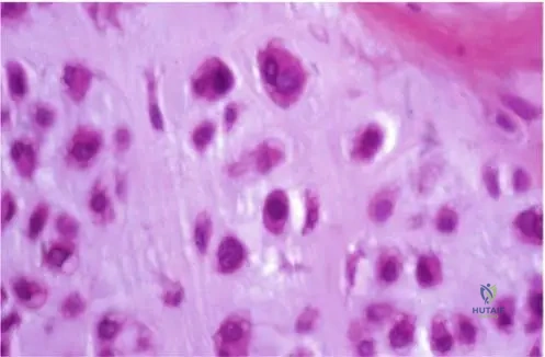
Histological evaluation of a curettage specimen from a benign hand enchondroma is most likely to demonstrate which of the following?
Explanation
Question 92
A patient with a presumed cartilaginous bone tumor undergoes molecular testing, revealing an H3F3B (Histone H3.3) mutation. This finding is virtually pathognomonic for which of the following tumors?
Explanation
Question 93
A 15-year-old male presents with a symptomatic surface lesion on his proximal humerus.
Imaging displays a 2 cm lesion causing saucerization of the outer cortex, with an intact sclerotic rim and without medullary extension. Which feature most reliably differentiates this lesion from a periosteal osteosarcoma?
Explanation
Question 94
A 25-year-old female presents with severe asymmetric limb deformity and multiple bluish subcutaneous nodules. Radiographs reveal multiple expansile cartilaginous lesions in the phalanges and long bones. Which of the following is true regarding this patient's condition compared to Ollier disease?
Explanation
Question 95
A 22-year-old male presents with a slow-growing, painless mass on his proximal humerus. Radiographs demonstrate the surface lesion shown below, measuring 2.5 cm with underlying cortical saucerization.
Biopsy reveals hypercellular cartilage with no cytological atypia or host bone permeation. What is the recommended definitive treatment?
Explanation
Question 96
A 30-year-old man presents with acute hand pain after a minor low-energy twisting injury. Radiographs show a pathologic fracture through a well-circumscribed, lucent lesion with central calcifications in the proximal phalanx, consistent with an enchondroma.
What is the most appropriate initial management for this pathologic fracture?
Explanation
Question 97
A 45-year-old male complains of new-onset, deep aching pain in his right shoulder, primarily occurring at rest and at night. Radiographs demonstrate a 6 cm cartilaginous intramedullary lesion in the proximal humerus. Which of the following radiographic features most strongly differentiates a secondary low-grade chondrosarcoma from a benign enchondroma?
Explanation
Question 98
A 40-year-old male presents with chronic hip pain. Radiographs reveal a purely lytic, slightly expansile lesion in the femoral head epiphysis extending to the articular surface. Biopsy demonstrates cells with abundant clear cytoplasm, distinct cell membranes, and interspersed areas of hyaline cartilage. What is the most likely diagnosis?
Explanation
Question 99
Somatic mosaic mutations in the IDH1 or IDH2 (isocitrate dehydrogenase) genes are most frequently associated with the pathogenesis of which of the following pairs of conditions?
Explanation
None
