Orthopaedic Board Exam Review: JIA, Bone Tumors, Syringomyelia & Charcot Joints | Part 8
Key Takeaway
This ABOS Orthopaedic Review covers critical topics for board exam preparation. Learn about Juvenile Idiopathic Arthritis (JIA) management, diverse bone tumor pathology including osteosarcoma and chondrosarcoma, and the diagnosis and treatment of syringomyelia with associated Charcot joint complications. Essential knowledge for orthopaedic specialists.
Orthopaedic Board Exam Review: JIA, Bone Tumors, Syringomyelia & Charcot Joints | Part 8
Comprehensive 100-Question Exam
00:00
Start Quiz
Question 1
A 4-year-old girl is diagnosed with oligoarticular juvenile idiopathic arthritis (JIA). Laboratory testing reveals a high titer of antinuclear antibodies (ANA). Which of the following is the most critical routine screening required for this patient?
Explanation
Question 2
A 35-year-old female with a long-standing history of polyarticular juvenile idiopathic arthritis is scheduled for a total hip arthroplasty. She has severe limitation in multiple joints. What is the most critical pre-operative imaging required prior to intubation?
Explanation
Question 3
A 12-year-old boy with systemic juvenile idiopathic arthritis (JIA) presents to the emergency department with acute high fever, hepatosplenomegaly, and altered mental status. Laboratory results show a sudden drop in ESR, thrombocytopenia, and hypofibrinogenemia. What is the most likely diagnosis?
Explanation
Question 4
Early-onset juvenile idiopathic arthritis commonly involves the temporomandibular joint (TMJ). Which of the following facial deformities is most characteristically seen as a consequence of this involvement?
Explanation
Question 5
A 16-year-old patient with JIA controlled on Etanercept is scheduled for a posterior spinal fusion for scoliosis. According to current perioperative guidelines, how should this biologic medication be managed?
Explanation
Question 6
A 15-year-old boy presents with knee pain and a palpable mass in the distal femur. Radiographs reveal a mixed lytic and sclerotic lesion with a 'sunburst' periosteal reaction. Core needle biopsy confirms high-grade, conventional osteosarcoma. What is the standard initial management?
Explanation
Question 7
A 10-year-old girl is diagnosed with Ewing sarcoma of the diaphysis of the humerus. Which specific chromosomal translocation is most classically associated with this malignancy?
Explanation
Question 8
A 30-year-old female presents with a large, lytic, expansile lesion in the proximal tibia extending to the subchondral bone. Biopsy demonstrates mononuclear cells interspersed with osteoclast-like multinucleated cells. Which targeted pharmacological agent can be used to downstage this tumor prior to surgery?
Explanation
Question 9
A 65-year-old man presents with a destructive lesion in the proximal humerus. Biopsy confirms metastatic clear cell renal cell carcinoma. He is scheduled for wide resection and endoprosthetic reconstruction. What is the most critical pre-operative step?
Explanation
Question 10
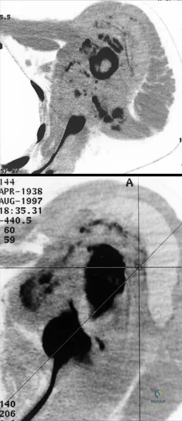
A 55-year-old man presents with dull pain in the proximal femur. Radiographs show a lytic lesion with 'rings and arcs' calcification. MRI reveals endosteal scalloping involving >2/3 of the cortical thickness. Biopsy confirms grade II chondrosarcoma. What is the primary modality of treatment?
Explanation
Question 11
A 13-year-old boy presents with a rapidly progressive, left-sided thoracic scoliosis and associated upper extremity weakness.
Given the underlying pathology seen on the MRI, what is the most appropriate initial management strategy?
Explanation
Question 12
A patient with syringomyelia presents with the classic 'cape-like' bilateral loss of pain and temperature sensation over the shoulders and upper extremities.
This specific neurological deficit is caused by the expanding syrinx compressing which structure within the spinal cord?
Explanation
Question 13
Syringomyelia, characterized by a fluid-filled cavity within the spinal cord, is most commonly associated with which of the following underlying congenital abnormalities?
Explanation
Question 14
When evaluating an adolescent for scoliosis, certain atypical features should prompt an entire spine MRI to rule out a neuroaxis abnormality such as syringomyelia. These 'red flag' features include all of the following EXCEPT:
Explanation
Question 15
A 45-year-old male with a history of a T12 burst fracture treated non-operatively 15 years ago presents with new-onset ascending spasticity, weakness, and sensory changes in his legs.
What is the most likely diagnosis shown on his current imaging?
Explanation
Question 16
A 55-year-old diabetic male presents with an acute, red, hot, swollen foot. Radiographs demonstrate bone fragmentation, subluxation, and periarticular debris around the midfoot. According to the Eichenholtz classification of Charcot arthropathy, which stage does this represent?
Explanation
Question 17
What is the most appropriate initial management for a patient presenting with an acute, non-ulcerated Eichenholtz Stage I Charcot arthropathy of the foot?
Explanation
Question 18
While modern neuropathic (Charcot) arthropathy is most commonly seen in the foot and ankle due to diabetes mellitus, historically, severe Charcot changes of the large joints (knee) and spine were classically associated with which condition?
Explanation
Question 19
A 4-year-old girl is diagnosed with ANA-positive oligoarticular Juvenile Idiopathic Arthritis (JIA). What is the most critical routine screening required for this patient?
Explanation
Question 20
A 45-year-old man presents with progressive, painless swelling and instability of his right shoulder over 6 months. Exam reveals loss of pain and temperature sensation in a cape-like distribution. X-rays show severe glenohumeral destruction with osseous debris. MRI of the spine will most likely show:
Explanation
Question 21
A 14-year-old boy has a high-grade intramedullary osteosarcoma of the distal femur. Following neoadjuvant chemotherapy and wide resection, what histological finding in the resected specimen is the most important prognostic factor for overall survival?
Explanation
Question 22
A 32-year-old woman presents with an expansive, purely lytic lesion in the distal radius extending to the subchondral bone. Biopsy confirms a Giant Cell Tumor (GCT). For an unresectable or recurrent GCT, which targeted medical therapy is most appropriate?
Explanation
Question 23
In a 12-year-old patient with long-standing, poorly controlled polyarticular Juvenile Idiopathic Arthritis (JIA), what is the most common pattern of cervical spine involvement?
Explanation
Question 24
A 55-year-old diabetic male presents with a swollen, erythematous, and warm left foot without open ulcerations. Radiographs show acute fragmentation and subluxation of the tarsometatarsal joints. What is the initial recommended orthopedic management during this Eichenholtz stage I phase?
Explanation
Question 25
A 30-year-old female presents with atypical scoliosis and weakness in her intrinsic hand muscles. Neurological exam demonstrates diminished pinprick sensation over her shoulders. Imaging confirms a cervical syrinx. What cranial anomaly is most frequently associated with this condition?
Explanation
Question 26
A 10-year-old boy presents with a diaphyseal lytic lesion of the fibula with an "onion-skin" periosteal reaction. Biopsy shows uniform small blue round cells. Which chromosomal translocation is most characteristic of this tumor?
Explanation
Question 27
A 55-year-old man is found to have a 6 cm calcified intramedullary lesion in his proximal humerus with endosteal scalloping. Biopsy confirms grade II (intermediate-grade) chondrosarcoma. What is the most appropriate definitive management?
Explanation
Question 28
A 9-year-old child with unilateral pauciarticular JIA presents with a 2 cm leg length discrepancy, with the arthritic limb being longer. She also has a mild knee flexion contracture. What is the primary pathophysiological cause of the limb overgrowth?
Explanation
Question 29
Which of the following best describes the 'neurovascular theory' in the pathogenesis of a Charcot neuropathic joint?
Explanation
Question 30
A 65-year-old man presents with severe back pain and a solitary lytic lesion in the L3 vertebral body. Laboratory testing reveals an M spike on serum protein electrophoresis. What imaging modality is considered the standard of care for staging skeletal involvement in this disease?
Explanation
Question 31
A 12-year-old girl is evaluated for scoliosis. She has an atypical left thoracic curve and absent abdominal reflexes. An MRI reveals a large syrinx in the thoracic spinal cord. What is the recommended sequence of treatment for her condition?
Explanation
Question 32
A 19-year-old male complains of severe, progressively worsening right thigh pain that occurs primarily at night and is dramatically relieved by NSAIDs. Imaging shows a radiolucent nidus surrounded by dense reactive sclerosis. What is the most definitive, minimally invasive treatment?
Explanation
Question 33
A 5-year-old boy presents with daily spiking fevers, a generalized salmon-pink macular rash, hepatosplenomegaly, and severe polyarthritis. Laboratory tests show markedly elevated ferritin, ESR, and CRP, but negative ANA and RF. Which of the following is the most likely diagnosis?
Explanation
Question 34
In a patient with diabetic neuroarthropathy of the foot, serial radiographs demonstrate absorption of fine debris, coalescence of large fracture fragments, and early sclerosis, alongside a clinical reduction in soft tissue edema. This corresponds to which stage of the Eichenholtz classification?
Explanation
Question 35
A 12-year-old boy presents with a left-sided thoracic scoliosis. Neurological examination reveals absent superficial abdominal reflexes and diminished pain and temperature sensation in both upper extremities. Which of the following is the most appropriate next step in evaluation?
Explanation
Question 36
Which subtype of Juvenile Idiopathic Arthritis (JIA) carries the highest risk for developing asymptomatic anterior uveitis, thereby requiring the most frequent ophthalmologic screening?
Explanation
Question 37
A 14-year-old boy presents with a painful diaphyseal mass in his right femur, low-grade fever, and an elevated erythrocyte sedimentation rate (ESR). Biopsy reveals sheets of uniform small round blue cells. Cytogenetic analysis of this tumor is most likely to demonstrate which of the following translocations?
Explanation
Question 38
A 55-year-old patient with long-standing, poorly controlled diabetes presents with a warm, swollen, erythematous, and painless foot. Radiographs demonstrate periarticular debris, bony fragmentation, and midfoot subluxation. According to the Eichenholtz classification, what is the current stage and best initial management?
Explanation
Question 39
A 45-year-old male presents with a massively swollen, painless shoulder joint. Radiographs show severe destruction of the humeral head with abundant bony debris. He mentions chronic neck stiffness and frequent unrecognized burns on his hands. What is the most likely underlying etiology of his shoulder pathology?
Explanation
Question 40
In the multimodal management of high-grade, conventional intramedullary osteosarcoma of the distal femur, which of the following is considered the most critical prognostic factor for long-term patient survival?
Explanation
Question 41
A 9-year-old girl with severe polyarticular JIA is scheduled for bilateral total hip arthroplasty due to debilitating pain and contractures. Prior to general anesthesia, which of the following imaging studies is absolutely mandatory?
Explanation
Question 42
A 60-year-old man presents with dull pain in his proximal humerus. Radiographs reveal a large lytic lesion with intralesional "popcorn" calcifications and endosteal scalloping >2/3 of the cortical thickness. Biopsy confirms grade II chondrosarcoma. What is the most appropriate definitive treatment?
Explanation
Question 43
The neurovascular theory of the pathogenesis of Charcot neuroarthropathy postulates that joint destruction is primarily initiated by which of the following mechanisms?
Explanation
Question 44
A 14-year-old girl presents with a rapidly progressive right thoracic scoliosis. MRI reveals a large fluid-filled cavity within the central spinal cord extending from C4 to T8. Which of the following cranial abnormalities is most commonly associated with this specific spinal finding?
Explanation
Question 45
A 6-year-old child with oligoarticular JIA presents with a chronic, persistent unilateral knee effusion. Clinical exam reveals a noticeable limb length discrepancy. What is the most common lower extremity deformity pattern observed in this specific clinical scenario?
Explanation
Question 46
A 32-year-old woman presents with knee pain. Radiographs reveal an eccentric, purely lytic, epiphyseal lesion extending to the subchondral bone of the proximal tibia. Biopsy confirms multinucleated giant cells in a background of mononuclear stromal cells. Which targeted therapy acts as a RANKL inhibitor and is indicated for advanced or unresectable forms of this tumor?
Explanation
Question 47
In a patient with diabetic Charcot neuroarthropathy, structural collapse of the longitudinal arch resulting in plantar prominence of the cuboid and navicular (rocker-bottom foot deformity) most frequently occurs during which Eichenholtz stage?
Explanation
Question 48
The pathogenesis of scoliosis in patients with syringomyelia is best explained by which of the following mechanisms?
Explanation
Question 49
An 18-year-old male presents with painful scoliosis. Imaging reveals a 2.5 cm radiolucent lesion with a mineralized matrix in the posterior elements of L3. The pain is persistent and is only partially relieved by nonsteroidal anti-inflammatory drugs (NSAIDs). What is the most likely diagnosis?
Explanation
Question 50
Which of the following best describes the characteristic facial morphology of a patient who develops temporomandibular joint (TMJ) ankylosis secondary to severe juvenile idiopathic arthritis (JIA) during early childhood?
Explanation
Question 51
A 62-year-old diabetic male with an Eichenholtz Stage 1 Charcot arthropathy of the midfoot presents with a non-healing, non-infected plantar ulcer directly overlying a massive cuboid prominence. What is the most appropriate surgical management?
Explanation
Question 52
A 65-year-old man presents with new-onset back pain, anemia, and an M-spike on serum protein electrophoresis. Radiographs show a compression fracture of T12 and punched-out lytic skull lesions. Which of the following imaging modalities is most sensitive for detecting additional early osseous lesions in this patient?
Explanation
Question 53
A 4-year-old girl is diagnosed with oligoarticular Juvenile Idiopathic Arthritis (JIA). Her ANA is positive and rheumatoid factor is negative. Which of the following is the most appropriate screening protocol for the most common extra-articular complication in this patient?
Explanation
Question 54
A 15-year-old boy presents with a painful mass in his distal femur. Radiographs show a "sunburst" periosteal reaction. He has a history of bilateral enucleation as an infant. Which mutated gene is most directly implicated in both of his neoplastic conditions?
Explanation
Question 55
A 35-year-old man presents with progressive weakness and painless burns on his hands. Neurological examination reveals loss of pain and temperature sensation in a cape-like distribution over his shoulders and upper extremities, with preserved light touch. MRI is shown.
What is the most common associated condition?
Explanation
Question 56
A 45-year-old patient develops a rapidly progressive, painless, and massively swollen shoulder joint. Radiographs show severe osseous destruction, debris, and subluxation. Which of the following underlying conditions is the most likely etiology for this presentation?
Explanation
Question 57
A 6-year-old boy presents with daily spiking fevers up to 39.5°C, a transient salmon-pink macular rash, and hepatosplenomegaly. He also has bilateral knee effusions. Laboratory tests show elevated ferritin and leukocytosis. What is the most likely diagnosis?
Explanation
Question 58
A 32-year-old woman presents with knee pain. Radiographs reveal an eccentric, expansile lytic lesion in the distal femoral epiphysis extending to the articular surface. Biopsy shows multinucleated giant cells in a stroma of mononuclear cells. Which of the following is the most appropriate targeted medical therapy?
Explanation
Question 59
A 55-year-old diabetic male presents with a warm, erythematous, and swollen foot. Radiographs show periarticular debris, fragmentation of the tarsal bones, and subluxation. According to the Eichenholtz classification, what is the current stage and most appropriate initial management?
Explanation
Question 60
A 12-year-old boy presents with an atypical, rapidly progressive left thoracic scoliotic curve. He also reports frequent occipital headaches and upper extremity clumsiness. An MRI confirms a syrinx.
What is the most appropriate initial management for his spinal deformity?
Explanation
Question 61
A 60-year-old man presents with dull, aching hip pain. Radiographs show a lytic lesion in the proximal femur with endosteal scalloping and "rings and arcs" calcification. Biopsy confirms a grade II chondrosarcoma. What is the mainstay of treatment?
Explanation
Question 62
A 14-year-old girl with severe, long-standing polyarticular JIA complains of neck pain, myelopathic symptoms, and a "clunking" sensation when nodding. Which of the following radiographic findings is most critical to evaluate before planning any elective surgical procedures requiring intubation?
Explanation
Question 63
A 65-year-old man presents with a massively enlarged, painless knee. He has a history of untreated syphilis in his youth. Radiographs reveal severe joint destruction and large osteophytes. Neurological exam shows loss of proprioception and a positive Romberg sign. Which of the following is the most likely pathophysiological mechanism for his joint destruction?
Explanation
Question 64
A 14-year-old girl presents with fever and localized pain in her mid-tibia. Radiographs show a permeative, diaphyseal lytic lesion with an "onion-skin" periosteal reaction. Histology reveals small round blue cells. Which of the following immunohistochemical markers and genetic translocations are characteristic of this tumor?
Explanation
Question 65
In a patient with early syringomyelia
experiencing isolated loss of pain and temperature sensation in the upper extremities, which specific spinal cord structure is primarily compressed by the expanding syrinx?
Explanation
Question 66
A 7-year-old boy with systemic JIA suddenly deteriorates, developing lethargy, unremitting fever, and mucosal bleeding. Laboratory findings show profound cytopenias, a rapidly falling erythrocyte sedimentation rate (ESR), hypofibrinogenemia, and markedly elevated serum ferritin. What is the most likely diagnosis?
Explanation
Question 67
A 65-year-old man presents with severe back pain and fatigue. Radiographs show multiple "punched-out" lytic lesions in his skull and vertebral bodies. Laboratory studies reveal hypercalcemia, anemia, and an M-spike on serum protein electrophoresis. The osteolytic lesions in this disease are primarily mediated by the upregulation of which factor?
Explanation
Question 68
The pathogenesis of Charcot arthropathy is classically described by two main theories. The "neurovascular theory" attributes the initial bone destruction and fragmentation to which of the following mechanisms?
Explanation
Question 69
A 22-year-old male presents with dull, aching pain in his mid-back that is constant, night-predominant, and only partially relieved by ibuprofen. CT scan shows a 3.5 cm expansile lytic lesion in the posterior elements of T8 with a distinct radiolucent nidus and surrounding sclerosis. What is the most likely diagnosis?
Explanation
Question 70
A 40-year-old woman with a history of severe spinal trauma 15 years ago presents with new-onset spasticity in her lower extremities and progressive sensory loss in her hands.
MRI of the cervical and thoracic spine reveals a large cystic cavitation within the spinal cord. What is the most likely diagnosis?
Explanation
Question 71
A 5-year-old girl with untreated monoarticular JIA of the right knee presents for evaluation. Her parents note a progressive gait abnormality. On examination, the right knee is warm and swollen with a 15-degree flexion contracture. Which of the following limb length discrepancies is most likely to be present?
Explanation
Question 72
A 4-year-old girl with ANA-positive oligoarticular Juvenile Idiopathic Arthritis (JIA) presents for routine follow-up. Which of the following represents the most critical routine screening recommendation for this patient?
Explanation
Question 73
A 25-year-old female with severe systemic JIA is undergoing bilateral total hip arthroplasties. Which of the following is the most likely intraoperative finding or technical challenge?
Explanation
Question 74
A 14-year-old boy is diagnosed with high-grade osteosarcoma of the distal femur. Following neoadjuvant chemotherapy, what percentage of tumor necrosis is the threshold defining a 'good histological response' according to the Huvos grading system?
Explanation
Question 75
A 32-year-old male presents with painless swelling and instability of his left shoulder. He has noted recent burns on his hands that he did not feel. Imaging shows severe joint destruction.
What is the most likely underlying pathophysiology?
Explanation
Question 76
A 55-year-old poorly controlled diabetic presents with an erythematous, warm, and swollen right foot. Radiographs show periarticular fragmentation and debris without signs of coalescence. According to the Eichenholtz classification, what is the appropriate management?
Explanation
Question 77
A 30-year-old female presents with a lytic lesion in the distal femur extending to the subchondral bone. Biopsy confirms a Giant Cell Tumor. Prior to extended intralesional curettage, the surgeon administers denosumab. What is the mechanism of action of denosumab?
Explanation
Question 78
A 12-year-old boy with polyarticular JIA complains of progressive neck pain and stiffness. Which of the following cervical spine abnormalities is most characteristic of this disease?
Explanation
Question 79
A 9-year-old boy presents with a diaphyseal lesion of the fibula with an 'onion skin' periosteal reaction. A biopsy reveals small blue round cells. Which chromosomal translocation is most commonly associated with this malignancy?
Explanation
Question 80
A 14-year-old girl is evaluated for a left-sided thoracic scoliotic curve of 45 degrees. Neurological exam reveals absent abdominal reflexes. An MRI confirms a cervical syrinx.
What is the most appropriate initial management for her spinal deformity?
Explanation
Question 81
The neurotraumatic and neurovascular theories are postulated for the development of Charcot arthropathy. Which of the following best describes the core principle of the neurovascular theory?
Explanation
Question 82
A 60-year-old male presents with dull right hip pain. Radiographs show a large lytic lesion with 'popcorn' calcifications in the right ilium. Biopsy confirms a grade II chondrosarcoma. What is the mainstay of treatment for this condition?
Explanation
Question 83
A 9-year-old boy with poorly controlled JIA presents with a progressive unilateral knee deformity. The affected knee exhibits overgrowth and a fixed flexion contracture. Which phenomenon best explains the observed limb length discrepancy?
Explanation
Question 84
A 16-year-old male complains of severe, aching thigh pain that is worse at night and dramatically relieved by NSAIDs. Radiographs demonstrate a 1-cm radiolucent nidus surrounded by dense sclerotic bone in the femoral diaphysis. What is the preferred definitive treatment if medical management fails?
Explanation
Question 85
A 28-year-old female with rapidly progressive scoliosis and intrinsic hand muscle wasting undergoes whole-spine MRI.
The MRI shows a CSF-filled cavity within the spinal cord. What brain malformation is most frequently associated with this finding?
Explanation
Question 86
A 50-year-old diabetic patient presents with a swollen, warm, and red foot without open ulcers. To differentiate an acute Eichenholtz Stage I Charcot arthropathy from acute osteomyelitis, which imaging modality is most specific?
Explanation
Question 87
In a child with polyarticular JIA who requires a distal femoral extension osteotomy to correct a severe fixed knee flexion contracture, what is a primary concern if performed before skeletal maturity?
Explanation
Question 88
A 65-year-old woman presents with severe back pain. Radiographs reveal multiple punched-out lytic lesions in the skull and a compression fracture of L2. Laboratory workup shows hypercalcemia and a monoclonal spike. What is the most common primary bone malignancy in this age group?
Explanation
Question 89
A 10-year-old boy presents with a left-sided thoracic scoliotic curve. Neurological examination reveals a loss of pain and temperature sensation over his bilateral shoulders and upper extremities with preserved light touch. Which of the following is the most appropriate next step in evaluation?
Explanation
Question 90
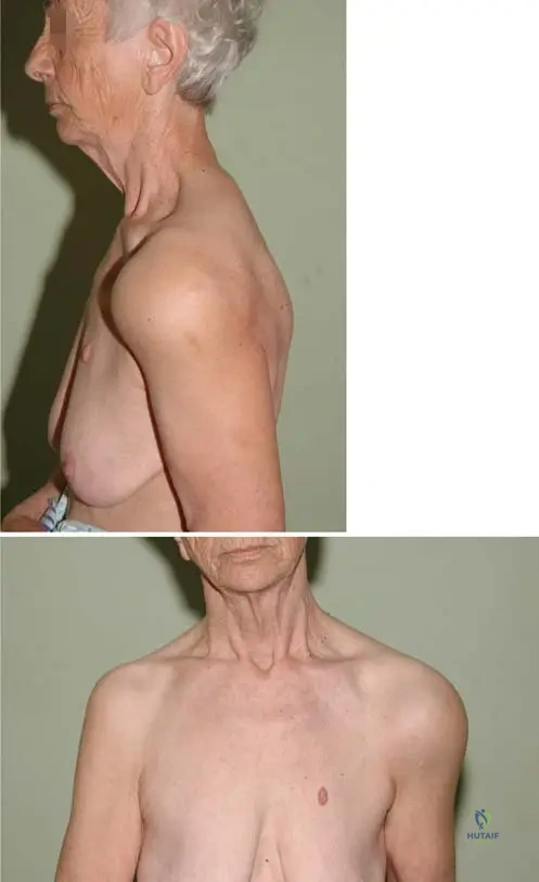
A 45-year-old male presents with painless swelling and severe crepitus in his right shoulder. He has a history of cervical syringomyelia. Radiographs show severe joint destruction, debris, and dislocation.
What is the primary pathophysiologic mechanism for this joint destruction?
Explanation
Question 91
A 14-year-old boy is diagnosed with conventional high-grade osteosarcoma of the distal femur. Genetic testing reveals a mutation in the TP53 gene. Which of the following syndromes is most closely associated with this patient's diagnosis?
Explanation
Question 92
A 60-year-old male presents with deep thigh pain. Radiographs reveal a large, permeative lytic lesion with "popcorn" calcifications in the proximal femur. Biopsy confirms grade III conventional chondrosarcoma. What is the most appropriate management?
Explanation
Question 93
A 16-year-old female with polyarticular JIA is scheduled for a bilateral total hip arthroplasty. She is currently managed with methotrexate and etanercept. To minimize the risk of postoperative infection while preventing a severe disease flare, what is the best perioperative medication management strategy?
Explanation
Question 94
A 55-year-old diabetic patient presents with a swollen, warm, and erythematous left foot. Radiographs show osteopenia, bony fragmentation, and joint subluxation at the midfoot. According to the Eichenholtz classification, what is the current stage and most appropriate initial management?
Explanation
Question 95
A 12-year-old girl presents with a destructive diaphyseal lesion of her tibia with an associated "onion-skin" periosteal reaction. A biopsy is performed, and molecular analysis demonstrates a t(11;22) chromosomal translocation. Which of the following fusion proteins is highly specific to this tumor?
Explanation
Question 96
An MRI of the cervical spine is performed on a 28-year-old female presenting with hand weakness and loss of temperature sensation. It demonstrates a large central intramedullary fluid collection extending from C2 to T1.
Which of the following cranial abnormalities is most commonly associated with this condition?
Explanation
Question 97
A 32-year-old woman presents with a large, lytic, epiphyseal-metaphyseal lesion of her distal radius. Biopsy confirms a Giant Cell Tumor of bone (GCTB). She is started on Denosumab prior to surgical intervention. What is the mechanism of action of this medication?
Explanation
Question 98
A 25-year-old male with a history of long-standing systemic juvenile idiopathic arthritis (JIA) presents with worsening neck stiffness. Radiographs of the cervical spine are most likely to demonstrate which of the following classical findings?
Explanation
Question 99
The pathogenesis of neuropathic (Charcot) arthropathy is traditionally described by two main theories: the neurotraumatic theory and the neurovascular theory. Which of the following best describes the core tenet of the neurovascular theory?
Explanation
Question 100
A 15-year-old male is suspected of having an osteosarcoma of the proximal tibia. A core needle biopsy is planned. To adhere to standard orthopedic oncology principles and avoid compromising definitive limb-salvage surgery, which of the following is an absolute requirement for the biopsy tract?
Explanation
None
