ABOS Part I & AAOS OITE Orthopaedic Surgery Review: Clinical Cases & Exam Questions | Part 22139
Key Takeaway
ABOS Part I Review modules offer advanced orthopedic multiple-choice questions mirroring the ABOS Part I and AAOS OITE examinations. Content is derived from high-yield clinical teaching cases, covering essential topics like shoulder dislocations, fracture fixation principles, and orthopedic screw biomechanics to ensure comprehensive exam preparation.
ABOS Part I & AAOS OITE Orthopaedic Surgery Review: Clinical Cases & Exam Questions | Part 22139
Comprehensive 100-Question Exam
00:00
Start Quiz
Question 1
A 24-year-old male presents to the emergency department after a football injury, complaining of severe right shoulder pain. His arm is held in slight abduction and external rotation. On inspection, the anterior aspect of his shoulder appears prominent, and there is a palpable void beneath the acromion. Which of the following physical exam findings is MOST concerning for an associated neurovascular injury in this patient?

Explanation
Correct Answer: D
An absent radial pulse is a critical finding indicating potential compromise of the brachial artery, which is a surgical emergency. While axillary nerve injury (loss of sensation over the lateral deltoid, weakness in abduction) is the most common nerve injury with anterior shoulder dislocations, it is rarely an acute limb-threatening condition unless it's a traction injury without spontaneous recovery. Weakness in wrist extension would suggest radial nerve involvement, which is less common. Ecchymosis is a common finding but not acutely life- or limb-threatening.
Question 2
A 35-year-old patient presents with a history of recurrent anterior shoulder dislocations. During your examination, you perform the Apprehension Test. Which of the following describes a positive test?

Explanation
Correct Answer: A
The Apprehension Test is performed by abducting the shoulder to 90 degrees and slowly externally rotating the arm. A positive test is indicated by the patient's feeling of impending dislocation (apprehension) or significant pain, often due to stretching of the anterior capsule. Options B and C describe findings related to rotator cuff or glenohumeral arthritis. Option D describes the Sulcus Sign, indicative of inferior or multidirectional instability. Option E describes a clunk, which could be related to labral pathology but is not the apprehension test.
Question 3
A 50-year-old patient presents with acute shoulder pain after a seizure. On examination, the arm is held in internal rotation, and the anterior shoulder appears flattened. External rotation is severely restricted. Which radiographic finding on an AP shoulder view is pathognomonic for a posterior shoulder dislocation?

Explanation
Correct Answer: C
The Trough line sign (or reverse Hill-Sachs lesion) is an impaction fracture on the anterior-medial aspect of the humeral head, often seen with posterior dislocations. The other options are incorrect: Hill-Sachs and Bankart lesions are typically associated with anterior dislocations. HAGL lesions are avulsions of the glenohumeral ligaments, often associated with anterior dislocations. Os acromiale is an anatomical variant.
Question 4
During the examination of a patient with suspected shoulder dislocation, you note a sulcus sign. What does this finding MOST commonly indicate?

Explanation
Correct Answer: C
The Sulcus Sign is elicited by applying inferior traction to the arm, causing a dimple or sulcus to appear below the acromion. It is indicative of inferior capsular laxity and is a hallmark of inferior or multidirectional glenohumeral instability. While multidirectional instability often includes an inferior component, the most direct interpretation of a sulcus sign is inferior instability.
Question 5
A 68-year-old woman falls directly onto her shoulder. She presents with severe pain and an inability to move her arm. On exam, the shoulder appears abducted, and a prominent hard mass is palpable inferior to the glenoid, consistent with a Luxatio Erecta. Which neurovascular structure is at highest risk of injury in this type of dislocation?

Explanation
Correct Answer: C
Luxatio Erecta (inferior dislocation) involves extreme abduction, forcing the humeral head inferiorly. The head can impinge upon or stretch the neurovascular bundle in the axilla. The axillary artery is at significant risk due to its proximity and the severe displacement. While the axillary nerve and brachial plexus are also at risk, arterial compromise (axillary artery) is a more acute and limb-threatening complication associated with the extreme force and direction of displacement in luxatio erecta, often leading to intimal tears or thrombosis.
Question 6
Following reduction of an anterior shoulder dislocation, a patient complains of persistent weakness in active shoulder abduction. Sensation over the lateral aspect of the deltoid is intact. Which of the following is the MOST likely cause of this isolated weakness?

Explanation
Correct Answer: B
If sensation over the lateral deltoid (axillary nerve sensory distribution) is intact, persistent isolated weakness in shoulder abduction, especially in an older patient or high-energy trauma, should raise suspicion for an associated rotator cuff tear (supraspinatus or deltoid dysfunction). Axillary nerve neuropraxia would typically present with sensory deficits in addition to motor weakness. Musculocutaneous nerve injury affects biceps and coracobrachialis, and lateral forearm sensation. Long thoracic nerve injury causes scapular winging. Brachial plexus avulsion would present with more widespread neurological deficits.
Question 7
A 22-year-old male presents with his first-time anterior shoulder dislocation. During the initial assessment, which of the following is a critical component of the examination PRIOR to any reduction attempts?

Explanation
Correct Answer: C
A thorough neurovascular examination of the affected extremity, including palpation of pulses and assessment of sensation and motor function, is paramount before any reduction attempts. This establishes a baseline and helps identify any pre-existing or acute neurovascular compromise that could be exacerbated by or misattributed to the reduction maneuver. Analgesia is important but secondary to neurovascular assessment. Contralateral shoulder ROM is not critical pre-reduction. Ice is for comfort. Family history is irrelevant in acute management.
Question 8
You are examining a patient with a suspected posterior shoulder dislocation. Which maneuver is most likely to confirm your suspicion on physical exam?

Explanation
Correct Answer: C
Posterior dislocations classically present with the arm held in internal rotation and adduction, with a significant block to external rotation. The anterior shoulder may appear flattened, and the coracoid process prominent. Apprehension with abduction and external rotation is characteristic of anterior instability. Limited internal rotation with intact external rotation is incorrect. Increased superior translation with anterior force is not directly indicative of posterior dislocation. A palpable defect below the coracoid is more suggestive of anterior dislocation.
Question 9
Which finding on a post-reduction physical exam of an anterior shoulder dislocation indicates successful reduction and suggests stability?

Explanation
Correct Answer: B
Successful reduction is indicated by the restoration of normal shoulder contour (loss of the anterior prominence of the humeral head), relief of severe pain, and the ability to achieve full or near-full passive range of motion without a 'block.' Persistent apprehension or instability signs (like a sulcus sign or continued apprehension with external rotation) suggest potential underlying pathology or incomplete reduction. Crepitus might indicate cartilage damage, and inability to actively abduct could suggest a rotator cuff tear or nerve injury, not necessarily unsuccessful reduction.
Question 10
A 70-year-old male sustains an anterior shoulder dislocation. After reduction, plain radiographs show a concomitant fracture. Which fracture is MOST commonly associated with anterior shoulder dislocation in this age group?

Explanation
Correct Answer: B
While Hill-Sachs and Bankart lesions are very common with anterior dislocations, in older patients, a greater tuberosity fracture is particularly common (up to 30% in some series) due to the weaker bone and the forces involved in the injury. The rotator cuff avulses a piece of the tuberosity during the dislocation. Surgical neck fracture is also possible but less frequent than greater tuberosity in direct association with dislocation. Clavicle fractures are less directly associated with glenohumeral dislocation mechanism.
Question 11
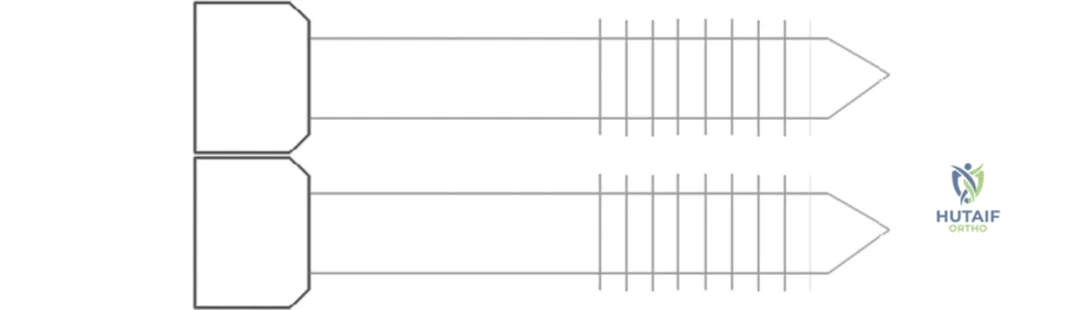
A resident is reviewing the basic anatomy of an orthopedic screw. Referring to the provided image, which labeled component is primarily responsible for providing a smooth, unthreaded link between the screw head and the threaded portion?
Explanation
Correct Answer: B
The case explicitly states, 'The shank provides a smooth link between the head and thread.' The image clearly labels the shank as the unthreaded portion immediately distal to the head. The head (A) provides a connection for a screwdriver and prevents sinking. The thread (C) engages with the bone. Pitch (D) is the distance between adjacent threads. Flutes (E) provide a route for bone debris removal.
Question 12
A 68-year-old female with osteoporotic bone requires internal fixation for a distal femur fracture. To maximize the pull-out strength of the screws in this challenging bone quality, which of the following biomechanical principles should the surgeon prioritize?
Explanation
Correct Answer: C
The case states that pull-out strength can be increased by 'increasing thread density' or by having a 'finer' pitch, which allows more turns of the spiral thread to engage in a given depth of cortex, creating greater resistance to pull-out. Options A and B would decrease the contact surface area or thread depth, thereby reducing pull-out strength. Option D directly contradicts the principle of increasing engaged threads. Option E (larger pilot hole) is a surgeon factor that reduces pull-out strength.
Question 13
During a complex ankle fracture fixation, a junior resident inadvertently makes a pilot hole slightly larger than recommended for the chosen screw. This technical error is most likely to result in which of the following?
Explanation
Correct Answer: C
The case explicitly lists 'making too large a pilot hole' as a surgeon factor that can reduce screw pull-out strength. A larger pilot hole reduces the contact area between the screw threads and the bone, diminishing the frictional and mechanical interlock necessary for strong fixation. Options A, B, D, and E are incorrect as a larger pilot hole would generally weaken the fixation, not strengthen it or improve insertion characteristics in a beneficial way.
Question 14
A surgeon is selecting a screw for metaphyseal fixation in a comminuted proximal humerus fracture, where bone quality may be compromised. To optimize the screw's resistance to axial dislodgement, which characteristic related to the screw's thread design would be most beneficial?
Explanation
Correct Answer: C
The case states, 'The 'finer' the pitch, the more turns of the spiral thread engage in a given depth of cortex, creating greater resistance to pull-out.' A finer pitch means more threads per unit length, increasing the contact surface area with the bone. A larger lead (A) or coarser pitch (B) would mean fewer threads engaged. A shallower thread depth (D) or smaller outer diameter (E) would reduce the contact surface area, thereby decreasing pull-out strength.
Question 15
In a scenario where a surgeon aims to maximize the pull-out strength of a non-locking screw in cortical bone, which combination of screw diameter characteristics would be most advantageous?
Explanation
Correct Answer: C
The case indicates that pull-out strength can be increased by 'increasing the contact surface area between screw threads and bone, either by increasing the outer diameter, decreasing the core diameter.' This combination (increased outer diameter and decreased core diameter) results in a greater thread depth, maximizing the volume of bone engaged by the screw threads and thus increasing the resistance to pull-out. Options A, B, and D would either reduce the thread depth or the overall screw-bone contact. Option E describes a smooth pin, not a screw.
Question 16
A 55-year-old male sustains a periprosthetic femur fracture around a total hip arthroplasty stem. The surgeon opts for plate and screw fixation. The use of locking screws in this context primarily enhances stability by:
Explanation
Correct Answer: B
The case states, 'The use of a locking screw can also create a monobloc effect for greater stability.' Locking screws thread into the plate, creating a fixed-angle construct that acts as a single unit (monobloc) with the bone, providing enhanced stability, especially in osteoporotic bone or comminuted fractures. They do not primarily rely on screw-plate friction (A) or provide dynamic compression (C) in the same way non-locking screws do. They increase, rather than reduce, the stiffness of the construct (D). Flutes, not locking screws, facilitate bone debris removal (E).
Question 17
During a revision surgery for a failed ankle fusion, a surgeon attempts to re-insert a screw into a previously drilled hole. After initial resistance, the screw feels loose and does not achieve adequate purchase. This situation is most likely due to:
Explanation
Correct Answer: C
The case identifies 'repeated withdrawal and reintroduction of a screw causing damage to the negative threads in the bone tissue' as a surgeon factor that can reduce screw pull-out strength. When a screw is removed and reinserted, the bone threads (negative threads) can be stripped or damaged, leading to poor purchase and reduced pull-out strength upon reinsertion. Insufficient pilot hole depth (A) would typically lead to difficulty inserting the screw initially, not looseness after reinsertion. Over-tightening (B) could strip the threads on the first attempt, but the scenario describes re-insertion. An excessively fine pitch (D) would increase pull-out strength, not decrease it. The type of screw (E) is not the primary cause of this specific failure mode.
Question 18
A biomechanical engineer is testing various orthopedic screw designs. The primary parameter being measured when assessing a screw's "pull-out strength" is the:
Explanation
Correct Answer: C
The case clearly defines pull-out strength: 'A screw's pull-out strength refers to the axial force required to remove a screw from bone.' Options A, B, D, and E describe other mechanical properties of screws (torsional strength, bending strength, fatigue strength, compressive strength) but not specifically pull-out strength, which is a measure of resistance to axial extraction.
Question 19
A 72-year-old patient with severe osteoporosis undergoes open reduction and internal fixation of a proximal tibia fracture. The surgeon is particularly concerned about screw pull-out. Which of the following intraoperative strategies would provide the greatest cumulative benefit in maximizing screw pull-out strength in this patient?
Explanation
Correct Answer: B
This option combines multiple strategies mentioned in the case to maximize pull-out strength. The case states that pull-out strength can be increased by 'increasing the number of threads engaged in the bone cortex' and by using a 'finer' pitch. An undersized pilot hole (the opposite of 'too large a pilot hole' which reduces strength) would maximize thread purchase. Option A describes characteristics that would decrease pull-out strength (larger core, coarser pitch). Option C suggests avoiding locking screws, which the case states 'create a monobloc effect for greater stability,' and micro-motion is generally undesirable for fixation. Option D (repeated insertion/removal) is explicitly listed as a factor that reduces pull-out strength. Option E (smaller outer diameter, larger lead) would reduce thread engagement and pull-out strength.
Question 20
Referring to the provided image, the component labeled 'Depth' is crucial for a screw's pull-out strength. According to the case, how does the 'Depth' of a screw thread primarily contribute to its resistance against axial removal from bone?
Explanation
Correct Answer: B
The case states, 'the depth determines the amount of contact with bone for resistance to pull-out.' A greater thread depth allows for more bone engagement and thus a larger contact surface area, which directly increases pull-out strength. Option A describes the lead. Option C describes the function of flutes. Option D describes the pitch. Option E describes the shank.
Question 21
A 45-year-old male sustains a comminuted mid-shaft femoral fracture (OTA/AO 32-C3) in a high-energy trauma. He is hemodynamically stable. Which of the following is the most appropriate initial surgical approach concerning reaming?

Explanation
Correct Answer: C
For a hemodynamically stable patient with a comminuted mid-shaft femoral fracture, immediate reamed intramedullary nailing is generally the preferred approach. Reaming clears the medullary canal, allowing for a larger diameter nail, which provides greater bending and torsional stiffness, leading to superior biomechanical stability and higher rates of union. While unreamed nailing might be considered in polytrauma patients who are unstable or have significant pulmonary compromise to reduce the risk of fat embolism, a stable patient benefits from reamed nailing. Staged procedures are often reserved for patients who are initially unstable. External fixation is typically a temporizing measure. Percutaneous plating is not the standard of care for a comminuted mid-shaft femoral fracture due to inferior load-sharing capabilities compared to IM nailing.
Question 22
Regarding the entry point for an antegrade femoral intramedullary nail, which statement is most accurate to prevent iatrogenic injury?

Explanation
Correct Answer: A
While both piriformis fossa and trochanteric entry points are utilized, the piriformis fossa entry point, when properly executed, is considered to minimize the risk of avascular necrosis of the femoral head by avoiding excessive penetration into the vascular watershed area of the superior retinacular vessels. However, it can be technically challenging and increase the risk of gluteal muscle damage. A trochanteric tip entry point may risk damage to the gluteus medius and piriformis tendons and can lead to lateral hip pain. A medial-based trochanteric entry point is more likely to cause iatrogenic fracture of the greater trochanter or varus malalignment due to impingement. The size of the nail is determined by the medullary canal, not the entry point directly. Hip pain is often multifactorial but can be higher with more lateral entry points.
Question 23
What is the primary biomechanical advantage of reamed compared to unreamed intramedullary nailing for diaphyseal fractures?

Explanation
Correct Answer: B
The primary biomechanical advantage of reamed intramedullary nailing is the ability to use a larger diameter nail. This significantly increases the nail's moment of inertia, which dramatically improves its bending and torsional stiffness. This enhanced stability is crucial for fracture healing, especially in comminuted or unstable fractures. While reaming does increase intramedullary pressure and transiently disrupts the endosteal blood supply, the long-term benefit of superior stability often outweighs these initial concerns. Reduced thermal necrosis is incorrect, as reaming generates heat. Faster insertion time is not a primary biomechanical advantage, and reaming typically increases insertion time. Preservation of endosteal vascularity is generally better with unreamed nailing.
Question 24
When performing antegrade humeral intramedullary nailing, which specific nerve is most at risk during the proximal locking screw placement?

Explanation
Correct Answer: A
During proximal locking screw placement for an antegrade humeral intramedullary nail, the axillary nerve is most vulnerable. It courses around the surgical neck of the humerus, deep to the deltoid, and is susceptible to injury, particularly with excessively long screws or imprecise drilling techniques in the superolateral aspect of the proximal humerus. The radial nerve is at risk more distally, especially with distal locking or in the spiral groove. The ulnar, musculocutaneous, and median nerves are typically not at high risk with proximal humeral locking screws.
Question 25
A 30-year-old male sustains an open Gustilo-Anderson Type IIIA tibia fracture. After debridement and irrigation, the most appropriate definitive fixation method is:

Explanation
Correct Answer: D
For open Gustilo-Anderson Type IIIA tibia fractures, immediate unreamed intramedullary nailing, after thorough debridement and irrigation, is generally considered the preferred definitive fixation method. Unreamed nailing reduces the theoretical risk of disseminating contaminants into the medullary canal compared to reamed nailing, while still providing stable fixation and promoting early weight-bearing. Reamed nailing in an open fracture setting carries a higher theoretical risk of infection. External fixation is often a temporizing measure for more severe open fractures (e.g., Type IIIB/C) or when soft tissue coverage is an immediate concern, but definitive IM nailing is superior for union rates and function. Plate fixation has higher infection rates in open tibia fractures. Casting is insufficient for an open, unstable tibia fracture.
Question 26
Which of the following conditions is considered a relative contraindication to reamed intramedullary nailing?

Explanation
Correct Answer: D
Severe pulmonary compromise, such as Acute Respiratory Distress Syndrome (ARDS), is a relative contraindication to reamed intramedullary nailing. Reaming can lead to increased intramedullary pressure, release of fat emboli, and inflammatory mediators into the systemic circulation, which can exacerbate existing pulmonary issues. In such cases, unreamed nailing or external fixation might be preferred. Age and obesity are not contraindications per se, though they can pose technical challenges. Active systemic infection is generally a contraindication to any implant surgery. Polytrauma with an ISS < 16 is typically not a contraindication, and IM nailing is often beneficial in these patients.
Question 27
What is the primary role of static locking in intramedullary nailing?

Explanation
Correct Answer: B
Static locking, achieved by placing locking screws both proximally and distally, is primarily used to prevent shortening and rotational instability of the fracture. This is particularly important in unstable or comminuted fractures where axial loading might otherwise lead to collapse. While some controlled micromotion is desirable for callus formation, static locking aims to control excessive motion. Dynamization (removing one set of locking screws) is done to achieve compression, which is the opposite of the initial goal of static locking. Static locking does not inherently reduce implant fatigue failure more than dynamic locking, as fatigue is often due to micromotion. Bending stiffness is generally high with IM nails, and static locking maintains length and rotation, not primarily enhancing bending stiffness over dynamic locking.
Question 28
A patient with a comminuted subtrochanteric femur fracture (AO/OTA 32-C1) is treated with a long cephalomedullary nail. Which reduction maneuver is often necessary to achieve adequate alignment before nail insertion?

Explanation
Correct Answer: D
Subtrochanteric fractures are notoriously difficult to reduce due to the strong deforming forces of the hip musculature (iliopsoas, gluteus medius/minimus, adductors). A femoral distractor or manual traction is often necessary to overcome the powerful adductor spasm and length discrepancy, allowing for proper reduction. Once length is restored, other maneuvers may be needed for rotational and angular control. Knee flexion is more relevant for distal femur fractures (gastrocsoleus pull). External rotation is often the deformity, so internal rotation may be needed. Direct manipulation with a Schanz pin can aid, but overcoming severe shortening/displacement usually requires traction first. Axial compression before achieving length and alignment is counterproductive.
Question 29
What is the most common iatrogenic complication associated with a piriformis fossa entry point for femoral intramedullary nailing?

Explanation
Correct Answer: A
The most common iatrogenic complication associated with a piriformis fossa entry point is postoperative hip pain, often attributed to gluteal tendon irritation or heterotopic ossification (HO) in the gluteal region. While avascular necrosis of the femoral head is a theoretical concern with excessive penetration or damage to the retinacular vessels, it is less common than hip pain/HO. Greater trochanteric fracture is more associated with a lateral entry point. Damage to the superior gluteal neurovascular bundle is possible but less frequent than HO. Varus malunion is more related to an excessively medial entry point or improper reduction, rather than the piriformis fossa specifically.
Question 30
A patient undergoes IM nailing for a femoral shaft fracture. Postoperatively, they develop chest pain, dyspnea, and petechial rash. Which complication is most likely?

Explanation
Correct Answer: C
The classic triad of symptoms – respiratory distress, neurological dysfunction, and a petechial rash – following long bone fracture fixation (especially IM nailing) is highly indicative of Fat Embolism Syndrome (FES). The pathophysiology involves the release of marrow fat into the circulation, leading to mechanical obstruction and inflammatory response in the lungs and other organs. While pulmonary embolism is a possibility after any surgery, the presence of the petechial rash makes FES the more likely diagnosis. Pneumonia and AMI would present differently, and ARDS is a potential severe manifestation of FES but FES is the primary diagnosis here.
Question 31
A 19-year-old college football player sustains a first-time anterior shoulder dislocation during a game. It is successfully reduced in the emergency department. Which of the following factors is the single greatest predictor of recurrent glenohumeral instability in this patient?
Explanation
Question 32
A 55-year-old female presents to the clinic two weeks after successfully undergoing closed reduction of an anterior shoulder dislocation. She complains of persistent pain and is unable to actively abduct her arm past 45 degrees. Passive range of motion is full. What is the most likely underlying diagnosis?
Explanation
Question 33
A 24-year-old male presents with recurrent anterior shoulder instability. Diagnostic arthroscopy reveals the anterior-inferior labrum is avulsed from the glenoid margin, but the scapular periosteum remains intact, allowing the labrum to roll medially and inferiorly down the glenoid neck. Which of the following describes this lesion?
Explanation
Question 34
A 32-year-old male with a history of multiple anterior shoulder dislocations presents for surgical evaluation. A 3D CT scan of the shoulder reveals 28% anterior glenoid bone loss. Which of the following is the most appropriate definitive surgical intervention?
Explanation
Question 35
During a Latarjet procedure, the coracoid process is osteotomized and transferred to the anterior glenoid. Which two nerves are at the highest risk of iatrogenic injury during the coracoid mobilization and subsequent screw fixation?
Explanation
Question 36
A 45-year-old male presents with severe shoulder pain following a generalized tonic-clonic seizure. On examination, his arm is locked in internal rotation. Radiographs demonstrate a locked posterior shoulder dislocation with a 35% anteromedial humeral head defect. What is the most appropriate surgical treatment?
Explanation
Question 37
A 28-year-old construction worker falls from a height and sustains an inferior shoulder dislocation. On clinical presentation, his arm is hyperabducted, and his hand is resting behind his head.
Which of the following vascular injuries is most commonly associated with this specific dislocation pattern?
Explanation
Question 38
Biomechanical studies have demonstrated that different bands of the glenohumeral ligaments act as primary restraints in varying shoulder positions. In which position is the anterior band of the inferior glenohumeral ligament (IGHL) the primary restraint to anterior humeral translation?
Explanation
Question 39
A 29-year-old competitive weightlifter presents with acute right shoulder pain and loss of the anterior axillary fold after attempting a heavy bench press. MRI confirms a complete pectoralis major rupture. Regarding the anatomy of the pectoralis major insertion, where does the sternocostal head attach relative to the clavicular head?
Explanation
Question 40
A 21-year-old collegiate volleyball player complains of vague posterior shoulder pain. Examination reveals isolated weakness in external rotation, with normal internal rotation and normal abduction strength. An MRI reveals a paralabral cyst. Where is the cyst most likely located?
Explanation
Question 41
A 35-year-old male sustained a midshaft clavicle fracture 6 months ago, treated non-operatively in a sling. He now presents with persistent pain and motion at the fracture site. Radiographs confirm a non-union. Which of the following initial injury characteristics was the most significant risk factor for this outcome?
Explanation
Question 42
A 24-year-old rugby player falls directly onto his shoulder and sustains an acute acromioclavicular (AC) joint separation. Radiographs reveal 150% superior displacement of the distal clavicle relative to the acromion. Which ligaments are completely disrupted in this Type III injury?
Explanation
Question 43
A 40-year-old male is struck from behind while skiing. He presents with severe chest pain, dyspnea, and a palpable void at the medial end of the right clavicle. What is the most appropriate next step in management?
Explanation
Question 44
A 55-year-old female presents with lateral scapular winging three weeks after undergoing a lymph node biopsy in the posterior triangle of her neck. She has difficulty elevating her arm above 90 degrees. Which nerve was most likely injured during the procedure?
Explanation
Question 45
According to recent quantitative anatomic studies regarding the proximal humerus, which vessel provides the predominant blood supply to the humeral head, challenging historical teachings?
Explanation
Question 46
A 26-year-old male presents with recurrent anterior shoulder instability but no evidence of a Bankart lesion on initial imaging. An MRI arthrogram is obtained, revealing extravasation of contrast inferiorly forming a classic "J-sign." What is the diagnosis?
Explanation
Question 47
A 33-year-old overhead athlete undergoes an arthroscopic labral repair. Post-operatively, he notes new-onset numbness over the lateral aspect of his deltoid. Which arthroscopic portal placement is most strongly associated with this specific iatrogenic nerve injury?
Explanation
Question 48
An 18-year-old baseball pitcher presents with deep shoulder pain and clicking. Examination reveals a positive active compression (O'Brien's) test. Diagnostic arthroscopy identifies a Type II SLAP tear. What defines a Type II SLAP lesion?
Explanation
Question 49
A 28-year-old weightlifter feels a sudden pop in his shoulder while performing heavy bench presses. On physical examination, he demonstrates weakness in internal rotation and an inability to lift his hand away from his lower back against resistance. Which of the following tendons is most likely injured?
Explanation
Question 50
A 22-year-old collegiate wrestler presents with a history of recurrent anterior shoulder dislocations. Advanced imaging demonstrates an engaging Hill-Sachs lesion and a 26% anterior glenoid bone loss. What is the most appropriate definitive surgical management?
Explanation
Question 51
A 45-year-old man presents to the emergency department complaining of severe right shoulder pain and an inability to rotate his arm outward following a generalized tonic-clonic seizure. Based on typical radiographic findings for this mechanism of injury, what is the most likely diagnosis?
Explanation
Question 52
An 18-year-old gymnast sustains an anterior shoulder dislocation. After successful closed reduction, she complains of decreased sensation over the lateral aspect of her deltoid muscle. Which of the following nerve roots primarily contributes to the injured nerve?
Explanation
Question 53
During a posterior approach to the shoulder, the surgeon must carefully navigate the quadrangular space to avoid injury to the axillary nerve and posterior circumflex humeral artery. Which muscle forms the superior border of this anatomic space?
Explanation
Question 54
A 30-year-old male sustains a severe hyperabduction injury to his shoulder. He presents to the trauma bay with his arm locked in an overhead position. Which complication is disproportionately associated with this specific type of shoulder dislocation compared to anterior dislocations?
Explanation
Question 55
In a patient with recurrent anterior shoulder instability, imaging reveals an engaging Hill-Sachs lesion with subcritical (10%) glenoid bone loss. The surgeon plans an arthroscopic Bankart repair. Which adjunctive procedure is most commonly performed to address the humeral head defect?
Explanation
Question 56
A 12-year-old obese male presents with a two-week history of a limp and poorly localized thigh and knee pain. Radiographs reveal a slipped capital femoral epiphysis (SCFE). Which of the following represents the primary blood supply to the femoral head that is at risk of disruption in this condition?
Explanation
Question 57
A 45-year-old female presents with severe paresthesias in her right thumb, index, and middle fingers that consistently awaken her at night. Examination reveals profound thenar atrophy. Which muscle is typically innervated by the recurrent motor branch of the median nerve in this condition?
Explanation
Question 58
A 60-year-old poorly controlled diabetic patient presents with a severely swollen, erythematous, but painless midfoot. Radiographs show extensive joint destruction, bone fragmentation, and subluxation. What is the primary underlying pathogenesis of this destructive joint disease?
Explanation
Question 59
A 25-year-old male sustains a comminuted midshaft tibia fracture. Four hours post-admission, he develops severe calf pain out of proportion to the injury, exacerbated by passive toe extension. Which measured parameter is the most reliable threshold for indicating emergent fasciotomy?
Explanation
Question 60
A 6-year-old boy is brought to the clinic with an acute onset of right hip pain and a limp. He had a brief upper respiratory infection two weeks prior. He is afebrile, and his WBC, ESR, and CRP are within normal limits. Ultrasound demonstrates a small joint effusion. What is the most appropriate initial management?
Explanation
Question 61
A 35-year-old female falls on an outstretched hand and sustains a closed, displaced fracture of the distal third of the radial shaft along with a dislocation of the distal radioulnar joint (DRUJ). What is the eponymous classification for this specific injury pattern?
Explanation
Question 62
A 55-year-old male presents to the emergency department with worsening lower back pain, bilateral lower extremity weakness, and saddle anesthesia. He reports recent episodes of urinary incontinence. Which of the following is the most appropriate immediate step in management?
Explanation
Question 63
During a posterior approach to the hip (Kocher-Langenbeck), the surgeon divides the short external rotators. Which specific tendinous structure is typically tagged and reflected posteriorly over the sciatic nerve to protect it during the procedure?
Explanation
Question 64
A 15-year-old male complains of a rapidly growing, painful mass around his distal femur. Radiographs display a destructive metaphyseal lesion with a 'sunburst' periosteal reaction and elevation of the periosteum forming a Codman triangle. What is the most likely diagnosis?
Explanation
Question 65
A 7-month-old infant is undergoing treatment with a Pavlik harness for developmental dysplasia of the hip (DDH). The mother notes the child is no longer actively extending the knee on the affected side. Upon examination, the quadriceps reflex is absent. Which nerve is most likely compressed by the harness?
Explanation
Question 66
A 32-year-old female falls onto her outstretched arm, sustaining a complex elbow injury. The orthopedic surgeon describes the injury as the 'terrible triad' of the elbow. Which of the following distinct injury components define this triad?
Explanation
Question 67
A 21-year-old football player sustains a non-contact pivoting injury to his knee. MRI reveals an isolated complete tear of the anterior cruciate ligament (ACL). When planning an anatomic reconstruction, the surgeon must identify the native femoral footprint of the ACL. Where is this located?
Explanation
Question 68
A 65-year-old female undergoes a total hip arthroplasty utilizing the direct anterior approach (Smith-Petersen interval). Postoperatively, she reports a distressing patch of numbness and burning pain over the anterolateral aspect of her proximal thigh. Which nerve was most likely stretched or injured during the exposure?
Explanation
Question 69
A 22-year-old male rugby player presents with recurrent anterior shoulder instability after multiple prior dislocations. A CT scan of the shoulder reveals a 27% anterior glenoid bone loss. What is the most appropriate surgical management for this patient?
Explanation
Question 70
Which of the following muscles is most directly affected by the nerve most commonly injured during an anterior shoulder dislocation?
Explanation
Question 71
A 40-year-old male is evaluated in the emergency department for severe shoulder pain and a locked internal rotation deformity following an electrocution injury. An AP radiograph demonstrates the "lightbulb sign." Which of the following associated lesions is most likely present?
Explanation
Question 72
A 28-year-old collegiate baseball pitcher presents with deep shoulder pain, mechanical clicking, and decreased throwing velocity. The pain is strongly reproduced with resisted forearm supination while the shoulder is flexed to 90 degrees. Which structure is most likely injured?
Explanation
Question 73
A 19-year-old female gymnast complains of bilateral shoulder pain and a subjective feeling of the shoulders "sliding out of place." Examination reveals a positive sulcus sign, positive apprehension at varying degrees, and generalized ligamentous laxity. What is the most appropriate initial management?
Explanation
Question 74
During a stabilization procedure for recurrent anterior shoulder instability, the surgeon elects to perform a "remplissage." This specific surgical technique is designed to address which of the following pathomorphological findings?
Explanation
Question 75
A 65-year-old female falls onto her outstretched hand and sustains the injury depicted below.
Based on modern anatomic perfusion studies, which vessel provides the predominant blood supply to the humeral head that is at risk in complex proximal humerus fractures?
Explanation
Question 76
A 25-year-old mountain biker falls directly onto the point of his shoulder. Radiographs demonstrate a 150% superior displacement of the distal clavicle relative to the acromion with an increased coracoclavicular distance. Which classification and typical management strategy correspond to this injury?
Explanation
Question 77
A 55-year-old female presents with an insidious onset of progressive global shoulder stiffness and poorly localized pain over the past 4 months. She has profound restriction of active and passive external rotation. Which of the following systemic conditions is most strongly associated with the development and refractoriness of this disorder?
Explanation
Question 78
A 32-year-old male bodybuilder feels a sudden, painful "pop" in his anterior axilla while performing a heavy bench press. Examination reveals loss of the normal anterior axillary fold contour and pronounced weakness with resisted internal rotation. At which specific anatomical location does this rupture most commonly occur?
Explanation
Question 79
A 29-year-old elite volleyball player presents with posterior shoulder pain and paresthesias over the lateral aspect of the deltoid. An MRI demonstrates isolated denervation and atrophy of the teres minor. Compression of a neurovascular structure in the quadrilateral space is suspected. What are the correct borders of this anatomic space?
Explanation
Question 80
Three weeks following an acute anterior shoulder dislocation, a 60-year-old male complains of continued weakness and an inability to bring his hand to his abdomen. You suspect a concomitant rotator cuff injury. Which of the following physical examination maneuvers would most specifically isolate the affected tendon in this scenario?
Explanation
Question 81
A 74-year-old female sustains a complex 3-part proximal humerus fracture involving the surgical neck and greater tuberosity. Which of the following patient or radiographic factors most strongly supports the decision to perform a primary reverse total shoulder arthroplasty (RTSA) instead of open reduction internal fixation (ORIF)?
Explanation
Question 82
A 16-year-old male is tackled forcefully during a football game and presents with shortness of breath, dysphagia, and a palpable defect at the medial aspect of the left clavicle. Which imaging modality is considered the gold standard for evaluating this specific injury?
Explanation
Question 83
A 35-year-old male cyclist sustains a completely displaced midshaft clavicle fracture. Which of the following initial radiographic or clinical characteristics is most highly predictive of nonunion if this fracture is treated non-operatively?
Explanation
Question 84
A 62-year-old male is incidentally found to have an asymptomatic, full-thickness, 1.5 cm supraspinatus tear on an MRI ordered for a suspected cervical spine issue. What is the most likely natural history of this rotator cuff tear if observed conservatively over the next 2 to 3 years?
Explanation
Question 85
A 22-year-old collegiate rugby player presents with recurrent anterior shoulder instability. He has had 5 dislocations over the past year. Advanced imaging reveals a 25% anterior glenoid bone loss. Which of the following surgical interventions is most appropriate to minimize his risk of recurrent dislocation?
Explanation
Question 86
A 45-year-old male with a history of poorly controlled epilepsy presents with right shoulder pain following a generalized tonic-clonic seizure. Examination reveals the arm locked in internal rotation. A CT scan confirms a locked posterior shoulder dislocation with an associated reverse Hill-Sachs lesion involving 35% of the articular surface. What is the most appropriate surgical management?
Explanation
Question 87
A 68-year-old female sustains a 4-part proximal humerus fracture. According to the Hertel criteria, which of the following radiographic findings is the strongest predictor of humeral head ischemia?
Explanation
Question 88
A 65-year-old male presents with worsening left shoulder pain. Radiographs demonstrate severe primary glenohumeral osteoarthritis. A preoperative CT scan classifies the glenoid as a Walch B2 type. Which of the following morphological features characterizes a Walch B2 glenoid?
Explanation
Question 89
A 32-year-old male competitive weightlifter feels a sudden "pop" in his axilla while performing a heavy bench press. Examination demonstrates loss of the anterior axillary fold and weakness with internal rotation. He is diagnosed with a pectoralis major rupture. Which of the following best describes the anatomic footprint of the sternal head of the pectoralis major on the humerus?
Explanation
Question 90
A 28-year-old male sustains a shoulder injury during a recreational wrestling match. Based on the likely diagnosis shown in the representative radiograph, which of the following is the most commonly associated nerve injury?
Explanation
Question 91
A 25-year-old cyclist falls directly onto his right shoulder. Clinical examination reveals profound superior prominence of the distal clavicle. Radiographs demonstrate the distal clavicle displaced superiorly by 150% relative to the acromion. Which of the following describes the injured structures in a Type V acromioclavicular (AC) joint separation?
Explanation
Question 92
A 74-year-old female presents with pseudoparalysis of the right shoulder and severe pain. Radiographs reveal an acromiohumeral interval of 3 mm and severe "bone-on-bone" arthritic changes. MRI confirms a massive, retracted rotator cuff tear with grade 4 fatty infiltration of the supraspinatus and infraspinatus. What is the most appropriate surgical intervention?
Explanation
Question 93
A 48-year-old female with insulin-dependent diabetes presents with a 4-month history of insidious onset right shoulder stiffness and pain. Radiographs are normal. Which physical examination finding is the hallmark diagnostic sign of adhesive capsulitis?
Explanation
Question 94
A 19-year-old male involved in a high-speed motor vehicle collision presents to the trauma bay with severe chest pain, stridor, and dysphagia. Examination shows a palpable depression at the right medial clavicle. Standard AP chest radiographs are inconclusive. What is the most appropriate next step in management?
Explanation
None
