ABOS Part I Review: Nail Unit Injuries & Congenital Thumb Hypoplasia Management | Part 22302
Key Takeaway
This ABOS Part I review module covers essential topics in orthopaedic hand surgery, focusing on the diagnosis, surgical management, and rehabilitation of nail unit injuries, including subungual hematomas, germinal matrix lacerations, and Seymour fractures. It also extensively details congenital thumb hypoplasia, utilizing the Blauth classification to guide management strategies such as web space deepening, opposition tendon transfers, and pollicization, alongside associated systemic conditions.
ABOS Part I Review: Nail Unit Injuries & Congenital Thumb Hypoplasia Management | Part 22302
Comprehensive 100-Question Exam
00:00
Start Quiz
Question 1
A patient presents with a crush injury to the fingertip resulting in a deep laceration extending into the proximal nail fold area, involving the structure primarily responsible for 90% of nail plate volume and thickness. Injury to this specific structure is most likely to result in which permanent nail deformity?
Explanation
Correct Answer: C
The case explicitly states under Surgical Anatomy and Biomechanics that the Germinal Matrix (Proximal Matrix) is responsible for producing approximately 90% of the nail plate volume. It further notes that 'Injury to the germinal matrix often results in permanent nail deformities such as a split nail or an absent nail.' Therefore, a split nail is the most likely permanent deformity from an injury to this structure.
Incorrect Options:
- A. Onycholysis: This is typically associated with injury to the sterile matrix, leading to non-adherence of the nail plate.
- B. Ridging/Dystrophy: While general nail dystrophy can occur from various injuries, ridging is more specifically linked to sterile matrix injury or chronic inflammation, not primarily germinal matrix laceration leading to a split.
- D. Hook nail (Pterygium Inversum): This is often associated with underlying bone malunion or specific nail bed scarring, not a direct consequence of germinal matrix laceration.
- E. Chronic paronychia: This is an inflammatory condition of the nail folds, often due to infection or irritation, not a direct deformity of the nail plate resulting from germinal matrix laceration.
Question 2
A 32-year-old carpenter sustains a crush injury to his right index finger. Examination reveals a painful subungual hematoma covering 60% of the nail plate, an intact eponychial fold, and no obvious nail plate avulsion. Radiographs show a non-displaced tuft fracture. Based on the provided case, what is the most appropriate initial management?
Explanation
Correct Answer: B
The Indications and Contraindications section, specifically under Operative Indications for Subungual Hematoma, states: 'Large, painful hematomas involving >50% of the nail plate. While simple trephination suffices for smaller, intact hematomas, larger collections often signify an underlying nail bed laceration, warranting nail plate removal and exploration.' The presence of a non-displaced tuft fracture further supports the need for exploration to ensure the nail bed is intact and to provide a stable foundation for healing. Nail plate removal allows for direct visualization and meticulous repair of any underlying nail bed laceration, which is crucial for preventing long-term deformities.
Incorrect Options:
- A. Simple trephination of the hematoma and splinting: This is indicated for hematomas <50% of the nail plate with an intact nail plate and eponychial fold. For >50%, underlying nail bed laceration is highly suspected, requiring exploration.
- C. Observation with pain control and close follow-up: This is insufficient for a large hematoma with a suspected underlying nail bed laceration and an associated fracture, as it risks permanent nail deformity and potential complications.
- D. Incision and drainage of the hematoma without nail plate removal: This is not a standard or effective approach for subungual hematomas, as it does not allow for proper visualization or repair of the nail bed. Trephination is the method for drainage without removal.
- E. Immediate referral for advanced imaging (CT scan) to assess fracture stability: While CT can provide more detail, plain radiographs are typically sufficient for initial assessment of tuft fractures. The primary concern here is the nail unit injury and its management, not just fracture stability, which is often managed in conjunction with nail bed repair.
Question 3
During meticulous repair of a germinal matrix laceration in a 28-year-old patient, which of the following suture materials and sizes is most appropriate for optimal anatomical restoration and minimal scarring?
Explanation
Correct Answer: C
Under the Detailed Surgical Approach and Technique section, specifically Nail Bed Repair, the case states: 'Using fine absorbable monofilament sutures (e.g., 6-0 or 7-0 Chromic gut, PDS, or Monocryl) on a fine ophthalmic needle, meticulously reapproximate the nail bed edges.' Chromic gut is an absorbable monofilament suture, and 6-0 is within the recommended fine range.
Incorrect Options:
- A. 4-0 Nylon, non-absorbable: 4-0 Nylon is too large and non-absorbable, which would require removal and could cause more tissue reaction in the delicate nail bed. Non-absorbable sutures (5-0 or 6-0 Nylon) are typically used for skin closure, not nail bed repair.
- B. 5-0 Prolene, non-absorbable: 5-0 Prolene is also too large and non-absorbable for nail bed repair.
- D. 3-0 Vicryl, absorbable: 3-0 Vicryl is much too large and typically braided, which is not ideal for delicate matrix repair where a smooth, monofilament suture is preferred to minimize tissue drag and reaction.
- E. 7-0 Silk, non-absorbable: While 7-0 is a fine size, silk is a braided, non-absorbable suture that can cause significant tissue reaction and is not recommended for nail bed repair.
Question 4
A 6-year-old child presents with a crush injury to the distal phalanx of the thumb, exhibiting a Salter-Harris Type I physeal fracture with an associated nail bed laceration and dorsal displacement of the distal fragment. This injury pattern, commonly known as a Seymour fracture, requires specific management to prevent long-term complications. Which of the following is the most critical step in its surgical management?
Explanation
Correct Answer: B
The Detailed Surgical Approach and Technique section, under Distal Phalanx Fracture Management, specifically addresses Seymour Fractures (Physeal Fractures): 'These require meticulous reduction and often involve direct repair of the nail bed tear (germinal matrix) and percutaneous K-wire fixation across the physis to stabilize the distal fragment.' This comprehensive approach is critical to prevent complications such as growth arrest and chronic nail deformity.
Incorrect Options:
- A. Closed reduction and splinting without nail bed exploration: This is inadequate for a Seymour fracture, as the nail bed laceration (germinal matrix tear) must be repaired to prevent chronic nail deformity and potential infection.
- C. Excision of the nail plate and simple trephination of any hematoma: While nail plate removal is often necessary for exploration, simple trephination is insufficient; the underlying germinal matrix tear requires meticulous repair.
- D. Application of a volar plate and screw construct for rigid fixation: Plate and screw fixation is generally not used for distal phalangeal physeal fractures in children due to the small size of the bone, the presence of the physis, and the risk of growth disturbance. K-wires are the standard.
- E. Primary amputation due to high risk of growth arrest: Primary amputation is an extreme measure and is not indicated for a Seymour fracture, which is typically salvageable with appropriate surgical management.
Question 5
Following a complex nail bed repair and distal phalanx fracture stabilization, the surgeon decides to use a splint to maintain the patency of the eponychial fold and provide a scaffold for new nail growth. If the original nail plate is too damaged, which of the following is the most appropriate alternative material and duration for this splint?
Explanation
Correct Answer: B
Under the Detailed Surgical Approach and Technique section, in the Nail Plate Replacement or Splinting subsection, it states: 'If the original nail plate is too damaged or contaminated, a non-adherent material (e.g., silicone sheeting, aluminum foil cut to shape, Xeroform gauze) can be used as a stent. It is similarly tucked into the eponychial fold and secured. This alternative splint should remain in place for 3-4 weeks.'
Incorrect Options:
- A. Xeroform gauze, removed at 1 week: While Xeroform gauze is listed as a possible material, 1 week is too short a duration to effectively maintain the eponychial fold patency and prevent synechiae.
- C. Absorbable gelatin sponge, left to resorb: This material is not typically used as a structural splint to maintain the eponychial fold.
- D. Cotton pledget, removed at 2 weeks: A cotton pledget is not ideal for maintaining the eponychial fold and 2 weeks is likely too short.
- E. Surgical glue, no removal needed: Surgical glue is not used as a splint to maintain the eponychial fold; it's for skin approximation.
Question 6
A 45-year-old patient, 1 year post-nail unit repair for a severe crush injury, presents with a persistent, symptomatic split nail (onychoschisis). Despite initial meticulous repair, this complication has significantly impacted their quality of life. According to the case, what is the most appropriate salvage strategy for this specific complication?
Explanation
Correct Answer: C
Under the Complications and Management section, in the table for Common Complications and Salvage Strategies, for 'Split Nail (Onychoschisis)', the salvage strategy is described as: 'Excision of scar tissue causing the split, meticulous primary repair of the nail bed defect (with local advancement flaps or free matrix grafts if needed), and sometimes permanent nail ablation (matricectomy) if recurrent and symptomatic.' Option C provides the most comprehensive and appropriate surgical salvage strategy before considering permanent ablation.
Incorrect Options:
- A. Observation and reassurance, as it often resolves spontaneously: While some minor issues may resolve, a persistent, symptomatic split nail 1 year post-injury is unlikely to resolve spontaneously and requires intervention.
- B. Regular trimming of the non-adherent portion of the nail: This is a management strategy for non-adherence/onycholyis, not a definitive salvage for a split nail caused by a scar.
- D. Application of topical corticosteroids to reduce inflammation: This might be used for inflammatory conditions but will not correct a structural split nail caused by scar tissue.
- E. Complete matricectomy with skin grafting as a first-line approach: While matricectomy is an option for recurrent and symptomatic cases, it is typically considered after attempts at reconstructive repair have failed, making it a last resort rather than a first-line approach.
Question 7
A patient sustains a laceration primarily affecting the sterile matrix of the nail unit. Based on the anatomical description, which of the following is the most likely long-term consequence of an inadequately repaired injury to this specific structure?
Explanation
Correct Answer: C
Under the Surgical Anatomy and Biomechanics section, in the Nail Bed subsection, it states: 'Injury to the sterile matrix can lead to nail plate dystrophy, non-adherence, or onycholysis.' The sterile matrix provides adherence to the nail plate and contributes to its shape and contour.
Incorrect Options:
- A. Complete absence of the nail plate: This is more likely with severe germinal matrix destruction.
- B. Permanent split nail: This is a classic consequence of germinal matrix injury.
- D. Pterygium formation: This is typically associated with eponychial fold injuries or severe scarring that causes the eponychium to adhere to the nail bed.
- E. Chronic paronychia: This is an inflammatory condition of the nail folds, not a direct consequence of sterile matrix laceration leading to nail plate deformity.
Question 8
During preoperative planning for a nail unit repair, the surgeon discusses anesthesia options. While a digital block with plain lidocaine is commonly used, the case mentions an evolving concept regarding the use of vasoconstrictors. Which statement accurately reflects the current understanding regarding epinephrine in digital blocks for nail unit injuries, as presented in the case?
Explanation
Correct Answer: C
Under the Summary of Key Literature and Guidelines section, in Evolving Concepts and Controversies, it states: 'The long-held dogma of avoiding epinephrine in digital blocks has been challenged by recent evidence. Multiple prospective studies and meta-analyses, particularly in emergency medicine and hand surgery literature, suggest that the judicious use of lidocaine with epinephrine is safe for digital blocks in non-compromised digits, offering prolonged anesthesia and a bloodless field. However, many orthopedic surgeons still prefer plain lidocaine for nail unit repair to mitigate any perceived, albeit low, risk.'
Incorrect Options:
- A. Epinephrine is absolutely contraindicated in all digital blocks due to high risk of digital ischemia: This reflects the outdated dogma, which has been challenged by recent evidence.
- B. Epinephrine is recommended for all digital blocks to ensure a bloodless field and prolonged anesthesia: While it offers these benefits, the case notes that 'many orthopedic surgeons still prefer plain lidocaine,' indicating it's not universally recommended for all cases.
- D. Epinephrine should only be used in pediatric patients to minimize pain during the procedure: The case does not specify its use only for pediatric patients or primarily for pain minimization, but rather for prolonged anesthesia and a bloodless field in non-compromised digits.
- E. The use of epinephrine in digital blocks is a new technique with no supporting evidence for safety: This is incorrect; the case explicitly mentions 'multiple prospective studies and meta-analyses' supporting its judicious use.
Question 9
A 30-year-old patient undergoes nail bed repair for a laceration without an associated distal phalanx fracture. In the early healing phase (Weeks 1-4), what is the most appropriate recommendation regarding mobilization of the injured digit?
Explanation
Correct Answer: C
Under the Post Operative Rehabilitation Protocols section, in the Early Healing Phase (Weeks 1-4), it states: 'Suture Removal: Skin sutures are typically removed at 10-14 days post-op.' And under 'Early Mobilization': 'If no fracture, or once fracture stability is adequate (typically 3-4 weeks for simple fractures), begin gentle active and passive range of motion exercises for the distal interphalangeal (DIP) joint.' For a case without a fracture, the constraint of waiting for fracture stability is removed, allowing for earlier mobilization once the skin sutures are out and the wound is stable. Therefore, starting gentle ROM at 10-14 days post-op (after suture removal) is the most appropriate early mobilization for a non-fracture case.
Incorrect Options:
- A. Complete immobilization of the digit for the entire 4-week period: Prolonged immobilization can lead to stiffness, especially without a fracture requiring rigid protection.
- B. Begin gentle active and passive range of motion exercises for the distal interphalangeal (DIP) joint once fracture stability is adequate (typically 3-4 weeks): This timing is for cases with fractures, not for a simple nail bed laceration without fracture.
- D. Initiate aggressive strengthening exercises immediately after suture removal: Aggressive strengthening is too early and could disrupt the healing nail bed. Gentle ROM precedes strengthening.
- E. Avoid all movement of the digit until the new nail plate has fully grown out (4-6 months): This would lead to severe stiffness and functional impairment. Early, gentle mobilization is crucial.
Question 10
Beyond protection, the nail unit serves crucial biomechanical roles. Which of the following best describes a primary biomechanical function of the nail plate in conjunction with the fingertip pulp?
Explanation
Correct Answer: B
Under the Surgical Anatomy and Biomechanics section, in the Biomechanical Considerations subsection, it states: 'The rigid nail plate provides dorsal support, acting as a counterforce to the tactile pulp, which is essential for precision pinch, grip, and fine motor manipulation.'
Incorrect Options:
- A. Facilitating thermoregulation of the digit: While the digit has vascularity for thermoregulation, this is not a primary biomechanical function of the nail plate itself.
- C. Producing synovial fluid for the distal interphalangeal joint: Synovial fluid is produced by the synovial membrane lining the joint capsule, not the nail unit.
- D. Anchoring the flexor digitorum profundus tendon: The flexor digitorum profundus tendon inserts into the volar base of the distal phalanx, not the nail unit.
- E. Enhancing proprioception through specialized mechanoreceptors: While the fingertip is highly innervated and sensitive, the primary biomechanical role described for the nail plate is as a counterforce, not primarily proprioception enhancement.
Question 11
A 1-year-old child presents with a hypoplastic thumb. On examination, the thumb is slightly shortened and narrowed, but all intrinsic and extrinsic muscles appear to be present and functional. The carpometacarpal (CMC) joint is stable, and the first web space is adequate, allowing for nearly full abduction and opposition. Radiographs confirm normal skeletal components, albeit slightly hypoplastic. The child's parents report mild difficulty with fine motor tasks requiring precise pinch. Based on the Blauth classification, what is the most appropriate initial management strategy?
Explanation
Correct Answer: C
Explanation:
The clinical presentation describes a Blauth Type I hypoplastic thumb: slight shortening and narrowing, intact intrinsic and extrinsic musculature, normal skeletal components, stable CMC joint, and adequate first web space. According to the case material, Blauth Type I thumbs typically have minimal functional deficit. The primary management for Blauth Type I is observation with regular follow-up and referral for occupational therapy to optimize existing function and monitor for any progression of functional impairment. Surgical intervention is generally not indicated for Type I unless significant functional limitations develop that cannot be addressed non-operatively.
- Option A (Immediate pollicization of the index finger): Pollicization is reserved for severe hypoplasia (Blauth Type IIIB, IV, V) or severe Type IIIA where reconstruction is deemed insufficient, not for Type I.
- Option B (Surgical deepening of the first web space with a Z-plasty and FDS tendon transfer): This is indicated for Blauth Type II or IIIA thumbs with a narrow web space and intrinsic muscle deficiency, which are not present in this case.
- Option D (CMC joint arthrodesis with K-wire stabilization): Arthrodesis is a salvage procedure for severe, intractable CMC instability, typically in older patients, and is not indicated for a stable CMC joint in a young child with Type I hypoplasia.
- Option E (Distraction osteogenesis for thumb lengthening): Lengthening procedures are less common for primary hypoplasia reconstruction and are not indicated for Type I, where the primary goal is functional optimization rather than significant length increase.
Question 12
A 6-month-old infant presents with a hypoplastic thumb. Physical examination reveals a narrow first web space, palpable but weak abductor pollicis brevis (APB) muscle, and a stable carpometacarpal (CMC) joint. Radiographs show a hypoplastic first metacarpal and phalanges, but all skeletal elements are present. The child is unable to achieve effective pinch or grasp. Which of the following surgical interventions is most appropriate for this presentation?
Explanation
Correct Answer: C
Explanation:
This clinical scenario describes a Blauth Type II hypoplastic thumb: moderate hypoplasia with a narrow first web space, mild intrinsic muscle deficiency (weak APB), and a stable CMC joint. The extrinsic muscles are generally present, and skeletal elements are diminished but present. The case material indicates that for Blauth Type II, the management involves first web space deepening (e.g., Z-plasty or dorsal rotation flap) often combined with an opposition tendon transfer if the APB is functionally deficient. The goal is to improve abduction and opposition.
- Option A (Pollicization of the index finger): Pollicization is typically reserved for more severe deficiencies (Blauth Type IIIB, IV, V) or severe Type IIIA where the existing thumb is not reconstructible to a functional level. This thumb has sufficient structures for reconstruction.
- Option B (Observation with serial casting): While observation is appropriate for Blauth Type I, this child has documented functional impairment and anatomical deficiencies (narrow web space, weak APB) that require surgical correction, not just casting.
- Option D (CMC joint arthrodesis): Arthrodesis is for severe CMC instability or failed reconstructions, not for a stable CMC joint in a young child.
- Option E (Intercalary bone grafting for thumb lengthening): While lengthening can be part of reconstruction, it's less common for primary hypoplasia and not the primary intervention for a Blauth Type II thumb where web space and opposition are the main issues.
Question 13
During a surgical reconstruction for a Blauth Type IIIA hypoplastic thumb, the surgeon performs a radical release of the adductor pollicis muscle to deepen the first web space. Which of the following neurovascular structures is at greatest risk of iatrogenic injury during this specific step?
Explanation
Correct Answer: C
Explanation:
The case material explicitly states under 'Detailed Surgical Approach / Technique' for 'Deep Soft Tissue Release': 'Care must be taken to protect the deep palmar arch and the ulnar nerve motor branch' during Adductor Pollicis release. The Adductor Pollicis muscle lies deep in the palm, and its release requires careful dissection in proximity to these vital structures.
- Option A (Superficial radial nerve): This nerve provides sensation to the dorsoradial hand and thumb. It is typically not in the immediate surgical field during a deep palmar adductor release.
- Option B (Median nerve motor branch to the thenar muscles): While the median nerve innervates most thenar muscles (APB, OP, superficial FPB), its motor branch typically courses more radially and superficially than the deep palmar arch and ulnar nerve motor branch during adductor pollicis release.
- Option D (Radial artery in the anatomical snuffbox): The radial artery passes through the snuffbox to form the deep palmar arch, but the direct release of the adductor pollicis muscle is more distal and palmar, placing the deep palmar arch itself and its branches at risk, rather than the artery within the snuffbox.
- Option E (Anterior interosseous nerve): This nerve innervates the FPL, FDP to index/middle, and pronator quadratus. It is located in the forearm and is not at risk during a hand-based adductor pollicis release.
Question 14
A 2-year-old patient undergoes reconstruction for a Blauth Type IIIA hypoplastic thumb, including CMC joint stabilization using an FCR slip and an FDS ring finger tendon transfer for opposition. Post-operatively, the thumb is immobilized in a long arm thumb spica cast. Which of the following positions is critical for initial immobilization to protect the reconstruction and optimize long-term function?
Explanation
Correct Answer: C
Explanation:
The 'Post-Operative Rehabilitation Protocols' section explicitly details the immediate post-operative immobilization: 'The wrist is typically held in 20-30 degrees of extension, the thumb is maximally abducted, the CMC joint is stabilized in abduction and slight pronation (often with K-wires), the MCP joint is in 15-20 degrees of flexion, and the IP joint is in slight flexion or neutral. This position protects the web space, CMC stabilization, and tendon transfer.'
- Option A: Adduction and full extension would place stress on the web space reconstruction and tendon transfer, leading to contracture and failure.
- Option B: Wrist flexion and full MCP/IP flexion would overstretch the extensor mechanisms and potentially compromise the tendon transfer.
- Option D: Full wrist extension and neutral thumb position would not adequately protect the web space or the opposition transfer.
- Option E: Ulnar deviation and thumb adduction would directly oppose the goals of the surgery, leading to recurrent adduction contracture.
Question 15
A 10-month-old patient presents with a hypoplastic thumb characterized by a rudimentary digit connected to the hand only by a neurovascular pedicle, complete absence of intrinsic musculature, and a non-articulating metacarpal. Radiographs confirm severe skeletal deficiency. The parents are seeking the most functional long-term outcome. Based on the Blauth classification and the case's recommendations, what is the definitive management for this condition?
Explanation
Correct Answer: D
Explanation:
The description of a 'rudimentary digit connected to the hand only by a neurovascular pedicle, with complete absence of intrinsic musculature and a non-articulating metacarpal' perfectly matches the definition of a Blauth Type IIIB 'floating thumb' as described in the case material. The case explicitly states: 'For the scope of "rebuilding the first web space," we focus on scenarios where the existing thumb is salvageable. While reconstruction is technically possible, pollicization (transferring another digit, usually the index finger, to the thumb position) is often the preferred option due to superior long-term functional and aesthetic outcomes for these severe deficiencies.' It further clarifies under 'Contraindications for Surgical Intervention (Web Space Reconstruction)': 'Blauth Type IV and V: Complete absence or a rudimentary "floating" thumb without sufficient skeletal or neurovascular structures to reconstruct a functional digit. Pollicization is the definitive management.'
- Option A (First web space deepening with a dorsal rotation flap and CMC capsulodesis): These are techniques for reconstructing a salvageable thumb (Blauth Type II/IIIA) with sufficient skeletal elements, not for a floating thumb.
- Option B (FDS ring finger tendon transfer for opposition with K-wire stabilization): This is an opposition reconstruction technique for salvageable thumbs, not applicable when the entire thumb structure is rudimentary and non-articulating.
- Option C (Observation with serial occupational therapy): This is appropriate for Blauth Type I, not for a severe, functionally non-existent thumb.
- Option E (Distraction osteogenesis of the rudimentary metacarpal): While distraction osteogenesis can lengthen bone, it cannot create a functional thumb from a rudimentary, non-articulating structure lacking intrinsic musculature and a stable base. Pollicization offers a far superior functional outcome.
Question 16
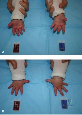
A 3-year-old patient undergoes reconstruction for a Blauth Type IIIA hypoplastic thumb. The procedure includes a comprehensive web space deepening, CMC joint stabilization, and an FDS ring finger tendon transfer for opposition. The image shows the thumb positioned during the critical step of tensioning the tendon transfer. What is the primary reason for positioning the thumb in full abduction, full flexion, and full pronation during this step?
Explanation
Correct Answer: C
Explanation:
The case material, under 'Detailed Surgical Approach / Technique' in the 'Opposition Reconstruction (Tendon Transfer)' section, specifically states: 'To achieve the pronation component of opposition, the tendon is often directed more towards the ulnar aspect of the thumb metacarpal or the insertion site on the APB tendon is created more ulnarly. The tendon is secured under appropriate tension using a Pulvertaft weave or a direct tendon-to-bone suture technique... The thumb should be positioned in full abduction, full flexion, and full pronation during tensioning. This is a critical step in restoring thumb function.' This position ensures that the transferred tendon is tensioned to provide all three components of opposition (abduction, flexion, and pronation) effectively.
- Option A (To prevent post-operative stiffness of the interphalangeal joint): While preventing stiffness is a general goal, this specific positioning during tendon tensioning is not primarily for IP joint stiffness.
- Option B (To ensure adequate blood supply to the transferred tendon): Tendon tensioning does not directly ensure blood supply; careful dissection and preservation of the pedicle (if applicable) or general vascularity are key.
- Option D (To facilitate easier skin closure of the web space): Skin closure is typically performed after the deep structures and tendon transfers are completed, and while thumb position affects web space tension, this specific position is for tendon tensioning, not primarily skin closure.
- Option E (To minimize tension on the CMC joint capsulodesis): While the position of abduction helps protect the CMC stabilization, the pronation and flexion components are specifically for optimizing the opposition transfer, not solely for CMC tension.
Question 17
A 4-year-old child who underwent reconstruction for a Blauth Type IIIA hypoplastic thumb 6 months ago presents with a progressive inability to abduct the thumb away from the palm, despite diligent post-operative therapy. Examination reveals a tight, contracted first web space and limited active opposition. The CMC joint remains stable. What is the most likely complication and its appropriate initial salvage strategy?
Explanation
Correct Answer: C
Explanation:
The patient's symptoms of 'progressive inability to abduct the thumb away from the palm' and a 'tight, contracted first web space' despite therapy are classic signs of recurrent adduction contracture. The case material lists 'Recurrent Adduction Contracture' as a common complication (10-30% incidence) and states its salvage strategy: 'Aggressive post-operative splinting/therapy. Serial casting. If established, revision web space deepening (repeat Z-plasty, dorsal rotation flap), full-thickness skin grafting, or release of deeper structures (adductor pollicis tenotomy/myotomy).' Since the contracture is established and progressive, revision web space deepening is the most appropriate surgical salvage strategy.
- Option A (Tendon transfer rupture; re-exploration and repair): While tendon transfer failure can cause limited opposition, the primary issue described is a tight web space and inability to abduct, pointing more to contracture than rupture.
- Option B (CMC joint instability; revision capsulodesis): The vignette states the CMC joint remains stable, ruling out this complication.
- Option D (Infection; intravenous antibiotics and debridement): There are no signs or symptoms of infection (e.g., redness, swelling, fever, purulent discharge).
- Option E (Neurovascular injury; surgical neurolysis): There are no signs of nerve or vascular compromise (e.g., paresthesias, numbness, pallor, diminished pulses).
Question 18
A 1-year-old patient is scheduled for reconstruction of a Blauth Type IIIA hypoplastic thumb. Pre-operative imaging includes standard AP, lateral, and oblique radiographs of the hand and wrist. Which of the following specific anatomical features is most critical to assess on these radiographs to guide the decision for reconstruction versus pollicization?
Explanation
Correct Answer: C
Explanation:
The case material emphasizes the importance of skeletal anatomy in guiding management. Under 'Pre-Operative Planning & Patient Positioning' -> 'Imaging Studies' -> 'Radiographs', it states: 'Specifically assess the trapezium, first metacarpal, and phalanges for size, shape, and articulation.' The stability and presence of sufficient skeletal elements, particularly the CMC joint formed by the trapezium and first metacarpal, are paramount for a functional reconstruction. If these are severely deficient or non-articulating (as in Blauth Type IIIB/IV), pollicization becomes the preferred option.
- Option A (The presence and ossification of the carpal bones): While carpal bone ossification centers can aid in estimating skeletal maturity, they are not the primary determinant for the reconstruction vs. pollicization decision in hypoplastic thumb.
- Option B (The integrity of the deep transverse metacarpal ligament): This ligament connects the metacarpal heads and is less directly involved in the primary pathology of thumb hypoplasia or the decision for reconstruction vs. pollicization.
- Option D (The presence of a Palmaris Longus tendon): The Palmaris Longus is a potential donor for tendon transfer, but its presence or absence does not dictate the fundamental choice between reconstruction and pollicization. Other donor tendons are available.
- Option E (The vascularity of the princeps pollicis artery): While crucial for any thumb surgery, vascularity is typically assessed clinically (capillary refill, pulses) or with specialized studies (arteriogram) if anomalies are suspected, not primarily on standard radiographs.
Question 19
A 5-year-old patient with a Blauth Type IIIA hypoplastic thumb undergoes an FDS ring finger tendon transfer for opposition. The surgeon plans to route the tendon. According to the case material, which routing method is most commonly employed for this procedure?
Explanation
Correct Answer: C
Explanation:
Under 'Detailed Surgical Approach / Technique' -> 'Opposition Reconstruction (Tendon Transfer)' -> 'FDS Ring Finger Transfer' -> 'Routing', the case states: 'Subcutaneous Route (most common): A subcutaneous tunnel is created across the palm, from the point of FDS delivery to the ulnar side of the thumb metacarpal. This route minimizes scar tissue adherence to deep structures.'
- Option A (Through the interosseous membrane to the dorsal aspect of the thumb): This routing is rarely used for opposition transfers and is more typical for extrinsic extensor transfers.
- Option B (Through the carpal tunnel, then radially to the thumb): While possible, the case notes this 'requires carpal tunnel release' and is not described as the 'most common' route for FDS opposition transfer.
- Option D (Deep to the adductor pollicis muscle): Routing deep to the adductor pollicis would be anatomically challenging, risk neurovascular structures, and not provide the optimal line of pull for opposition.
- Option E (Around the radial artery in the anatomical snuffbox): This is not a standard or recommended route for an FDS opposition transfer, which originates volarly.
Question 20
A 7-year-old patient with a history of Blauth Type IIIA hypoplastic thumb reconstruction 3 years prior presents with persistent, severe instability of the carpometacarpal (CMC) joint, significantly compromising pinch strength. Previous capsulodesis and K-wire fixation failed to provide lasting stability. The patient is otherwise healthy. What is the most appropriate surgical option for this patient's CMC joint instability?
Explanation
Correct Answer: B
Explanation:
The case material, under 'Detailed Surgical Approach / Technique' -> 'Carpometacarpal (CMC) Joint Stabilization' -> 'Arthrodesis', states: 'Indication: Reserved for severe, intractable CMC instability in older patients, failed previous reconstructions, or arthritic changes. Not typically a primary procedure in children due to concerns about growth and joint mobility.' Given the patient is 7 years old, has persistent severe instability after a failed capsulodesis, and is experiencing functional compromise, CMC arthrodesis is the most appropriate definitive solution to provide a stable base for pinch, despite the patient being a child. The benefits of stability often outweigh the loss of motion in such refractory cases.
- Option A (Repeat capsulodesis with more prolonged K-wire fixation): While a reasonable first step for initial failure, given the 'persistent, severe instability' after a previous attempt, a more definitive solution is warranted.
- Option C (Distraction osteogenesis of the first metacarpal): This procedure is for lengthening, not primarily for stabilizing a severely unstable joint.
- Option D (Pollicization of the index finger): Pollicization is for severe thumb aplasia or hypoplasia where the existing thumb is not reconstructible. This patient has an existing thumb that has undergone reconstruction, and the primary issue is CMC instability, not the overall viability of the thumb.
- Option E (Excision of the trapezium): Trapezium excision (trapeziectomy) is typically performed for CMC osteoarthritis in adults to relieve pain, not to stabilize a congenitally unstable joint in a child. It would further destabilize the thumb.
Question 21
A 1-year-old patient is diagnosed with a Blauth Type IIIA hypoplastic thumb. During the pre-operative workup, the orthopedic surgeon orders a thorough diagnostic evaluation to identify potential systemic associations. Which of the following syndromes or conditions is most commonly associated with congenital thumb hypoplasia and should be specifically screened for?
Explanation
Correct Answer: C
Explanation:
The 'Introduction & Epidemiology' section of the case explicitly states: 'It can occur as an isolated anomaly or, more commonly, in association with other syndromes and conditions, including VACTERL association (vertebral defects, anal atresia, cardiac defects, tracheoesophageal fistula, renal anomalies, limb defects) or VARCATL syndrome... Other associated conditions include Fanconi's anemia, Holt-Oram syndrome, and thrombocytopenia-absent radius (TAR) syndrome. A thorough diagnostic workup is essential to identify these systemic associations.' VACTERL is listed as one of the most common associations.
- Option A (Marfan Syndrome): Primarily affects connective tissue, leading to skeletal, ocular, and cardiovascular abnormalities, but is not commonly associated with congenital thumb hypoplasia.
- Option B (Ehlers-Danlos Syndrome): A group of connective tissue disorders characterized by joint hypermobility, skin hyperextensibility, and tissue fragility, not typically associated with thumb hypoplasia.
- Option D (Osteogenesis Imperfecta): Characterized by brittle bones, not primarily by congenital limb deficiencies like thumb hypoplasia.
- Option E (Achondroplasia): A form of short-limbed dwarfism, but not a common association with isolated or syndromic thumb hypoplasia.
Question 22
A newborn presents with a hypoplastic thumb. Radiographs reveal an absent proximal half of the first metacarpal and an absent carpometacarpal joint. What is the most appropriate definitive management?
Explanation
Question 23
An infant presents with a hypoplastic thumb. Exam reveals an absent thenar eminence, a narrowed first web space, and an unstable carpometacarpal (CMC) joint. Radiographs show partial absence of the first metacarpal. According to the Blauth classification, what is the most appropriate definitive management?
Explanation
Question 24
A 14-month-old child presents with a hypoplastic thumb. Examination reveals absent thenar musculature, an adducted first web space, and an unstable carpometacarpal joint. Radiographs show partial absence of the first metacarpal base. What is the most appropriate surgical management?
Explanation
Question 25
A newborn presents with bilateral absent thumbs and severe radial hypoplasia. Echocardiogram reveals an atrial septal defect. Genetic testing is most likely to show a mutation in which of the following genes?
Explanation
Question 26
The nail unit consists of several specialized structures. Which of the following best describes the primary function and characteristic of the sterile matrix?
Explanation
Question 27
A 9-year-old boy sustains a hyperflexion injury to his long finger, resulting in a clinically displaced Salter-Harris I fracture of the distal phalanx and an associated laceration at the proximal nail fold with the nail plate resting superficial to the eponychium. What is the most critical step in initial management?
Explanation
Question 28
During an index finger pollicization for Blauth Type IIIB thumb hypoplasia, the extrinsic and intrinsic muscles of the index finger are reassigned to replicate thumb function. The first dorsal interosseous muscle is typically transferred to function as which of the following?
Explanation
Question 29
A patient undergoes a revision amputation of the distal phalanx following a severe crush injury. Postoperatively, the patient develops a symptomatic hook nail deformity. Which of the following surgical errors most likely contributed to this complication?
Explanation
Question 30
A 3-year-old child presents with a hypoplastic thumb. Examination reveals a narrowed first web space, hypoplasia of the thenar muscles, and instability of the metacarpophalangeal (MCP) joint, but the carpometacarpal joint is clinically and radiographically stable. Which combination of procedures is most appropriate?
Explanation
Question 31
A 24-year-old machinist sustains an avulsion injury to the thumb nail unit, resulting in a 60% loss of the sterile matrix. The germinal matrix is intact. Bone is completely covered by a thin layer of healthy tissue. What is the most appropriate source for reconstruction of this defect?
Explanation
Question 32
A 1-year-old boy with a Blauth Type IV thumb hypoplasia is being evaluated for pollicization. He is noted to have short stature and café-au-lait spots. Which of the following laboratory tests is mandatory before proceeding with elective surgery?
Explanation
Question 33
Following the repair of a severe nail bed laceration with proximal avulsion, a stent is placed beneath the eponychial fold. What is the primary purpose of this step?
Explanation
Question 34
During a Buck-Gramcko pollicization of the index finger, careful mobilization of the neurovascular bundles is required. To allow adequate mobilization and rotation of the index finger, which vascular structure is typically ligated?
Explanation
Question 35
A neonate is evaluated in the nursery for bilateral upper extremity anomalies. Examination reveals bilateral absent radii, but the thumbs are present and appear normal. What systemic hematologic abnormality is classically associated with this specific presentation?
Explanation
Question 36
In an index finger pollicization procedure, the extensor digitorum communis (EDC) and extensor indicis proprius (EIP) tendons are reassigned. The EDC tendon of the index finger is typically sectioned and attached to the base of the new first metacarpal (former proximal phalanx) to function as which muscle?
Explanation
Question 37
What clinical or radiographic finding definitively separates Blauth Type IIIA from Type IIIB congenital thumb hypoplasia, thereby dictating the need for index finger pollicization rather than reconstruction?
Explanation
Question 38
During a Buck-Gramcko index finger pollicization for Blauth Type IV thumb hypoplasia, the first dorsal interosseous (FDI) muscle of the index finger is typically transferred to function as which thumb muscle?
Explanation
Question 39
Which of the following statements best describes the primary function of the sterile matrix in the human nail unit?
Explanation
Question 40
A 2-year-old child presents with a Blauth Type II hypoplastic thumb. Which combination of surgical procedures is most typically indicated for optimal functional restoration?
Explanation
Question 41
A patient presents with a progressive "hook nail" deformity 6 months after sustaining a distal fingertip amputation. What is the most likely biomechanical cause of this specific deformity?
Explanation
Question 42
When performing an index finger pollicization, preservation of which of the following structures is most critical for the viability of the transferred digit?
Explanation
Question 43
A 7-year-old boy catches his finger in a door, sustaining an injury where the proximal nail plate rests superficial to the eponychium.
Radiographs confirm a displaced distal phalanx physeal fracture. What is the most appropriate management?
Explanation
Question 44
A 1-year-old child with severe congenital thumb hypoplasia and radial longitudinal deficiency is suspected of having Fanconi anemia. Which diagnostic test is the gold standard for confirming this syndrome?
Explanation
Question 45
A 32-year-old presents with a painful subungual hematoma covering 40% of the nail plate following a crush injury. The nail margins are intact, and radiographs show no underlying fracture. What is the most appropriate treatment?
Explanation
Question 46
In the Buck-Gramcko index finger pollicization technique, the transferred digit is fixed in approximately what degree of palmar abduction and pronation to optimize functional thumb opposition?
Explanation
Question 47
A patient presents with an infection localizing to the junction of the distal nail bed and the fingertip skin. This specific anatomic area, which normally acts as a waterproof barrier preventing pathogens from entering the subungual space, is known as the:
Explanation
Question 48
In patients with Blauth Type II or IIIA thumb hypoplasia, the flexor pollicis longus (FPL) tendon frequently exhibits an aberrant anatomical characteristic known as "pollex abductus." What is the defining feature of this anomaly?
Explanation
Question 49
Following the meticulous repair of a severe nail matrix laceration, what is the primary rationale for replacing the native nail plate (or placing a synthetic stent) into the proximal eponychial fold?
Explanation
Question 50
A newborn infant presents with complete absence of the thumb, including all phalanges and the first metacarpal. Based on the Blauth classification, what is the most appropriate definitive management for this deformity?
Explanation
Question 51
A patient complains of a chronic longitudinal split in their nail plate following an old crush injury. This specific deformity is most frequently caused by persistent scarring in which of the following structures?
Explanation
Question 52
During reconstruction of a Blauth Type II thumb hypoplasia, an opponensplasty (Huber transfer) is planned to restore opposition and thenar bulk. Which of the following muscles is transferred in this specific procedure?
Explanation
Question 53
A 35-year-old machinist suffers a crush-avulsion injury resulting in complete loss of the eponychial fold and exposure of the proximal nail matrix. What is the most reliable method for reconstructing the eponychial fold to prevent severe future nail deformity?
Explanation
Question 54
When counseling parents regarding an index finger pollicization for their child's Blauth Type IV thumb hypoplasia, what preoperative factor should be emphasized as the strongest predictor of limited functional outcome in the newly constructed thumb?
Explanation
Question 55
A patient develops a severe acute paronychia that has progressed to a fluctuant abscess extending completely under the proximal eponychial fold. What is the standard surgical approach to adequately drain this without causing permanent nail deformity?
Explanation
None
