العربية
Comprehensive Master Guide · Medically Reviewed

Plates and Screws After Fracture: When to Consider Removal

Bone fractures are one of the most common injuries in car accidents, falls, and sports, and they may require surgical fixation using metal plates and screws to…

144 Detailed Chapters
15 min read
Updated: مارس 2026
Dr. Mohammed Hutaif
Medically Reviewed by
Prof. Dr. Mohammed Hutaif
Verified Content Expert Reviewed

Quick Medical Answer

This article provides essential research regarding Plates and Screws After Fracture: When to Consider Removal. The removal of orthopedic plates and screws after fracture healing is decided by weighing potential benefits against surgical risks. Removal is typically recommended for pain, infection, device failure, or allergic reactions. However, this second surgery carries risks such as bleeding, new infections, or weakening of the bone. If no issues arise, plates and screws are often safely retained.

Should plates and screws be removed after bone fractures heal?

An in-depth exploration of the considerations surrounding the removal of surgical plates and screws following the healing of a bone fracture. This comprehensive guide will delve into the benefits, risks, decision-making process, surgical and recovery phases, costs, and patient experiences, providing a pillar of content for those contemplating this procedure.


The Ultimate Guide to Hardware Removal Surgery After a Fracture: Should Your Plates and Screws Stay or Go?

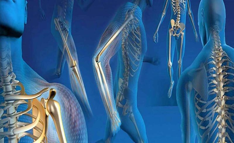
A sudden fall, a collision on the sports field, or a car accident can change your life in an instant, often resulting in a bone fracture. For many complex breaks, surgery is required to realign the bone and secure it with metal plates, screws, rods, or pins. This procedure, known as open reduction and internal fixation (ORIF), is a modern miracle of orthopedic medicine, providing the stability necessary for bones to heal correctly.

But once the bone has mended and you've completed months of rehabilitation, a new question emerges: What about the hardware inside? Does it need to come out? This question marks the beginning of a complex decision-making process for millions of people. While the hardware has served its purpose, its continued presence can be a source of debate and concern.

This guide is designed to be your comprehensive resource, a pillar of information to help you navigate this decision. We will explore every facet of orthopedic hardware removal, moving beyond simple pros and cons to provide a deep, evidence-based understanding of the entire journey. From the biological process of bone healing to the financial implications of a second surgery, we will arm you with the knowledge needed to have an informed conversation with your surgeon and make the choice that is right for you.

Chapter 1: A Foundation in Bone Healing and Surgical Fixation

Before we can decide whether to remove the hardware, it's essential to understand why it was put there in the first place and how the bone marvelously repairs itself.

The Miraculous Process of Bone Healing

When a bone fractures, the body initiates a remarkable four-stage healing cascade:

  1. Inflammation and Hematoma Formation: Immediately after the fracture, blood vessels rupture, causing a blood clot, or hematoma, to form at the site. This signals the start of the inflammatory response, bringing in specialized cells to begin the cleanup and repair process.
  2. Soft Callus Formation: Within a few days, the hematoma is gradually replaced by a soft callus made of fibrocartilage. This acts as a natural, flexible splint, providing initial stability to the fracture.
  3. Hard Callus Formation: Over the next several weeks, the soft callus is converted into a hard, bony callus. Osteoblasts, the bone-building cells, work tirelessly to create new bone, bridging the gap between the fractured ends. This is the stage where the bone begins to feel solid again.
  4. Bone Remodeling: This final stage can last for months or even years. The bulky hard callus is slowly reshaped and remodeled by the body into strong, compact bone. This process allows the bone to adapt to the loads and stresses placed upon it, eventually returning it to its original shape and strength.

The Role of Orthopedic Hardware

In a simple, well-aligned fracture, a cast may be all that is needed. However, in more severe fractures, the bone fragments can be displaced. This is where surgical fixation becomes critical. The primary goal of internal fixation with plates and screws is to achieve and maintain anatomical reduction—placing the bone fragments back into their precise, original alignment—to allow the healing process to occur correctly.

This hardware is crafted from biocompatible materials designed to be strong, durable, and non-reactive with the body. The most common materials are:

  • Medical-Grade Stainless Steel: A strong and cost-effective option.
  • Titanium and its Alloys: Known for their excellent strength-to-weight ratio, corrosion resistance, and superior biocompatibility. Titanium is also more compatible with MRI scans, causing less distortion of the image.

These devices act as an internal splint, holding everything in place while the body performs its natural healing magic.

Chapter 2: The Great Debate: To Remove or Not to Remove?

This is the central question. For some, the decision is clear-cut, driven by significant symptoms. For most, it's a gray area, requiring a careful weighing of the potential benefits against the undeniable risks of a second operation.

Part A: Compelling Reasons for Hardware Removal

While many people live comfortably with their hardware for a lifetime, there are several well-established reasons why removal surgery, or implant removal, becomes a necessary or highly desirable option.

1. Pain, Discomfort, and Irritation

This is the most common reason for hardware removal. The pain can manifest in several ways:
* Prominence Under the Skin: In areas with little soft tissue coverage, like the ankle, collarbone (clavicle), elbow, and wrist, hardware can be prominent. It can rub against clothing, shoes, or straps, causing persistent irritation.
* Tendon and Soft Tissue Irritation: Plates and screw heads can irritate overlying tendons as they glide back and forth, leading to tendonitis or a painful snapping sensation.
* Temperature Sensitivity: Some individuals report an aching pain at the hardware site in cold weather. While the exact mechanism is debated, it is a frequently reported symptom that often resolves after removal.

2. Infection

While uncommon, an infection can develop around the orthopedic implant.
* Early Infection: Occurs shortly after the initial surgery.
* Late-Onset Infection: Can appear months or even years later, sometimes after a minor illness or injury. Bacteria can form a protective layer on the implant called a biofilm, which makes them highly resistant to antibiotics alone. In cases of chronic, deep infection (osteomyelitis), removal of the hardware is almost always required to eradicate the infection.

3. Functional Impairment and Limited Range of Motion

Sometimes, the hardware itself can mechanically block a joint's full range of motion. A plate located near the elbow or ankle joint, for instance, might impede full flexion or extension, limiting a person's ability to perform daily activities or participate in sports.

4. Hardware Failure or Loosening

Though designed to be incredibly strong, orthopedic hardware can fail under repetitive stress. This can include the loosening or backing out of screws, or, in rare cases, the breaking of a plate. This is more likely to happen if the bone fails to heal properly (a "nonunion"), placing the full load on the hardware indefinitely. The signs of hardware failure include new pain, a popping or clicking sound, or a visible deformity, which would necessitate removal.

5. Allergic Reactions and Hypersensitivity

A small percentage of the population has allergies to metals like nickel, cobalt, or chromium, which can be components of stainless steel implants. An allergic reaction can cause a localized skin rash, persistent pain, swelling, and poor wound healing. If a metal allergy is suspected, a doctor may perform a patch test, and if positive, hardware removal is recommended.

6. Patient Preference and Psychological Factors

For some, the knowledge that they have a "foreign object" in their body is psychologically unsettling. Others may be concerned about the aesthetics of a prominent implant or a visible scar. For high-level athletes, the decision to remove hardware may be driven by a desire to return to their sport without any potential for implant-related complications during a future injury.

7. Long-Term Concerns

  • Stress Shielding: A very rigid plate can "shield" the underlying bone from the normal stresses of daily life. According to Wolff's law, bone remodels and strengthens in response to the loads placed upon it. If the plate carries too much of the load, the bone beneath it can gradually lose density and become weaker (osteopenia). Removing the plate allows the bone to be subjected to normal physiological stress, which can improve its density over time.
  • Interference with Imaging: Hardware can create significant artifacts on MRI and CT scans, making it difficult to diagnose future problems in that area.

Part B: The Case for Leaving Hardware In Place

If the hardware isn't causing any problems, the argument to "let sleeping dogs lie" is a powerful one. Every surgery, no matter how routine, carries inherent risks.

1. Avoiding the Risks of a Second Surgery

This is the most significant reason to leave asymptomatic hardware in place. The potential complications are serious and must be considered:
* Risk of Infection: Any time the skin is cut, there is a risk of introducing bacteria, which could lead to a serious infection.
* Nerve and Blood Vessel Damage: The hardware is often close to important nerves and blood vessels. During the removal surgery, these structures can be bruised, stretched, or even severed, potentially leading to numbness, weakness, or permanent paralysis in the affected area. The risk is particularly notable in areas like the upper arm (radial nerve) and lower leg (peroneal nerve).
* Anesthesia Complications: Whether general or regional, anesthesia carries its own set of risks, ranging from minor nausea to rare but severe reactions.
* Refracture: After the hardware is removed, the bone is left with empty screw holes. These holes act as "stress risers," creating weak points in the bone. There is a small but significant risk of the bone re-fracturing through one of these holes, especially if a patient returns to high-impact activities too soon.

2. The Difficulty of the Procedure

Hardware removal is not always a simple reversal of the initial surgery. Over time, bone can grow over and around the plates and screws, encasing them and making them difficult to extract. Screw heads can become stripped, or a phenomenon known as "cold welding" can occur, essentially fusing the screw to the plate. A difficult extraction can increase surgical time and raise the risk of complications like nerve damage or fracture.

3. The Second Recovery Period

Hardware removal is not a minor procedure. It requires another period of recovery, which can involve:
* Time off from work.
* A period of restricted weight-bearing or activity.
* Additional physical therapy.
* Pain and swelling.

For many, enduring another rehabilitation period is a significant deterrent.

Chapter 3: The Decision-Making Process: A Personalized Approach

There is no universal answer to the hardware removal question. The decision is highly individualized and should be made in close consultation with your orthopedic surgeon. Here are the key factors that will guide the conversation.

A Factor-Based Checklist

  • Anatomical Location: This is paramount. Hardware in some locations is more likely to be symptomatic and is more frequently removed.
    • High Removal Rates: Ankle (fibula), collarbone (clavicle), elbow (olecranon). These areas have thin skin and the hardware often causes irritation.
    • Moderate Removal Rates: Wrist/Forearm (radius/ulna), hand, foot. Removal is typically only for specific symptoms like tendon irritation.
    • Low Removal Rates: Upper arm (humerus), thigh (femur), shin (tibia). The significant risks (e.g., to the radial nerve in the humerus) and the fact that the hardware is buried deep under large muscles mean it is often left in place unless there is a compelling reason for removal. Intramedullary nails placed down the center of long bones are almost always left in permanently.
  • Patient Age and Activity Level:
    • Younger, Active Patients & Athletes: These individuals are more likely to opt for removal. They have a longer lifespan ahead of them, are more likely to experience issues during high-impact sports, and may benefit from addressing long-term concerns like stress shielding.
    • Older, More Sedentary Patients: For this group, the risks of a second surgery and another round of anesthesia and rehabilitation often outweigh the potential benefits, especially if the hardware is asymptomatic.
  • Timing is Everything: Hardware should never be removed until the fracture is completely and solidly healed. X-rays must confirm this. Most surgeons recommend waiting at least 12 to 18 months after the initial surgery to allow for full bone consolidation and remodeling.
  • Symptoms are the Main Driver: The presence and severity of pain, irritation, or functional limitation remain the number one reason to proceed with removal.

Your Consultation with the Surgeon: Questions to Ask

This decision is a partnership between you and your doctor. Be prepared for your appointment with a list of questions:
* "Given the specific location of my hardware, what are the primary risks of this removal surgery for me?"
* "In your experience, what percentage of patients with my injury and hardware find relief from their symptoms after removal?"
* "What are the chances that my pain is not from the hardware and will persist even after the surgery?"
* "What would the recovery process look like? How long until I can return to work/sports?"
* "What happens if the hardware is difficult to remove? Is it possible you might have to leave a piece behind?"
* "What is the risk of refracture, and how can I minimize it?"

Chapter 4: The Journey: Surgery and Recovery

If you and your surgeon decide to proceed, understanding the process can help alleviate anxiety and set you up for a successful outcome.

Preparing for Your Surgery

Preparation typically involves a pre-operative physical, bloodwork, and a consultation with the anesthesiologist to determine the safest method of anesthesia for you. You will need to arrange for time off from work and for help at home during the initial recovery period.

The Surgical Procedure

In many cases, the surgeon can use the original incision line, which helps to minimize additional scarring. The steps generally include:
1. Incision and Exposure: The surgeon carefully dissects through the layers of soft tissue to expose the plate and screws.
2. Hardware Removal: Each screw is carefully removed, followed by the plate itself. This is the most delicate part of the operation, requiring patience to avoid damaging surrounding structures.
3. Wound Closure: The incision is thoroughly cleaned and closed in layers.

The Road to Full Recovery

Recovery from hardware removal is typically faster and less painful than the original fracture surgery, but it still requires care and patience.
* The First Two Weeks: The focus is on pain management, rest, and keeping the incision clean and dry. Depending on the location, you may have weight-bearing restrictions to protect the bone.
* Weeks Two to Six: Your sutures or staples will be removed. You will likely begin a course of physical therapy to restore range of motion and strength.
* Beyond Six Weeks: As you regain function, you will be gradually cleared for more activity. However, it's crucial to remember the screw holes. It can take six months to a year or more for the bone to fully remodel and fill in these gaps. During this time, you must avoid high-impact activities or contact sports to prevent a refracture. Physical therapy is essential to ensure a complete and successful recovery.

Chapter 5: Unpacking the Costs and Navigating Insurance

The financial aspect of hardware removal surgery cannot be overlooked. Understanding the distinction between "medically necessary" and "elective" procedures is key.
* Medically Necessary: If you are having the hardware removed due to documented pain, infection, functional limitation, or hardware failure, insurance is very likely to cover the procedure.
* Elective: If the removal is for purely cosmetic reasons or a personal desire without symptoms, it may be deemed elective, and your insurance provider may deny coverage.

The total cost can vary dramatically based on your location, hospital, and insurance plan, but it includes the surgeon's fee, the anesthesiologist's fee, and the hospital or surgery center facility fees. Always obtain pre-authorization from your insurance company before scheduling the surgery to understand what will be covered and what your out-of-pocket expenses will be.

Chapter 6: Patient Stories and Finding the Right Expert

While data provides the facts, personal stories provide perspective.
* Case Study 1: The Runner's Ankle. A 30-year-old marathon runner had a plate and screws placed for a fibula fracture. A year later, while the bone was healed, the screw heads constantly rubbed against her running shoes, causing debilitating pain. For her, the decision was easy. After removal and a three-month recovery, she was back to pain-free running.
* Case Study 2: The Office Worker's Arm. A 55-year-old man had a plate on his humerus (upper arm bone). It caused him no pain or issues in his daily life. After discussing the significant risk to his radial nerve with his surgeon, he confidently decided against removal, deeming the risks far too great for a problem that didn't exist.

These stories highlight the spectrum of experiences. To navigate your own journey, it's vital to find a qualified surgeon. Look for a board-certified orthopedic surgeon with experience in trauma and, specifically, in hardware removal procedures.

Conclusion: A Decision Uniquely Yours

The question of whether to remove orthopedic hardware is not a simple one. There is no right or wrong answer, only the answer that is right for you. It is a deeply personal decision that requires a thorough understanding of the potential rewards and the unavoidable risks.

By understanding the mechanics of your injury, the function of your hardware, and the nuances of the removal process, you can move from a place of uncertainty to one of empowerment. Engage in an open and honest dialogue with your surgeon, weigh the evidence, listen to your body, and consider your lifestyle. This comprehensive approach will ensure that you make a confident, well-informed choice that best supports your long-term health and well-being.


Frequently Asked Questions (FAQ)

  1. How long does hardware removal surgery typically take?

    • It varies depending on the location and complexity, but most procedures take between 30 and 90 minutes.
  2. Will I have a bigger scar?

    • Surgeons almost always try to use the original incision line, so while the scar will be re-opened, it shouldn't be significantly larger.
  3. What happens to the empty screw holes in the bone?

    • The body's natural remodeling process will gradually fill in the holes with new bone over many months.
  4. Will my bone be weaker after the hardware is removed?

    • Initially, yes, due to the screw holes. However, once the holes have remodeled and the bone is no longer subject to stress shielding, it may actually regain its natural strength and density.
  5. Can I keep my hardware after it's removed?

    • Yes, in most cases. You can ask your surgeon to have the hardware cleaned and given to you after the procedure.
  6. Can I go through airport security with orthopedic hardware?

    • Yes. Modern scanners are very advanced. While your implant may be detected, it is a common occurrence, and security personnel are trained to handle it. You can carry a doctor's note, but it is usually not necessary.
  7. Is it true that hardware hurts more in cold weather?

    • Many patients report this phenomenon. While not fully understood scientifically, theories suggest that the metal, which conducts temperature differently than bone, may cause the surrounding tissues to contract, leading to a sensation of aching or pain.

Detailed Chapters & Topics

Dive deeper into specialized chapters regarding should-plates-and-screws-be-removed-after-bone-fractures-heal

144 Chapters
01
Chapter 1 10 min

Fracture Healing Understanding: How Bones Repair & What Treatments Help

Fracture Healing 1. What are the 3 stages of fracture healing? Show Answer Show Explanation 2. How do bone stimulators …

02
Chapter 2 24 min

Femoral Shaft Fractures: Comprehensive Management & Surgical Principles

Explore femoral shaft fractures: epidemiology, ATLS diagnosis (X-ray, CT), and surgical treatment, primarily with IM na…

03
Chapter 3 26 min

Navigating Femoral Shaft Fractures: Diagnosis to Successful Recovery

Understanding Femoral Shaft Fractures: Anatomy, Diagnosis, and Treatment Understanding Femoral Shaft Fractures: Anatomy…

04
Chapter 4 22 min

Diaphyseal Tibial Fractures: What You Must Know for Recovery

Diaphyseal Tibial Fractures: Diagnosis, Treatment, and Prognosis Diaphyseal Tibial Fractures Diaphyseal tibial fracture…

05
Chapter 5 28 min

Pelvic Ring Fractures: Your Guide to Diagnosis, Treatment & Recovery

Pelvic Ring Fractures: Diagnosis, Treatment, and Prognosis Pelvic Ring Fractures Pelvic ring fractures are high energy …

06
Chapter 6 50 min

Master All Types of Screws in Orthopedics: FRCS Exam Guide

This resource explores the different types of screws used in orthopedics, including corcal and cancellous screws, fully…

07
Chapter 7 57 min

Orth Oral Examination: Plates – Ace Every Examiner Question

In this FRCS (Tr & Orth) Oral Examination concept review, the candidate is asked to identify and describe a laminated p…

08
Chapter 8 62 min

Orthopedic Lag Screws: Biomechanics, Principles, and FRCS Exam Prep MCQs

Master lag screw biomechanics for orthopedic FRCS & board exams. Understand fracture fixation principles, interfragment…

09
Chapter 9 10 min

Sternoclavicular Joint Injury: Identify, Treat & Recover

The sternoclavicular (SC) joint is the only true joint that connects the upper arm and the chest. It is located at the …

10
Chapter 10 29 min

Orthopedic Splinting Mastery: Treating a Condition Where Damage Occurs

Splinting is a nonsurgical procedure that involves applying a rigid or semi-rigid device to an injured limb to immobili…

11
Chapter 11 9 min

Closed Reduction of Fractures: Master the Techniques & Principles

Fractures are common injuries that affect the skeletal system and require appropriate management to restore function an…

12
Chapter 12 9 min

Multiple Trauma Patients: Essential Guide to Care & Recovery

Multiple trauma is a serious condition that occurs when a person sustains two or more injuries in a single event. Learn…

13
Chapter 13 26 min

Proximal Femoral Fractures: Comprehensive Guide to Epidemiology, Anatomy, & Biomechanics

Explore proximal femoral fractures (PFFs): epidemiology, key classifications (Garden, Pauwels, AO/OTA), and vital surgi…

14
Chapter 14 21 min

What Causes Femoral Fractures? Essential Guide & Prevention Tips

Proximal femoral fractures, also known as hip fractures, are common injuries that affect older adults, especially women…

15
Chapter 15 30 min

Comprehensive Guide to Bone Fractures: Epidemiology, Classification, & Surgical Anatomy

Comprehensive guide to bone fractures for orthopedic residents & surgeons. Covers epidemiology, etiologies, and detaile…

16
Chapter 16 10 min

Mastering Proximal Humeral Fractures: Key Insights & Treatment

Summary Proximal humeral fractures are the third most common fractures and affect both young and elderly patients. Neer…

17
Chapter 17 12 min

Complex Tibial Shaft Fracture: Plate Fixation for Plafond Extension

Tibial shaft fracture extending into the plafond—plate fixation Case description A 26-year-old man fell from a height o…

18
Chapter 18 20 min

Holstein–Lewis Fracture: Comprehensive Clinical Management & Radial Nerve Injury

A comprehensive guide to Holstein–Lewis fracture, a distal humeral injury highly associated with radial nerve palsy (10…

19
Chapter 19 10 min

Partial Articular Fractureplate Fixation: Restore Anatomy

Partial articular fracture—plate fixation Case description A 53-year old man injured his right ankle in a motor vehicle…

20
Chapter 20 7 min

Distal Tibia Fracture Repair: Mastering Plating and Screws

Distal Tibia Medial plating and screws Case description A 65-year-old man fell down a flight of stairs and presented to…

21
Chapter 21 26 min

Greater Tuberosity Fracture: Epidemiology, Surgical Anatomy, Biomechanics, and Management

Learn about greater tuberosity fractures: their epidemiology, common causes (trauma, avulsion), surgical anatomy (rotat…

22
Chapter 22 8 min

Tibial Fracture Extending into the Plafond: Intramedullary Secrets

Tibial shaft fracture extending into the plafond—intramedullary fixation Case description a A 25-year-old woman landed …

23
Chapter 23 13 min

Decoding Pilon Fractures: Anterolateral and Medial Fragments Revealed

Metaphyseal fracture with joint involvement Case description A 38-year-old woman was involved in a motor vehicle accide…

24
Chapter 24 26 min

Ipsilateral Femoral Neck & Shaft Fractures: A Comprehensive Management Guide

Navigate the complexities of ipsilateral femoral neck & shaft fractures. This guide details epidemiology, diagnostic pi…

25
Chapter 25 21 min

Young Adult Femoral Shaft Fractures: Comprehensive Surgical Management & Outcomes

Detailed guide to femoral shaft fractures in young adults. Learn about epidemiology, surgical anatomy, biomechanics, op…

26
Chapter 26 17 min

Periprosthetic Femoral Fracture with Aseptic Stem Loosening: A Comprehensive Case Study

Explore a detailed case study of a 78-year-old with periprosthetic femoral fracture post-THA. Covers presentation, clin…

27
Chapter 27 17 min

High-Energy Pelvic Ring Injury: A Comprehensive Case Study of APC III with Vertical Shear

Explore a detailed case of a high-energy APC III pelvic ring injury with vertical shear and sacral fracture. Covers cli…

28
Chapter 28 27 min

Pathological Fractures: Etiology, Diagnosis, Biomechanics & Mirels' Score

Explore pathological fractures: definition, common etiologies like metastatic bone disease & tumors, diagnosis via imag…

29
Chapter 29 3 min

Comprehensive Pelvic Fracture Case Study: Interactive Module for Orthopedic Viva Exam Preparation

Master pelvic fracture diagnosis & management with our interactive case study. Ideal for orthopedic residents, test you…

30
Chapter 30 26 min

Tibial Plateau Fractures: Comprehensive Guide to Epidemiology, Classification & Biomechanics

Explore tibial plateau fractures, complex intra-articular knee injuries. Understand their epidemiology, detailed Schatz…

31
Chapter 31 24 min

Distal Femoral Fractures: Epidemiology, Anatomy, Biomechanics, and Surgical Principles

Explore comprehensive insights into distal femoral fractures (DFX): epidemiology, AO/OTA classification, surgical anato…

32
Chapter 32 22 min

Comprehensive Guide to Bone Ossification: Fetal Development, Growth Plates & Pediatric Orthopedic Implications

Delve into bone ossification, covering fetal primary & postnatal secondary centers, and growth plate mechanics. Essenti…

33
Chapter 33 8 min

Pathologic Fractures: Understand Causes, Identify Risks & Get Care

PATHOLOGIC FRACTURES DEFINITION A pathologic fracture is one that occurs when the normal integrity and strength of bone…

34
Chapter 34 59 min

Transverse Ligament Rupture: Recognizing & Treating This Serious Injury

Transverse Ligament Rupture (Traumatic C1–C2 Instability) This rare, usually fatal, injury is seen mostly in older age …

35
Chapter 35 14 min

Understanding Femoral Shaft Fractures: From Epidemiology to Treatment

FEMORAL SHAFT Fractures EPIDEMIOLOGY The highest age- and gender-specific incidences of femoral shaft fracture are seen…

36
Chapter 36 13 min

Tibiafibula Shaft Fractures: Causes, Symptoms & Care

TIBIA/FIBULA SHAFT Fractures EPIDEMIOLOGY Fractures of the tibia and fibula shaft are the most common long bone fractur…

37
Chapter 37 10 min

Humeral Shaft Fractures: Expert Insights on Diagnosis & Treatment

HUMERAL SHAFT FRACTURES EPIDEMIOLOGY It is a common injury, representing 3% to 5% of all fractures. Incidence is 14.5 p…

38
Chapter 38 29 min

Odontoid Process (Dens) Fractures: Epidemiology, Anatomy, Biomechanics & Classification

Delve into odontoid process (dens) fractures: epidemiology (bimodal distribution, trauma), intricate surgical anatomy o…

39
Chapter 39 17 min

Atlas (C1) Fractures: Advanced Insights into Pathophysiology, Anatomy & Clinical Management

Understand Atlas (C1) fractures: epidemiology, high-energy mechanisms, and Levine classification. Explore C1-C2 stabili…

40
Chapter 40 19 min

C2 Lateral Mass Fractures: A Comprehensive Academic Review of Diagnosis & Management

Detailed guide to C2 lateral mass fractures: epidemiology, surgical anatomy, C1-C2 biomechanics, and diagnostic insight…

41
Chapter 41 27 min

Sternoclavicular Joint Injury: A Comprehensive Academic Review of Epidemiology, Anatomy, and Biomechanics

Explore sternoclavicular (SC) joint injuries, their rare epidemiology, high-energy trauma causes, and critical surgical…

42
Chapter 42 8 min

Decoding Femoral Neck Fractures: Risks, Anatomy & What to Know

FEMORAL NECK FRACTURES EPIDEMIOLOGY More than 250,000 hip fractures occur in the United States each year (50% involve t…

43
Chapter 43 25 min

Orthopedic Gunshot Wounds: Ballistics, Energy Transfer & Injury Biomechanics

Delve into orthopedic gunshot wound biomechanics, epidemiology, and energy transfer. Understand low/high-velocity proje…

44
Chapter 44 10 min

Periprosthetic Fractures: Causes, Risks, & Effective Treatment

PERIPROSTHETIC FRACTURES TOTAL HIP ARTHROPLASTY Femoral Shaft Fractures Epidemiology Intraoperative: There is a 0.3% up…

45
Chapter 45 13 min

Multiple Trauma: A Complete Guide to Multiple Injury Management

MULTIPLE TRAUMA INTRODUCTION High-velocity trauma is the number one cause of death in the 18- to 44-year-old age group …

46
Chapter 46 10 min

Mastering Type IV Fractures: Essential Periprosthetic Strategies

PERIPROSTHETIC FRACTURES TOTAL HIP ARTHROPLASTY Femoral Shaft Fractures Epidemiology Intraoperative: There is a 0.3% up…

47
Chapter 47 18 min

Proximal Tibial Diaphyseal Fracture: High-Energy Trauma Case Study, Clinical & Imaging Diagnostics

Explore a detailed high-energy motorcycle accident case of a proximal tibial diaphyseal fracture. Learn about patient p…

48
Chapter 48 16 min

High-Energy Comminuted Tibial Diaphyseal Fracture: Comprehensive Patient Presentation, Clinical Examination & Diagnostic Imaging

Detailed case study of a high-energy comminuted tibial diaphyseal fracture in a 32-year-old. Covers patient presentatio…

49
Chapter 49 18 min

Acute Compartment Syndrome Diagnosis in Open Tibial Fractures: A Clinical Case Study

Explore a detailed case of acute compartment syndrome following an open tibial shaft fracture. Learn to identify 'red f…

50
Chapter 50 18 min

Complex Lower Extremity Trauma: A Case Study of Gustilo IIIC Open Tibia-Fibula Fracture & Critical Ischemia

Comprehensive case study on a Gustilo IIIC open tibia-fibula fracture with critical limb ischemia. Details patient pres…

51
Chapter 51 23 min

High-Energy Diaphyseal Tibia Fractures: Epidemiology, Anatomy & Biomechanics Guide

Explore high-energy diaphyseal tibia fractures: epidemiology, risk factors, and common complications. Delve into surgic…

52
Chapter 52 23 min

Intramedullary Nailing for Proximal Tibia Fractures: Mastering Reduction & Preventing Deformities

Explore intramedullary nailing (IMN) for proximal tibia fractures. Understand unique challenges like malreduction (valg…

53
Chapter 53 79 min

Femoral Neck and Shaft Fractures: Epidemiology, Surgical Anatomy & Biomechanics of Isolated & Ipsilateral Injuries

Explore femoral neck and shaft fractures, including epidemiology, detailed surgical anatomy, biomechanics, and the crit…

54
Chapter 54 27 min

Surgical Plates in Orthopedic Trauma: Biomechanics, Functions, and Clinical Application

Explore surgical plates in orthopedic trauma: their evolution, materials (titanium, steel), biomechanics, and functions…

55
Chapter 55 24 min

Terrible Triad Injury of the Elbow: Epidemiology, Anatomy, Biomechanics & Surgical Indications

Master the terrible triad elbow injury: a severe elbow instability with radial head fracture, coronoid fracture, and LU…

56
Chapter 56 19 min

Complex Calcaneal Fracture: Clinical Presentation, Advanced Imaging & Sanders IIIBC Classification Case Study

Explore a detailed calcaneal fracture case: 38M after a 10ft fall. Covers patient presentation, clinical exam, X-ray & …

57
Chapter 57 8 min

Pilon Fracture Case: Critical Steps for High-Energy Injury

Describe the radiographs pictured above. These are AP and lateral radiographs showing a comminuted and displaced pilon …

58
Chapter 58 18 min

Geriatric Complex Acetabular Fractures: A Detailed Clinical Case Study

Explore a detailed case study of a 78-year-old with a complex acetabular fracture. Covers patient history, clinical exa…

59
Chapter 59 20 min

Acute Compartment Syndrome Post-Trauma: A Comprehensive Clinical Case Study

Clinical case: Acute compartment syndrome post-motorcycle trauma. Covers history, exam (tense compartments, nerve defic…

60
Chapter 60 21 min

Managing Complex Open Tibia Fractures with Neurovascular Injury: A Case Study

Explore a detailed case study of a 35-year-old male with a Gustilo IIIC open tibia fracture from high-energy trauma. Le…

61
Chapter 61 18 min

Orthopedic Trauma: A Comprehensive Guide to Low and High-Velocity Gunshot Wound Management

Understand the critical distinction between low and high-velocity gunshot wounds in orthopedic trauma. This guide detai…

62
Chapter 62 24 min

Gusillo–Anderson Classification System: A Comprehensive Review of Pilon Fracture Management

Gusillo–Anderson (G-A) classification for distal tibia pilon fractures. Details articular, metaphyseal, and soft tissue…

63
Chapter 63 15 min

Open Fractures: Why Continuous Pressure Monitoring is Crucial

1. Describe what you see and your initial management in the emergency department. This is a clinical photograph reveali…

64
Chapter 64 19 min

High-Energy Syndesmotic Ankle Injury: Clinical Presentation & Diagnostic Insights

Learn to diagnose high-energy syndesmotic ankle injuries with this detailed case study. Covers clinical presentation, s…

65
Chapter 65 23 min

Coronoid Fractures: Comprehensive Guide to Anatomy, Biomechanics, & Elbow Instability

Coronoid fractures are critical for elbow stability. This guide explores their epidemiology, detailed anatomy, biomecha…

66
Chapter 66 23 min

Nonunion Fractures: Orthopedic Epidemiology, Biomechanics, & Classification

Explore nonunion fractures, defined by delayed healing, affecting 5-10% of fractures. Understand risk factors, surgical…

67
Chapter 67 23 min

Orthopedic Management of Gunshot Wounds: Ballistics, Anatomy & Surgical Indications

Explore comprehensive orthopedic management of gunshot wounds (GSWs), detailing epidemiology, ballistics, biomechanics,…

68
Chapter 68 27 min

Early Total Care in Orthopedic Trauma: Principles, Anatomy, and Biomechanics

Delve into Early Total Care (ETC) in orthopedic trauma: its definition, goals, epidemiology, and critical surgical anat…

69
Chapter 69 23 min

Advanced Trauma Life Support (ATLS): Principles, Anatomy & Biomechanics for Orthopedic Trauma

Explore Advanced Trauma Life Support (ATLS) principles, epidemiology, and critical anatomical systems (ABCDE) for rapid…

70
Chapter 70 51 min

Humeral Shaft Fracture: Optimal Treatment & Orthopedic Board Prep MCQs

Master optimal treatment for humeral shaft fractures with our orthopedic board prep MCQs. Review non-operative manageme…

71
Chapter 71 23 min

Radial Head Fractures: Comprehensive Epidemiology, Classification, Anatomy & Biomechanics

Gain a deep understanding of radial head fractures, including epidemiology, Mason-Hotchkiss classification, associated …

72
Chapter 72 24 min

Adolescent Distal Tibial Tillaux & Triplane Fractures: Pathophysiology, Diagnosis, and Management

Master Tillaux & Triplane fractures: unique intra-articular distal tibia injuries in adolescents. Understand complex mo…

73
Chapter 73 26 min

Periprosthetic Fractures: Epidemiology, Biomechanics, and Advanced ORIF Principles

Explore periprosthetic fractures: epidemiology, surgical anatomy, and ORIF principles. Understand Vancouver & OTA class…

74
Chapter 74 18 min

Mastering Supramalleolar Osteotomy with Internal Fixation

DEFINITION A supramalleolar osteotomy is an osteotomy at the level of the distal tibia with or without osteotomy of the…

75
Chapter 75 18 min

Percutaneous Pedicle Screw: Minimally Invasive Trauma Fixation

DEFINITION The advancement of minimally invasive techniques in spinal surgery, specifically percutaneous pedicle screw …

76
Chapter 76 21 min

Optimizing Internal Fixation of Sesamoid Fractures

DEFINITION Hallux sesamoid bone fracture is a break through the sesamoid bone or cartilage. Medial sesamoid bone fractu…

77
Chapter 77 27 min

Secure Sacropelvic Fixation: Guiding Screws Toward the AIIS

DEFINITION Sacropelvic fixation is a term used to describe instrumentation into the sacrum and pelvis. The most common …

78
Chapter 78 22 min

Mastering Supracondylar & Intercondylar Fracture Fixation

PATIENT HISTORY AND PHYSICAL FINDINGS Distal humerus fractures occur in two age groups: Younger patients who sustain hi…

79
Chapter 79 23 min

Mastering ORIF for Capitellartrochlear Shear Fractures

DEFINITION Capitellar fractures are uncommon, accounting for less than 1% of all elbow fractures and 6% of all distal h…

80
Chapter 80 23 min

Radial Head & Neck Fractures: Mastering ORIF for Best Results

DEFINITION Radial head and neck fractures are the most common elbow fractures in adults representing 33% of elbow fract…

81
Chapter 81 18 min

Optimizing Plate Fixation of Humeral Shaft Fractures

DEFINITION Humeral shaft fractures, which account for about 3% of adult fractures, usually result from a direct blow or…

82
Chapter 82 27 min

Release of Posttraumatic Metacarpophalangeal and Proximal Interphalangeal Joint Contractures

DEFINITION Posttraumatic metacarpophalangeal (MCP) joint and proximal interphalangeal (PIP) contractures may devel…

83
Chapter 83 33 min

Lateral to Medial Nailing: Optimizing Humeral Fracture Fixation

r DEFINITION Incidence: 3% to 5% of all fractures 12 The AO/ASIF classification of humeral shaft fractures is based on …

84
Chapter 84 23 min

Navigating Capitellum and Capitellar-Trochlear Shear Fracture Repair

DEFINITION Capitellar fractures are uncommon, accounting for less than 1% of all elbow fractures and 6% of all distal h…

85
Chapter 85 21 min

Microfracture Chondroplasty: Repairing Cartilage with Microfracture

DEFINITION Chondral defects in the knee are common. The lesions may be partial or full thickness ( FIG 1 ) , through al…

86
Chapter 86 24 min

Chronic Exertional Compartment Syndrome: Causes, Symptoms & Relief

DEFINITION Compartment syndrome can be either acute or chronic. Acute compartment syndrome is usually due to trauma to,…

87
Chapter 87 22 min

Beyond Plates: Intramedullary Fixation for Displaced Midshaft Clavicle

DEFINITION The clavicle is one of the most commonly fractured bones. The site on the clavicle most often fractured is t…

88
Chapter 88 19 min

Intramedullary Nailing: Mastering Proximal or Distal Tibial Fractures

DEFINITION A fracture of the proximal or distal tibial metaphysis can occur from a variety of high- and low-energy trau…

89
Chapter 89 30 min

Optimal ORIF for Diaphyseal Forearm Fractures: Your Guide

DEFINITION Diaphyseal forearm fractures include isolated or combined radial and ulnar fractures (“both-bone fractures”)…

90
Chapter 90 29 min

Achieve Stable Fixation of the Symphysis: Open Reduction

DEFINITION The pubic symphysis comprises a fibrocartilaginous disc between the bodies of the two pubic bones. A diastas…

91
Chapter 91 39 min

Optimizing Intramedullary Nailing for Tibial Shaft Fractures

DEFINITION Intramedullary nailing techniques are typically used for closed and open displaced diaphyseal tibial fractur…

92
Chapter 92 38 min

Acute Compartment Syndrome: Timely Fasciotomy Explained

DEFINITION Compartment syndrome remains one of the most devastating orthopaedic conditions if not treated appropriately…

93
Chapter 93 23 min

Restore Stability: Fixation of Radial Head & Neck Fractures

DEFINITION Radial head and neck fractures are the most common elbow fractures in adults representing 33% of elbow fract…

94
Chapter 94 18 min

Enhance Recovery: Humeral Plate Fixation for Brachialis & Brachioradialis

DEFINITION Humeral shaft fractures, which account for about 3% of adult fractures, usually result from a direct blow or…

95
Chapter 95 17 min

Femoral Head Fixation: Crucial Details for Effective Treatment

DEFINITION Fractures of the femoral head are rare and occur almost exclusively with associated high-energy hip dislocat…

96
Chapter 96 31 min

Open Reduction and Internal Fixation of Diaphyseal Forearm Fractures

Open Reduction and Internal Fixation of Diaphyseal Forearm Fractures ## DEFINITION Diaphyseal forearm fracture…

97
Chapter 97 23 min

Open Reduction and Internal Fixation of Radial Head and Neck Fractures

Open Reduction and Internal Fixation of Radial Head and Neck Fractures ## DEFINITION Radial head and neck frac…

98
Chapter 98 28 min

Closed, Percutaneous, Intramedullary, and Open Reduction of Radial Head and Neck Fractures

Closed, Percutaneous, Intramedullary, and Open Reduction of Radial Head and Neck Fractures ## DEFINITION Radia…

99
Chapter 99 45 min

Open Reduction and Internal Fixation of Fractures of the Medial Epicondyle

Open Reduction and Internal Fixation of Fractures of the Medial Epicondyle Introduction & Epidemiology Medi…

100
Chapter 100 25 min

Intramedullary Fixation of Forearm Shaft Fractures

Intramedullary Fixation of Forearm Shaft Fractures ## DEFINITION Forearm shaft fractures represent the third m…

101
Chapter 101 23 min

Open Reduction and Internal Fixation of Supracondylar and Intercondylar Fractures

Open Reduction and Internal Fixation of Supracondylar and Intercondylar Fractures ## PATIENT HISTORY AND PHYSICAL…

102
Chapter 102 23 min

Open Reduction and Internal Fixation of Capitellum and Capitellar-Trochlear Shear Fractures

Open Reduction and Internal Fixation of Capitellum and Capitellar-Trochlear Shear Fractures ## DEFINITION Capi…

103
Chapter 103 14 min

Cervical Fusion: Enhancing Stability with Lateral Mass Screws

Posterior Cervical Fusion with Lateral Mass Screws DEFINITION The lateral mass is a quadrangular area of bone lateral t…

104
Chapter 104 34 min

Less Pain, More Function: Fassier-Duval Nailing for Osteogenesis Imperfecta

Multiple Percutaneous Osteotomies and Fassier-Duval Telescoping Nailing of Long Bones in Osteogenesis Imperfecta r DEFI…

105
Chapter 105 23 min

Mastering Fixation of Capitellar Fractures: Techniques & Outcomes

Chapter 14 Open Reduction and Internal Fixation of Capitellum and Capitellar-Trochlear Shear Fractures Asif M. Ilyas Mi…

106
Chapter 106 24 min

Tired of Exercise Pain? Uncover Chronic Compartment Syndrome Relief

Chronic Exertional Compartment Syndrome DEFINITION Compartment syndrome can be either acute or chronic. Acute compartme…

107
Chapter 107 19 min

Mastering Intramedullary Nailing of Metaphyseal Tibia Fractures

Chapter 33 Intramedullary Nailing of Metaphyseal Proximal and Distal Fractures of the Mature Tibia Robert Ostrum Michae…

108
Chapter 108 11 min

Optimal Open Reduction & Internal Fixation of Clavicular Fractures

Chapter 20 Open Reduction and Internal Fixation of Clavicular Fractures J. Todd R. Lawrence R. Justin Mistovich DEFINIT…

109
Chapter 109 14 min

Adolescent Tibial Tuberosity Fractures: What Parents Need to Know

Chapter 34 Tibial Tuberosity Fractures Eric W. Edmonds DEFINITION Tibial tuberosity fractures are rare fractures that p…

110
Chapter 110 40 min

Advanced Nailing of the Mature Tibia: Overcoming Complexities

Chapter 32 Intramedullary Nailing of the Mature Tibia Mark A. Lee Jonathan G. Eastman Brett Crist DEFINITION Intramedul…

111
Chapter 111 15 min

Mastering Femoral Shaft Fixation: How a Screw Is Inserted

Chapter 24 Closed Reduction and External Fixation of Femoral Shaft Fractures Afamefuna M. Nduaguba John M. Flynn DEFINI…

112
Chapter 112 23 min

Mastering Radial Head ORIF: Approximating Perfect Fracture Repair

Chapter 11 Open Reduction and Internal Fixation of Radial Head and Neck Fractures Yung Han George Frederick Hatch III J…

113
Chapter 113 29 min

How to Reduce Radial Neck Fractures: A Comprehensive Overview

Chapter 10 Closed, Percutaneous, Intramedullary, and Open Reduction of Radial Head and Neck Fractures Roger Cornwall DE…

114
Chapter 114 16 min

Sternoclavicular Fracture Injury: Prompt Diagnosis for Critical Cases

Chapter 21 Sternoclavicular Fracture Injury R. Jay Lee Afamefuna M. Nduaguba David A. Spiegel DEFINITION Sternoclavicul…

115
Chapter 115 16 min

Distal Femoral Physeal Fractures: Key to Preventing Long-Term Issues

Chapter 28 Distal Femoral Physeal Fractures Martin J. Herman DEFINITION Fractures of the distal femoral physis are thos…

116
Chapter 116 31 min

Mastering Forearm OR/IF: Lateral Antebrachial Cutaneous Nerve Safety

Chapter 4 Open Reduction and Internal Fixation of Diaphyseal Forearm Fractures Lee M. Reichel John R. Dawson DEFINITION…

117
Chapter 117 25 min

Mastering Intramedullary Fixation of Forearm Fractures

Chapter 3 Intramedullary Fixation of Forearm Shaft Fractures Charles T. Mehlman DEFINITION Forearm shaft fractures repr…

118
Chapter 118 19 min

Plate Fixation for Humeral Shafts: Optimal Stability via Six Cortices

Chapter 17 Plate Fixation of Humeral Shaft Fractures Matthew J. Garberina Charles L. Getz DEFINITION Humeral shaft frac…

119
Chapter 119 19 min

Treat SCFE with Percutaneous Screws: Richard S. Davidson's Guide

Chapter 79 Percutaneous In Situ Cannulated Screw Fixation of the Slipped Capital Femoral Epiphysis Richard S. Davidson …

120
Chapter 120 28 min

Multiple Percutaneous Osteotomies & Nailing for OI Function

Chapter 37 Multiple Percutaneous Osteotomies and Fassier-Duval Telescoping Nailing of Long Bones in Osteogenesis Imperf…

121
Chapter 121 13 min

Titanium Elastic Nails: Effective Femoral Fracture Nailing

Chapter 13 Flexible Intramedullary Nailing of Femoral Shaft Fractures Gilbert Chan and John M. Flynn DEFINITION Femoral…

122
Chapter 122 10 min

ORIF for Medial Epicondyle Fractures: Restoring Full Range of Motion

Chapter 3 Open Reduction and Internal Fixation of Fractures of the Medial Epicondyle Brian G. Smith and Kristan A. Pier…

123
Chapter 123 11 min

Submuscular Plating of Femoral Shafts: Superior Biologic Healing

Chapter 14 Submuscular Plating of Femoral Shaft Fractures Ernest L. Sink SURGICAL MANAGEMENT Plate osteosynthesis of pe…

124
Chapter 124 10 min

Mastering Open Reduction & Internal Fixation of Tibial Tuberosity Fractures

Chapter 17 Open Reduction and Internal Fixation of Tibial Tuberosity Fractures Ernest L. Sink DEFINITION Tibial tuberos…

125
Chapter 125 18 min

Mastering Joystick & Intramedullary for Radial Neck Fractures

Chapter 7 Percutaneous Joystick and Intramedullary Reduction (Metaizeau) Techniques for Radial Neck Fractures Unni G. N…

126
Chapter 126 21 min

Mastering Percutaneous & Open Reduction for Radial Head & Neck Fractures

Chapter 6 Closed, Percutaneous, and Open Reduction of Radial Head and Neck Fractures Jenny M. Frances and Roger Cornwal…

127
Chapter 127 24 min

Mastering Intramedullary Fixation for Biceps & Supinator Fractures

Chapter 1 ‌ Intramedullary Fixation of Forearm Shaft Fractures Charles T. Mehlman DEFINITION Forearm shaft fractures re…

128
Chapter 128 10 min

Understanding Physeal Fractures Distal: Prevent Growth Problems

Chapter 15 Distal Femoral Physeal Fractures R. Dale Blasier DEFINITION Distal femoral physeal fractures involve the fem…

129
Chapter 129 16 min

Achieve Optimal Results: Femoral Shaft Reduction & External Fixation

Chapter 12 Closed Reduction and External Fixation of Femoral Shaft Fractures Stuart M. Myers and John M. Flynn DEFINITI…

130
Chapter 130 29 min

Effective Fixation of the Tibia: Navigating Challenging Fracture Cases

DEFINITION Indications for external fixation of the tibial shaft in trauma applications include the treatment of open f…

131
Chapter 131 29 min

Mastering Femoral Neck Fractures: Reduction & Percutaneous Fixation

Chapter 6 Open Reduction and Internal Fixation and Closed Reduction and Percutaneous Fixation of Femoral Neck Fractures…

132
Chapter 132 17 min

Mastering Femoral Head Fracture Repair at the Base of the Femoral

Chapter 5 Open Reduction and Internal Fixation of Femoral Head Fractures Darin Friess and Thomas Ellis DEFINITION Fract…

133
Chapter 133 14 min

Mastering Jones Fracture Fixation: Open Reduction & Internal Fixation

Open Reduction and Internal Fixation of Jones Fracture DEFINITION A Jones fracture represents an acute injury at the me…

134
Chapter 134 44 min

Optimal Nailing of the Tibia: Techniques for Challenging Fractures

Chapter 16 Intramedullary Nailing of the Tibia Mark A. Lee and Brett D. Crist DEFINITION Intramedullary nailing (IMN) t…

135
Chapter 135 29 min

Symphysis ORIF: Optimize Planning with Crucial Inlet & Outlet Views

Chapter 2 Open Reduction and Internal Fixation of the Symphysis Michael S. H. Kain and Paul Tornetta III DEFINITION The…

136
Chapter 136 17 min

Proximal Hamstring Injury: Understand & Recover Effectively

Chapter 30 Proximal Hamstring Injury Robert T. Sullivan and William E. Garrett DEFINITION Stretch-induced proximal hams…

137
Chapter 137 17 min

Proximal Hamstring Injury: Your Expert Guide to Recovery

Chapter 30 Proximal Hamstring Injury Robert T. Sullivan and William E. Garrett DEFINITION Stretch-induced proximal hams…

138
Chapter 138 21 min

Microfracture Chondroplasty: How Tiny Holes Are Made to Fix Cartilage

Chapter 36 Microfracture Chondroplasty J. Richard Steadman and William G. Rodkey DEFINITION Chondral defects in the kne…

139
Chapter 139 23 min

Chronic Exertional Compartment Syndrome: Causes, Symptoms & Relief

Chronic Exertional Compartment Syndrom DEFINITION Compartment syndrome can be either acute or chronic. Acute compartmen…

140
Chapter 140 17 min

Proximal Hamstring Injury Module: Insights courtesy of Gary

Proximal Hamstring Injury Proximal Hamstring Injury DEFINITION Stretch-induced proximal hamstring injury is common amon…

141
Chapter 141 131 min

Ace Your Trauma Oral: How to Section the Trauma for Success

Section 7: The trauma oral 23. Trauma oral topics 401 23. T rauma or al topics 403 Abayomi Animasha wun and Paul A . Ba…

142
Chapter 142 21 min

Mastering Minimally Invasive Tibial Nailing: Principles & Techniques

Explore a comprehensive guide to mastering minimally invasive intramedullary nailing for tibial shaft fractures. Learn …

143
Chapter 143 20 min

Surgical Anatomy: Approaches for Decompression of Leg Compartment Syndrome

Applied Surgical Anatomy of the Leg— Approaches for Decompression of a Compartment Syndrome Overview The tibia and fibu…

144
Chapter 144 21 min

Mastering the Treatment of Forearm Compartment Syndrome

Anterior and Posterior Approaches for the Treatment of Forearm Compartment Syndrome The forearm contains muscle compart…

Dr. Mohammed Hutaif
Medically Verified Content by
Prof. Dr. Mohammed Hutaif
Consultant Orthopedic & Spine Surgeon