Partial Articular Fractureplate Fixation: Restore Anatomy
Key Takeaway
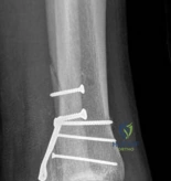
We review everything you need to understand about Partial Articular Fractureplate Fixation: Restore Anatomy. Partial articular fractureplate fixation addresses displaced intraarticular fractures, particularly those of the posterior distal tibia, to restore anatomical congruity and joint function. This open reduction internal fixation procedure often employs buttress plates to stabilize the fracture, ensuring proper length, alignment, and rotation for weight-bearing joints. This approach is crucial for complex ankle injuries.
A 45-year-old patient presents following a high-energy fall from a ladder. Radiographs suggest a partial articular fracture of the lateral tibial plateau. How would you justify your decision-making regarding the requirement for surgical intervention in this case?
Candidate: I would assess the displacement of the fracture. If the step-off or gap is greater than 2mm, I would recommend surgery. I’d also look at the stability of the joint and check for soft tissue injuries. If it's a closed fracture with minimal swelling, I’d plan for ORIF using screws and a plate.
Focusing purely on the 2mm threshold without mentioning the "first hit" effect or biomechanical rationale. Failing to mention soft tissue assessment or the "wrinkle sign" as a prerequisite for surgery. Not classifying the injury within the AO system framework.
Structure the answer by parameters: 1. Articular Congruity: Mention the 2mm threshold and the exponential increase in contact pressures leading to chondrocyte apoptosis. 2. Mechanical Stability: Assess clinical instability or subluxation. 3. Soft Tissue Status: Explicitly discuss the "wrinkle sign" and the danger of operating through compromised envelopes. 4. Patient Factors: Emphasize the goal of preventing early post-traumatic arthrosis through absolute stability and primary bone healing.
You have decided to proceed with open reduction and internal fixation. Explain the biomechanical rationale for the use of lag screws versus buttress plating in this setting.
Candidate: Lag screws are used to compress the fracture fragments together to create friction. The buttress plate is used to support the bone so it doesn't shift again under load.
Treating "lag screw" as a type of screw rather than a technique (gliding vs. thread hole). Failing to explain that the plate converts shear force into compressive force across the fracture plane.
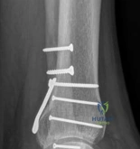
Distinguish clearly: Lag screw: A technique to generate interfragmentary compression, providing absolute stability and direct bone healing by neutralizing shear. Buttress plate: Acts as a physical barrier applied to the apex of the fracture. It counteracts axial, shear, and torsional forces. Note the importance of slight under-contouring of the plate to create an "active" buttress effect against the metaphyseal flare.
The patient's CT scan shows a significant central articular depression. How do you address this during your surgical approach?

Candidate: I would make a window in the side of the bone, lift the depressed part up until it looks flat on the X-ray, and then fill the hole with bone graft to keep it from sinking back down.
Vague terminology ("lifting it up"). Failing to mention the use of a bone tamp, the need for fluoroscopic verification, and the crucial distinction between different types of void fillers (autograft vs. synthetic vs. allograft).
Detail the algorithmic approach: 1. Cortical Window: Create a window in the metaphyseal region. 2. Elevation: Utilize a bone tamp to elevate the articular fragment under direct visualization and fluoroscopic control. 3. Structural Support: The created subchondral void MUST be filled with autograft, allograft, or a structural substitute (e.g., calcium phosphate cement) to prevent secondary subsidence. 4. Fixation: Secure the articular block with subchondral "rafting" screws or a locking plate construct.
