Pilon Fracture Case: Critical Steps for High-Energy Injury
Key Takeaway
Learn more about Pilon Fracture Case: Critical Steps for High-Energy Injury and how to manage it. A case pilon fracture involves a comminuted and displaced intra-articular injury of the distal tibia. Typically resulting from a high-energy axial load, like a fall or MVA, it often presents with articular and metaphyseal comminution. Initial management prioritizes soft tissue assessment, neurovascular integrity, and closed reduction, followed by a CT scan for surgical planning.
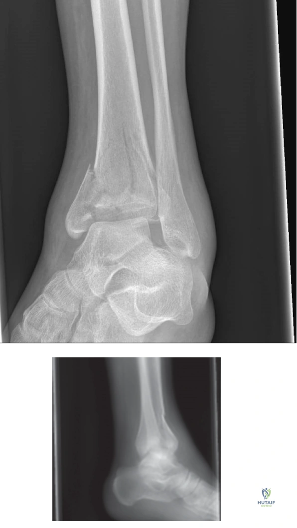
A 42-year-old construction worker presents after a 15-foot fall. You are presented with these initial radiographs and clinical findings: severe edema, tension, and hemorrhagic blisters over the anterior ankle. Describe your immediate management priorities and the rationale behind your strategy.
Candidate: I would immediately admit the patient, elevate the limb, and monitor for compartment syndrome. Given the high-energy nature and Tscherne grade III soft tissue injury, I would plan for staged management: "Span, Scan, and Plan." This involves applying a spanning external fixator, followed by a CT scan once the soft tissues settle, and definitive ORIF in 10-21 days when the wrinkle sign returns.
Candidates often fail to explicitly mention the assessment of the "soft tissue envelope" as the primary driver of decision-making. Failing to mention the specific risk of hemorrhagic blisters—which preclude incision—or omitting the delta pressure assessment for compartment syndrome demonstrates a lack of clinical nuance.
The candidate should categorize the answer: 1) Immediate Life/Limb priority: Rule out neurovascular compromise and acute compartment syndrome (delta pressure measurement). 2) Soft Tissue Assessment: Document the Tscherne grade and identify hemorrhagic blisters as contraindications to early surgical incisions. 3) Damage Control: Apply a spanning external fixator to restore length, alignment, and rotation, which helps mitigate compartment pressure. 4) Staged Strategy: Define "Span, Scan, and Plan," emphasizing that the definitive ORIF is delayed until the "wrinkle sign" confirms the resolution of interstitial edema, typically at 10-21 days.
You have decided to proceed with definitive ORIF. The CT scan confirms a 43-C3 pilon fracture. You are planning a dual-incision approach. What are the anatomical considerations, and how do you ensure the viability of the skin bridge between your incisions?
Candidate: I would use an anterolateral and a posteromedial approach. I must maintain at least a 7 cm skin bridge between the incisions to protect the angiosomes. I would carefully protect the superficial peroneal nerve in the anterolateral approach and the saphenous nerve and vein in the posteromedial approach.
Ignoring the "7 cm rule" or failing to mention specific structures at risk (like the superficial peroneal nerve) is a major oversight. Additionally, some candidates forget that fibular fixation might be required and must be integrated into the soft tissue plan, as adding a third lateral incision for the fibula can compromise the skin bridge even further.
Structure the response by Anatomical Landmarks and Soft Tissue Biology. Mention the "7 cm skin bridge" rule as an absolute imperative. Detail the internervous planes: Anterolateral (between the EDL and Peroneus Tertius) and Posteromedial (between the PTT and FDL). Emphasize the protection of the neurovascular bundles in both approaches and discuss how the lateral fibular approach must be planned such that it doesn't violate the zone of injury or compromise the vascularity of the remaining skin bridge.
During the definitive surgery, you find a significant metaphyseal void after elevating the central die-punch fragment. How do you manage this bone void, and what is your rationale?
Candidate: I would fill the metaphyseal void with bone graft. This provides structural support to the elevated articular surface, preventing secondary subsidence, and promotes osteoconduction for healing.
Not mentioning *why* the graft is needed (prevention of secondary subsidence) or suggesting autograft without acknowledging the morbidity of harvest sites (e.g., iliac crest) versus the convenience of synthetic bone graft substitutes.
Discuss the hierarchy of bone graft options: 1) Autologous bone graft (iliac crest) remains the gold standard for osteoinductivity/osteoconductivity. 2) Bone graft substitutes (e.g., Calcium Phosphate or Calcium Sulfate) are excellent for their compressive strength, which is vital in preventing the "die-punch" fragment from collapsing back into the void. State that the goal is to provide a mechanical scaffold to maintain the anatomical reduction achieved on the articular surface while the metaphyseal bone remodels.
