Complex Tibial Shaft Fracture: Plate Fixation for Plafond Extension
Key Takeaway
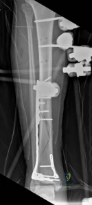
We review everything you need to understand about Complex Tibial Shaft Fracture: Plate Fixation for Plafond Extension. A **tibial shaft fracture** extending into the plafond is a complex injury characterized by a displaced, often multifragmentary break in the midshaft of the tibia that also involves the ankle joint's articular surface. These severe fractures commonly require immediate fasciotomy if compartment syndrome is present, followed by open reduction and internal fixation (ORIF) to restore anatomical alignment and joint function.
A 26-year-old male presents following a 3-meter fall. He has a deformed, tense, and swollen distal leg with a fracture extending into the tibial plafond. Radiographs show a multifragmentary articular injury with metaphyseal comminution. He is currently in the ED with a tense leg and diminished sensation in the first dorsal web space. What is your immediate priority, and how would you manage this patient's initial stabilization?
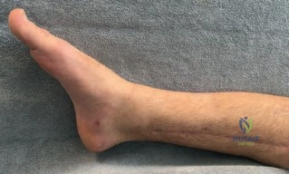
Candidate: I would immediately assess for acute compartment syndrome. Given the tense leg and nerve findings, I would perform a four-compartment fasciotomy. After stabilization of the patient, I would apply a spanning external fixator to restore limb length and allow the soft tissue to heal before planning definitive ORIF.
Candidates often focus exclusively on the bony fracture, neglecting the clinical diagnosis of compartment syndrome. Failing to explicitly mention the dual-incision technique for all four compartments or ignoring the need for urgent neurovascular assessment demonstrates a lack of "damage control" maturity.
The gold standard answer identifies this as a surgical emergency. The candidate should state: "This is a clinical diagnosis of acute compartment syndrome. I would immediately perform a four-compartment fasciotomy via dual incisions to decompress the anterior, lateral, superficial, and deep posterior compartments. Following fasciotomy, I would apply a spanning external fixator from the distal femur/proximal tibia to the calcaneus, respecting the neurovascular bundles, to restore length and alignment. This facilitates 'damage control'—allowing the soft tissue envelope to recover before secondary definitive fixation."
The patient has now reached the 14-day mark post-injury. The fasciotomy wounds are granulating, and the "wrinkle sign" is present. You are planning for definitive internal fixation of this AO/OTA 43C3.3 pilon fracture. What specific imaging and pre-operative planning steps are essential before you step into the theatre?
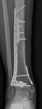
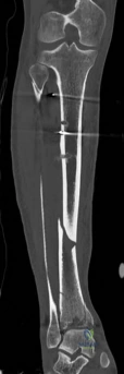
Candidate: I would order a CT scan with 2D multiplanar reformats and 3D reconstruction. I need to map the Chaput, Volkmann, and medial malleolus fragments. I would then template the length of the plate and plan the placement of locking screws to create a subchondral raft.
Failing to mention the assessment of the "central die-punch" impaction. Candidates who only look at the peripheral fragments often miss the need for bone graft or structural support for the central plafond.
The perfect answer demonstrates a structured approach: 1. Imaging: Mandatory fine-cut CT to identify primary articular column fragments and quantify central impaction. 2. Sequencing: Plan the reduction of the fibula (if needed for column length) followed by the articular block reconstruction. 3. Implants: Select a distal tibia locking plate, ensuring the distal screws can provide a subchondral raft. 4. Bone Void: Confirm availability of autograft/bone substitute for the metaphyseal defect created after disimpacting the articular segments.
During surgery, you have reconstructed the articular surface. You are now addressing the diaphyseal extension. Explain the biomechanical rationale for your choice of fixation in this transition zone from metaphysis to diaphysis.
Candidate: I need absolute stability for the articular surface and relative stability for the diaphysis. I will use lag screws for the joint and a long locking plate as a bridge construct for the shaft.
Neglecting the "working length" of the plate. Failing to mention that locking screws provide angular stability rather than compression to the periosteum is a common mark-loser in higher-level vivas.
A sophisticated answer emphasizes: "The goal is absolute stability at the articular surface using lag screws to create interfragmentary compression. Conversely, the diaphyseal zone requires relative stability to promote secondary bone healing via callus. By utilizing a long locking plate with a calibrated 'working length'—leaving holes empty over the comminuted zone—I preserve the extraosseous blood supply and allow for controlled micromotion, reducing the risk of construct failure and nonunion."
