Complex Pilon Fracture Case: High-Energy Trauma, Diagnosis & Initial Management
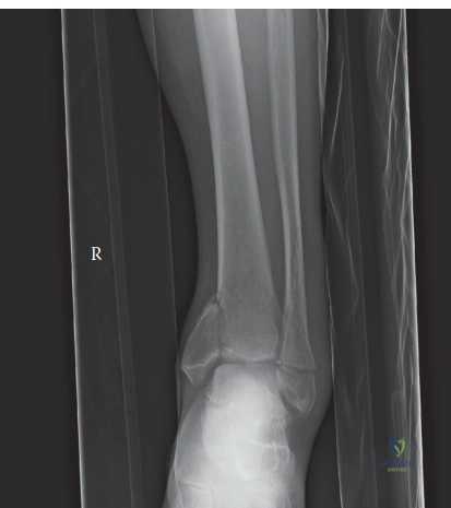
Key Takeaway
A Ruedi-Allgöwer Type III pilon fracture is a complex, high-energy distal tibial injury involving severe articular comminution and impaction. Diagnosis mandates detailed clinical examination and initial radiographs. Crucially, a CT scan with 3D reconstruction provides definitive classification, precise articular fragment mapping, and comprehensive data essential for surgical planning.
A 38-year-old cyclist presents with this high-energy injury after a fall from a height of 4 meters. On examination, the ankle is grossly deformed with significant swelling. Describe your immediate management strategy upon arrival in the Emergency Department.
Candidate: I would perform an ATLS primary survey to rule out life-threatening injuries. Specifically for the ankle, I would check neurovascular status, apply a splint, and order X-rays. Given the mechanism and clinical appearance, I would suspect a pilon fracture, request a CT scan for surgical planning, and likely manage it with a staged approach involving an external fixator followed by ORIF when the soft tissues allow.
Candidates often jump straight to "I would operate on this fracture" or "I would use a locking plate." Failing to prioritize the soft tissue envelope and the "staged" nature of high-energy pilon management is a major error. Also, failing to explicitly mention "neurovascular assessment" and "compartment syndrome monitoring" in a high-energy injury is a critical oversight.
A structured response is key: 1. Safety First: ATLS survey to rule out associated injuries. 2. Immediate Limb Assessment: Neurovascular status, looking for open fracture signs, and monitoring for evolving compartment syndrome. 3. Immediate Resuscitation/Stabilization: Gross reduction if there is skin tenting (to prevent necrosis), splinting, and elevation to control edema. 4. Imaging: Plain radiographs followed by a CT scan with 3D reconstruction for articular mapping. 5. The "Rule of Thumb": Emphasize the soft tissue envelope, stating I would apply a spanning external fixator for stabilization, elevation, and wait for the "wrinkle sign" before considering definitive ORIF.
The patient has been in an external fixator for 7 days. He asks, "Why can't we operate on this today?" What are the specific surgical risks you are trying to mitigate by delaying definitive fixation?
Candidate: I would explain that the high-energy nature of the injury causes severe soft tissue trauma. Operating while the soft tissues are swollen risks wound dehiscence, skin necrosis, deep surgical site infection, and potential hardware exposure. Waiting for the 'wrinkle sign' ensures the soft tissue envelope is viable enough to heal after the incision.
Simply saying "it's too swollen" is insufficient. A high-scoring candidate demonstrates an understanding of the biology—mentioning that the skin is the "limiting factor" and specifically noting that premature incision through edematous tissue leads to catastrophic complications like osteomyelitis or implant failure.
Acknowledge that this is a "biological" rather than "technical" decision. Explain the pathophysiology of the "zone of injury" in high-energy pilon fractures. State that we are waiting for the inflammatory phase to subside and the soft tissue to regain its vascularity. Use the buzzword "skin wrinkling" as the clinical endpoint indicating resolution of edema, which drastically reduces the risk of post-operative infectious complications.
During the definitive surgery, you have reduced the tibial plafond. You now need to address the syndesmosis. What are the principles for syndesmotic fixation in this type of complex injury?
Candidate: I would test the syndesmosis with a hook test or external rotation stress view. If unstable, I would fix it. I typically use a 3.5mm screw or a dynamic device. I ensure the ankle is in a neutral or dorsiflexed position so I don't overtighten the mortise, and I generally aim for the tibia at about 30 degrees anterior to the coronal plane.
Forgetting to mention the position of the ankle (plantarflexion leads to narrowing of the mortise and malreduction). Failing to discuss the level of the screw (usually 2cm proximal to the joint) or the number of cortices (typically 3 or 4) makes the answer sound inexperienced.
Systematically cover the three pillars of syndesmotic fixation: 1. Verification: Perform an intraoperative stress test (e.g., Cotton hook test) after the lateral and medial columns are stabilized. 2. Technique: Use a 3.5mm screw engaging 3-4 cortices, placed parallel to the joint line roughly 2cm proximal, oriented 30 degrees anteriorly. Emphasize fixing the ankle in neutral/dorsiflexion to prevent narrowing of the mortise. 3. Options: Mention the choice between rigid (screws) and dynamic (suture-button/TightRope) fixation, noting that dynamic fixation may allow for more physiological motion and avoid the need for mandatory second-look hardware removal.
