High-Energy Comminuted Tibial Diaphyseal Fracture: Comprehensive Patient Presentation, Clinical Examination & Diagnostic Imaging
Key Takeaway
Diagnosing a high-energy comminuted tibial diaphyseal fracture involves patient history and thorough clinical examination, assessing deformity and neurovascular status. Initial radiographs identify the fracture pattern and angulation. A CT scan then precisely characterizes comminution, rotational deformity, and joint involvement, crucial for surgical planning.
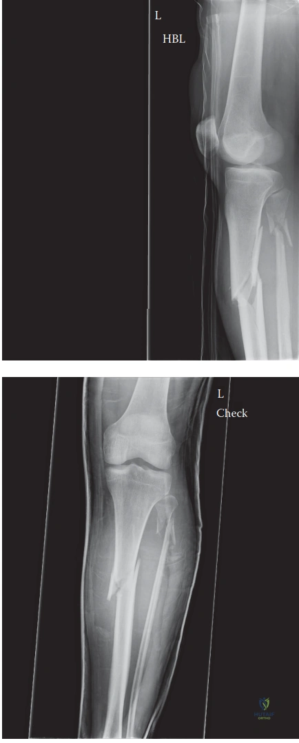
A 32-year-old male presents following a high-energy motorcycle accident. He has a closed, displaced, mid-diaphyseal tibial fracture. Please examine this radiograph and describe your initial assessment and the potential challenges this specific pattern presents.
Candidate: The radiograph shows a closed, comminuted, mid-diaphyseal tibial fracture. It is displaced with apex anterior and valgus angulation. The fibula is intact. The main challenge here is the comminution and the "splinting" effect of the intact fibula, which may make closed reduction difficult. I would also maintain a high index of suspicion for compartment syndrome given the high-energy mechanism.
Candidates often focus solely on the fracture geometry and neglect the soft tissue envelope. Failing to mention the risk of compartment syndrome, the importance of neurovascular status, or the impact of the intact fibula on reduction is a significant oversight for an FRCS candidate.
A superior answer begins with a structured description: AO/OTA classification (42-C2), displacement, and angulation. It then highlights the biomechanical environment: "The intact fibula acts as a lateral strut, potentially preventing anatomical reduction of the tibial length and alignment. I would assess for signs of impending compartment syndrome, confirm neurovascular integrity, and discuss the plan for a reamed intramedullary nail, potentially considering a prophylactic fibular osteotomy if closed reduction is unattainable."
You decide to proceed with intramedullary nailing. What specific intraoperative steps will you take to ensure you avoid the most common complications associated with this procedure?
Candidate: I would prioritize an accurate entry point, slightly medial to the lateral border of the patellar tendon, to avoid valgus malalignment. I would use blocking screws (Poller screws) to assist with reduction in the comminuted zone. I would also pay careful attention to rotational alignment by comparing the relationship of the foot and patella to the contralateral side and checking the fibular overlap under fluoroscopy.
Focusing only on the nail insertion without discussing reduction tools. A failing candidate often ignores the use of blocking screws or forgets that rotational alignment is the most common cause of long-term patient dissatisfaction.
A structured response includes: 1) Entry portal precision (avoiding "starting too lateral/anterior"). 2) Use of Poller screws to manage the butterfly fragment and correct angulation. 3) Multi-modal rotational assessment (clinical, fluoroscopic, and contralateral comparison). 4) Mention of the "Suprapatellar approach" as an modern alternative to mitigate knee pain and aid alignment. 5) Mention of reaming to optimize the biological environment for union in a comminuted fracture.
