Ipsilateral Femoral Neck & Shaft Fractures: A Comprehensive Management Guide
Key Takeaway
Ipsilateral femoral neck and shaft fractures are complex, high-energy injuries. The femoral neck fracture is often missed, risking avascular necrosis. Optimal management involves prompt anatomical reduction and stable fixation of both components. This requires a deep understanding of surgical anatomy and biomechanics to preserve femoral head vascularity and ensure successful patient outcomes.
A 32-year-old male presents to the Emergency Department following a high-energy motorcycle collision. He is haemodynamically stable. Examination reveals significant swelling and deformity of the right thigh. Radiographs demonstrate an obvious displaced mid-shaft femoral fracture. How would you approach the clinical assessment and initial workup for this patient?
Candidate: I would follow ATLS protocols to ensure the patient is stable. After resuscitation, I would obtain AP pelvis and full-length femur radiographs. I would then perform a CT scan of the hip and femur to rule out an associated femoral neck fracture, as these are often missed.
Failing to emphasize the high index of suspicion for an occult ipsilateral femoral neck fracture. Candidates who only request femur films and move straight to planning shaft surgery often miss the neck injury, which is the most critical component for long-term morbidity (AVN).
The candidate must articulate: 1. ATLS/polytrauma clearance. 2. A high index of suspicion for "missed" ipsilateral femoral neck fractures (19-50% reported). 3. Mandatory imaging: AP Pelvis and dedicated traction-internal rotation radiographs of the hip. 4. Crucially, the necessity of a CT scan of the hip and entire femur (1-2mm thin cuts) to definitively rule out sub-clinical neck pathology.
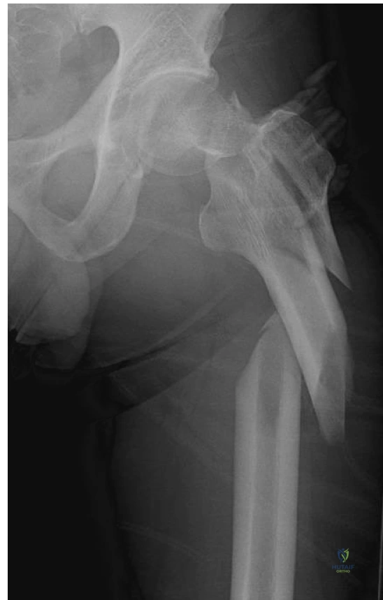
The CT scan confirms a displaced femoral neck fracture in addition to the mid-shaft fracture. Look at the provided imaging. What are the key surgical considerations regarding implant selection and the sequence of fixation for this injury?
Candidate: I would prioritize anatomical reduction and internal fixation of the femoral neck first, as it is the most critical for vascularity. I would use a cephalomedullary nail or separate cannulated screws with an IM nail. I need to ensure the hardware doesn't collide and avoid stress risers.
Assuming "shaft first" is always acceptable. Failing to mention that if closed reduction of the neck fails, an open approach is mandatory to achieve anatomical alignment before the shaft is nailed, to avoid irreversible AVN.
The candidate should structure the answer by: 1. Sequence: Prioritize neck reduction/fixation to protect retinacular vessels. 2. Implant choice: Discuss the benefits of Cephalomedullary (Recon) nails (unified construct) vs. IM Nail + Cannulated Screws (independent reduction). 3. Technical Pearls: Highlight the importance of the entry point (too lateral = varus; too medial = iatrogenic fracture). Mention avoiding stress risers by careful hardware spacing and the absolute requirement for anatomical (not just acceptable) reduction of the neck.
Despite a technically successful procedure, what are the primary complications you must counsel the patient about, and how would you manage the follow-up?
Candidate: The biggest risks are AVN of the femoral head and nonunion of the femoral neck. I would counsel the patient on these risks. Follow-up would involve serial radiographs to check for union and monitoring for signs of head collapse or hardware failure.
Listing only AVN. A high-scoring candidate must also mention the risks of shaft malunion/malrotation, infection, and the long-term impact on rehabilitation, such as delayed weight-bearing and the need for prolonged functional therapy.
The candidate should categorize complications into: 1. Biological: AVN (10-25% for displaced necks) and Nonunion. 2. Mechanical: Hardware failure, malrotation of the shaft, and periprosthetic fractures. 3. Systemic: DVT/PE and infectious risks. Follow-up strategy must include radiographic evaluation at 6-8 weeks and 12 weeks for bridging callus, alongside clinical assessment for functional milestones and neurological integrity.
