Complex Lower Extremity Trauma: A Case Study of Gustilo IIIC Open Tibia-Fibula Fracture & Critical Ischemia
Key Takeaway
A Gustilo IIIC open tibia-fibula fracture with critical limb ischemia presents with extensive soft tissue loss, gross contamination, and neurovascular compromise, typically from high-energy trauma. Diagnosis involves detailed clinical exam, including assessing pulses and sensation, alongside urgent plain radiographs and CT angiography to define skeletal and arterial injuries.
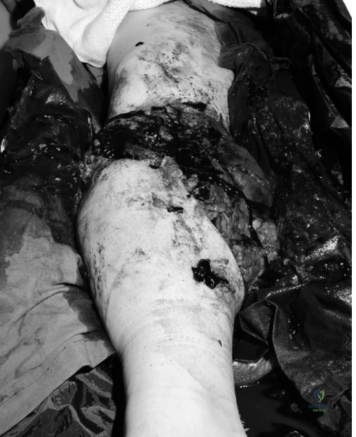
A 42-year-old male presents following an industrial crush injury. You have just completed the primary survey. On examination of the right leg, you note significant soft tissue loss, exposed cortical bone, and absent pulses. The limb is cool and mottled. How do you classify this injury, and what is your immediate management priority?
Candidate: This is a Gustilo-Anderson Type IIIC open tibia fracture. My priority is to save the patient's life first by continuing ATLS protocols. I would then need to address the vascular injury, perform aggressive debridement, and stabilize the fracture with an external fixator.
Candidates often jump straight to the classification or surgical technique without emphasizing the systemic status. Failing to explicitly mention the time-sensitive nature of "warm ischemia" or neglecting to call for a multidisciplinary team (Vascular/Plastics) suggests poor clinical leadership in a high-stakes trauma setting.
Start by confirming the patient's systemic stability (ATLS). Classify this as a Gustilo-Anderson Type IIIC injury due to the high-energy mechanism, severe soft tissue compromise, and, most importantly, the presence of critical arterial injury. State that the priority is the "Time-Tissue" relationship: urgent surgical debridement and restoration of perfusion (potentially via temporary vascular shunting) to minimize warm ischemia time. Finally, state you are activating the multidisciplinary team (Orthopedics, Vascular, Plastics) for definitive limb salvage planning vs. primary amputation assessment.
You have decided to proceed with attempted limb salvage. The vascular team is ready. What specific surgical steps are essential for the skeletal and soft tissue management at this stage?
Candidate: I would perform an extensive debridement of all non-viable tissue using the 4 C's criteria. I would stabilize the fracture with a spanning external fixator. Finally, I would perform prophylactic fasciotomies of all four compartments.
Missing the nuance of "damage control." Candidates often forget to mention the serial nature of debridement (don't close yet!) or fail to mention that the external fixator is chosen specifically to protect the vascular repair and allow for future soft tissue coverage.
Structure the answer into three phases: 1. Aggressive Radical Debridement: Use the "4 C's" (Color, Consistency, Contractility, Capacity to bleed) and excise all non-viable bone/soft tissue until the "paprika sign" is achieved. 2. Damage Control Stabilization: Use a spanning external fixator to protect the neurovascular repair, prioritizing access for future soft tissue transfer. 3. Compartment Management: Perform mandatory four-compartment fasciotomies given the ischemia-reperfusion risk, leaving the wounds open for delayed primary closure or flap coverage, managed with Negative Pressure Wound Therapy (NPWT).
The patient is 7 days post-injury. The vascular repair is patent, and the limb is viable. You have a 6.5cm segmental bone defect. Describe your definitive management strategy for this bone gap.
Candidate: I would use the Masquelet technique, also known as the induced membrane technique. I would place an antibiotic cement spacer into the defect and then perform a bone graft in a second stage once the membrane has formed.
Candidates failing to mention the importance of soft tissue stability before bone reconstruction. The Masquelet technique fails if the soft tissue envelope is not sound. You must also emphasize that this is a staged procedure.
Acknowledge that biological bone reconstruction requires a stable soft tissue envelope. Propose a two-stage approach: Stage 1 (Induced Membrane): Placement of an antibiotic-impregnated polymethylmethacrylate (PMMA) spacer to maintain length, prevent soft tissue collapse, and induce a vascularized pseudosynovial membrane. Stage 2 (Definitive Reconstruction): After 6-8 weeks, once the membrane is mature, carefully excise the spacer and fill the defect with autologous cancellous bone graft (e.g., via RIA technique from the iliac crests), ensuring rigid internal or circular frame fixation.
