Intramedullary Nailing for Proximal Tibia Fractures: Mastering Reduction & Preventing Deformities

Key Takeaway
Intramedullary nailing (IMN) for proximal tibia fractures faces challenges due to the proximal tibia's flared metaphysis and natural anterior bow. Common malreductions include valgus and procurvatum (apex anterior angulation), driven by deforming forces like patellar tendon tension and muscle pull. Meticulous planning and understanding biomechanics are crucial for preventing these iatrogenic deformities.
You are presented with this patient in the emergency department following a high-energy motor vehicle accident. The patient has a displaced proximal third tibial shaft fracture. Looking at the radiographs, what is the most significant anatomical challenge associated with treating this specific injury with an intramedullary nail, and how would you address it intra-operatively?
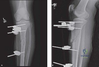
Candidate: The biggest issue is the malalignment, specifically procurvatum or apex anterior angulation. This happens because of the nail's straight design trying to pass through the proximal tibia's natural anterior bow. To fix this, I would use a suprapatellar approach to get a more central and posterior entry point, and I might use Poller screws to guide the nail.
Candidates often fail to describe the mechanism of the deformity. Simply stating "it causes malalignment" is insufficient. They often forget to mention the role of the patellar tendon pull or fail to specify that they would use a Poller screw specifically in the proximal fragment to force the nail posteriorly.
The primary challenge is procurvatum deformity (apex anterior angulation) combined with valgus malalignment. This is due to the mismatch between the straight intramedullary nail and the proximal tibial anatomy.
Solution:
1. Suprapatellar Approach: Provides a more anatomical, posterior entry point, reducing the "tendency to straighten" the proximal tibia.
2. Blocking (Poller) Screws: An anterior-to-posterior blocking screw placed in the proximal fragment forces the nail posteriorly (correcting procurvatum). A lateral-to-medial blocking screw can be used if there is valgus tendency.
3. Reduction Aids: Using a femoral distractor or percutaneous reduction clamps to maintain the reduction before nail insertion.
You have performed the nailing. During your final fluoroscopic assessment, you note a slight valgus malalignment of the proximal fragment. Looking at this intra-operative image, how would you classify this specific construct, and what was the intended function of the added screw?
Candidate: This is an intramedullary nail supplemented with a Poller screw, also known as a blocking screw. It is placed eccentrically to help guide the nail trajectory and maintain the bone's anatomical alignment by "blocking" the nail from migrating into the void created by the fracture.
Candidates often call it a "locking screw" or an "anti-rotation screw." These terms are incorrect in this context. A blocking screw serves a different biomechanical purpose than a locking screw (which attaches the nail to the bone).
This is a Poller screw (blocking screw). It is not a locking screw; it does not secure the nail to the bone. Its purpose is to function as a mechanical bumper. By narrowing the effective medullary canal, it prevents the nail from drifting into the wider metaphyseal flare, thereby forcing the nail to follow the desired anatomical path and correcting or preventing angular deformity (valgus/varus or procurvatum/recurvatum depending on the plane of insertion).
