Young Adult Femoral Shaft Fractures: Comprehensive Surgical Management & Outcomes

Key Takeaway
Femoral shaft fractures in young adults, typically from high-energy trauma, are predominantly managed surgically. Early operative stabilization ensures anatomical reduction, stable fixation, and restored limb length. This approach is critical for minimizing long-term disability, reducing pain, facilitating early mobilization, and preventing complications, especially in polytrauma patients.
A 28-year-old male presents following a high-velocity motorcycle accident. He has an isolated, closed, mid-diaphyseal femoral shaft fracture. He is haemodynamically stable. What is the definitive management of choice, and what are the critical biomechanical considerations for the implant selection?
Candidate: The gold standard is an intramedullary nail (IMN). I would perform a closed, reamed antegrade IMN. Biomechanically, the nail acts as a load-sharing device, which is superior to a plate for this diaphyseal location. It is essential to choose a nail that accounts for the anterior bow of the femur to avoid iatrogenic anterior cortical perforation.
Candidates often focus solely on the "how" (e.g., describing reaming or locking) while ignoring the "why." They fail to explicitly mention the "load-sharing" biomechanical advantage of an IMN over plating in the diaphysis, or they neglect to mention the anatomical anterior bow—a classic point of failure in templating.
The gold standard is a reamed antegrade IMN. Biomechanically, it provides load-sharing fixation, which minimizes stress shielding and promotes callus formation. For the implant, I must ensure the nail’s radius of curvature matches the patient's femoral anterior bow—a failure to account for this leads to anterior cortical impingement or fracture. Furthermore, I would emphasize restoring femoral anteversion and length by referencing the contralateral side or using the 'lesser trochanter profile' on fluoroscopy to prevent rotational malunion.
Look at this intraoperative fluoroscopic image. What is the significance of the guide wire position, and what specific steps must be taken to avoid a catastrophic complication related to the entry point?
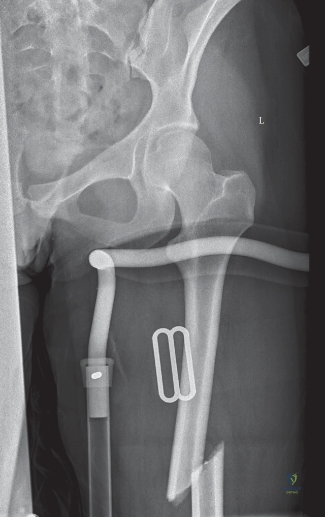
Candidate: The guide wire must be central in both the AP and lateral views. The critical complication related to the entry point is iatrogenic avascular necrosis (AVN) of the femoral head or a fracture of the femoral neck. We now prefer the tip of the greater trochanter as the entry point to avoid the piriformis fossa, which is associated with higher risks of damaging the blood supply to the femoral head.
Candidates often forget to mention the specific vascular anatomy (superior gluteal artery branches) that is endangered by a medial piriformis entry. They also miss the importance of the guide wire being centered on the *lateral* fluoroscopic view, which is the most common plane for missing a malaligned entry.
The guide wire must be collinear with the medullary canal. The entry point choice is decisive: a piriformis fossa entry point is medial and risks injuring the ascending branch of the medial circumflex femoral artery, potentially leading to AVN. Modern practice favors the tip of the greater trochanter entry portal. Crucially, on the lateral view, the entry must be slightly anterior to avoid the posterior cortical wall of the neck, which reduces the risk of iatrogenic comminution or 'fracture neck' propagation.
We are dealing with a polytrauma patient. Can you define the transition from 'Damage Control Orthopaedics' (DCO) to 'Early Appropriate Care' (EAC), and what clinical criteria dictate this shift?
Candidate: DCO is for unstable patients; we use external fixation to stabilize the femur quickly. Once the patient is physiologically resuscitated, we convert to definitive fixation. EAC suggests that if the patient is stable, early definitive fixation—ideally within 24 hours—leads to lower rates of pulmonary complications like fat embolism.
Failing to mention the 'second hit' phenomenon. Borderline candidates describe the timing but don't explain the physiological monitoring (lactate, base deficit, coagulopathy) that defines the 'unstable' patient who requires damage control.
DCO is employed in 'unstable' or 'borderline' patients—those with coagulopathy, acidosis (base deficit > 6, lactate > 4), or ongoing transfusion requirements—to avoid the 'second hit' of prolonged surgery. We use an external fixator to gain rapid skeletal stability. Once the patient is physiologically optimized (normalized pH/lactate), we convert to definitive IMN. EAC is the preferred strategy for 'stable' patients, where definitive fixation within 24-48 hours significantly reduces the risk of systemic inflammatory response and pulmonary complications such as ARDS/Fat Embolism Syndrome.
