Femoral Neck and Shaft Fractures: Epidemiology, Surgical Anatomy & Biomechanics of Isolated & Ipsilateral Injuries
Key Takeaway
Femoral neck and shaft fractures are distinct orthopedic injuries, often arising from high-energy trauma in young patients or low-energy falls in the elderly. Critical considerations include precise diagnosis due to high morbidity risk, detailed understanding of surgical anatomy and biomechanics, potential for ipsilateral occurrence, and managing complications like avascular necrosis or nonunion, guiding meticulous treatment strategies.
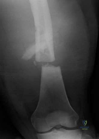
A 28-year-old male is brought to the trauma bay following a high-speed motorcycle collision. He has an obvious deformity of the mid-thigh. Initial radiographs confirm a closed, comminuted diaphyseal femoral shaft fracture. What is your immediate priority, and what diagnostic pitfall must you specifically exclude before proceeding to the operating theatre?
Candidate: My immediate priority is to follow ATLS protocol to stabilize the patient, as he likely has significant occult blood loss. Orthopaedically, I must ensure I don't miss an ipsilateral femoral neck fracture. I would order specific, dedicated views of the hip and, if there is any clinical doubt or difficulty in viewing the neck, a CT scan of the hip before I proceed to the theatre for an intramedullary nail.
Focusing solely on the femoral shaft fracture and jumping straight to "I will book him for a locked IM nail." A failing candidate fails to mention the high association (2-6%) of ipsilateral femoral neck fractures and the "hidden" nature of these injuries, often discovered post-operatively when the neck fracture displaces, leading to AVN.
Start with the ABCDE/ATLS assessment. Explicitly state the "Don't Miss It" imperative regarding the ipsilateral femoral neck fracture. Mention that in high-energy trauma, the femoral shaft is often the 'distracting' injury. Specify that plain radiographs (AP/Lateral femur) are often inadequate to rule out the neck injury and that a high-resolution CT of the hip is the investigation of choice if plain films are not definitive. Mention the need to sequence fixation—addressing the neck fracture first (or simultaneously) if present.
You have confirmed the absence of a neck fracture. You are planning an antegrade IM nail for the femoral shaft. Describe the key anatomical considerations for the entry portal and the potential complications associated with your chosen approach.
Candidate: I prefer a trochanteric entry portal. The entry point is at the tip of the greater trochanter. This reduces the risk of iatrogenic injury to the retinacular vessels, which is a concern with the piriformis fossa entry. Complications include subtrochanteric comminution if the portal is too lateral, or damage to the hip abductors if the approach is too extensive. I also monitor for sciatic nerve injury during reduction and ensure I achieve rotational alignment.
Failing to distinguish between the two portals. A poor candidate might not know why the piriformis entry is less favored now (risk of AVN) or fail to mention the specific risk of "varus malalignment" if the entry portal is positioned incorrectly.
Articulate the evolution from piriformis to trochanteric entry. Emphasize that piriformis entry is medial and risks AVN due to damage of the medial circumflex femoral artery branch. Contrast this with the trochanteric entry portal which is simpler, preserves abductor function better, and is less associated with AVN. Identify the key risk: an entry portal that is too lateral creates a valgus-directed force leading to varus malalignment of the proximal segment.
