Comprehensive Pelvic Fracture Case Study: Interactive Module for Orthopedic Viva Exam Preparation
Key Takeaway
This interactive pelvic fracture case study serves as a critical resource for orthopedic professionals. It provides an engaging platform to evaluate diagnostic skills, understand classification systems, and refine treatment planning for complex pelvic injuries. Featuring expandable answers, it's specifically designed to bolster knowledge and prepare effectively for challenging viva examinations.
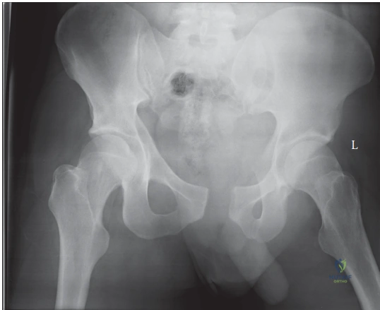
A 25-year-old male is brought into the ED following a high-speed motorcycle accident. He is haemodynamically stable after initial resuscitation. You are presented with his pelvic radiograph.
Describe the radiographic findings and classify the injury using the Young and Burgess system.
Candidate: The radiograph shows a widened pubic symphysis (greater than 2.5cm). The sacroiliac joints appear somewhat widened anteriorly but not posteriorly. This is an Anterior-Posterior Compression (APC) type II pelvic ring injury.
Failing to distinguish between APC II and III. Candidates often just say "open book fracture" without specifying the rotational/vertical stability, or they fail to mention the integrity of the posterior SI ligaments which defines the distinction between type II (stable vertically) and type III (unstable).
Systematically describe the findings: 1. Pubic symphysis diastasis (>2.5cm). 2. Assessment of the sacroiliac joint (anterior widening). 3. Classification as Young-Burgess APC II. Mention that this represents a rotationally unstable but vertically stable injury, as the posterior SI complex (specifically the interosseous and sacrotuberous/spinous ligaments) is disrupted anteriorly but the primary posterior tension band remains intact.
During your secondary survey, you suspect a possible urological injury. What are the clinical signs you are looking for, and what is your immediate management plan for a suspected urethral injury in this patient?
Candidate: I would look for blood at the meatus, a high-riding prostate on PR exam, and perineal bruising. I would perform one gentle attempt at catheterization; if that fails, I would stop, perform a retrograde urethrogram, and involve the urology team.
Being overly aggressive with the catheter or failing to mention the contraindication of inflating the balloon if resistance is met. Forgetting the multi-disciplinary involvement and rushing to attempt blind instrumentation.
Structure the answer: 1. Clinical indicators (High-riding prostate, blood at meatus, perineal hematoma). 2. Immediate management: Stop and consult. A single gentle attempt is only permissible if no gross signs are present. 3. If resistance or blood persists, stop, do not inflate the balloon, and request a retrograde urethrogram. 4. Emphasize that a suprapubic catheter should only be placed by the urology team, noting the potential impact on future surgical approaches to the pelvis.
The patient has now become hypotensive in the trauma bay despite initial fluid resuscitation. He is categorized as a "non-responder." Discuss the decision-making process for managing life-threatening pelvic hemorrhage.
Candidate: If the patient is a non-responder, I would stop resuscitation with fluids and initiate a massive transfusion protocol. I would assume the source is the pelvic venous plexus and consider pelvic packing over angiography as it is faster.
Suggesting immediate CT angiography for a patient who is not hemodynamically stable. The examiner expects you to prioritize physiologic stabilization (the "ABC" approach) over imaging.
Explain the hierarchy: 1. Continued resuscitation using blood products in a 1:1:1 ratio. 2. Recognize that "non-responders" require surgical intervention, not CT. 3. Identify that >80% of hemorrhage is venous; therefore, pelvic packing is the gold standard for rapid control in the unstable patient. 4. Contrast this with the role of interventional radiology (angio-embolization) which is reserved for the "responder" or "partial responder" where arterial bleeding (e.g., internal pudendal artery) is suspected after initial stabilization.
