ABOS Part I Orthopaedic Review: Mallet Finger & Skier's Thumb Diagnosis, Management & Treatment | Part 21589
Key Takeaway
This ABOS Part I Orthopaedic Review module offers 20 MCQs on mallet finger and skier's thumb. It covers mallet finger types, non-operative and surgical management, and rehabilitation. For skier's thumb, it details UCL injury mechanisms, Stener lesions, diagnosis via stress testing and MRI, and surgical repair techniques, crucial for board exam preparation.
ABOS Part I Orthopaedic Review: Mallet Finger & Skier's Thumb Diagnosis, Management & Treatment | Part 21589
Comprehensive 100-Question Exam
00:00
Start Quiz
Question 1
A 38-year-old recreational basketball player presents to your clinic 3 days after 'jamming' his left ring finger. He reports immediate pain and an inability to fully straighten the tip of his finger. On examination, his DIP joint rests in approximately 30 degrees of flexion, and he has a 30-degree active extensor lag. Passively, the DIP joint can be fully extended. Radiographs show no evidence of fracture or subluxation. According to the Doyle classification, what type of injury does this patient most likely have, and what is the most appropriate initial management?
Explanation
Correct Answer: C
The patient's presentation of a 'jammed' finger, inability to actively extend the DIP joint, a resting flexion deformity, and full passive extension, coupled with normal radiographs, is classic for a soft tissue mallet finger. According to the Doyle classification, this is a Type I injury, which involves a soft tissue avulsion of the terminal extensor tendon from its insertion on the distal phalanx without significant bony involvement. The case explicitly states that Type I is the primary focus of the review and that the vast majority of acute soft tissue mallet injuries are successfully managed non-operatively. The core principle of non-operative management is continuous immobilization of the DIP joint in full extension or slight hyperextension (0-10 degrees) for a prolonged period, typically 6 to 8 weeks, followed by a gradual weaning phase. The PIP joint should be left free to allow full range of motion.
Option A is incorrect because Type II involves a bony avulsion fracture of less than 30% of the articular surface, and surgical repair is not the initial management for an acute Type I injury.
Option B is incorrect because Type III involves a bony avulsion fracture of more than 30% of the articular surface, often with subluxation, which is not present here.
Option D is incorrect because Type IV is an epiphyseal fracture in children (Salter-Harris type I or II), which is not applicable to a 38-year-old adult.
Option E is incorrect because immediate active and passive DIP joint flexion would disrupt the healing tendon and lead to failure of treatment. Continuous immobilization is paramount during the initial healing phase.
Question 2
A 55-year-old female presents with a chronic soft tissue mallet finger of her right long finger, sustained 6 months prior. She initially attempted splinting but was non-compliant due to discomfort. She now has a persistent 35-degree extensor lag at the DIP joint and has developed a noticeable hyperextension deformity at her PIP joint. Which of the following anatomical structures is primarily responsible for the characteristic flexion deformity at the DIP joint in a soft tissue mallet finger?
Explanation
Correct Answer: D
The case explicitly states that in a soft tissue mallet injury (Type I), the terminal extensor tendon is ruptured from its insertion, disrupting the extensor moment arm at the DIP joint. This disruption leaves the DIP joint extension unopposed, and the powerful pull of the flexor digitorum profundus (FDP) tendon, which inserts on the volar aspect of the distal phalanx, creates the characteristic flexion deformity. The FDP is the sole flexor of the DIP joint.
Option A (Extensor Digitorum Communis) is an extrinsic extensor, but its terminal tendon is what is disrupted, leading to the deformity, not causing it.
Option B (Central slip of the extensor mechanism) primarily extends the PIP joint and is typically intact in a pure soft tissue mallet finger. Its overactivity can contribute to a swan neck deformity, but it does not cause DIP flexion.
Option C (Flexor Digitorum Superficialis) inserts on the middle phalanx and primarily flexes the PIP joint, not the DIP joint.
Option E (Lumbrical muscles) are intrinsic muscles that contribute to MCP joint flexion and PIP/DIP extension, but they are not the primary cause of the DIP flexion deformity in a mallet finger.
Question 3
A 28-year-old carpenter presents with a 4-month-old soft tissue mallet finger of his index finger. He initially tried splinting for 3 weeks but removed it due to work demands. He now has a fixed 25-degree extensor lag at the DIP joint and complains of difficulty picking up small objects. He is considering surgical intervention. Which of the following is a critical supporting structure of the extensor mechanism at the DIP joint that stabilizes the lateral bands and prevents their volar subluxation?
Explanation
Correct Answer: C
The 'Surgical Anatomy & Biomechanics' section clearly identifies the Triangular Ligament as a crucial supporting structure. It states: 'Located dorsally, it stabilizes the lateral bands, preventing their volar subluxation and maintaining their position for efficient DIP joint extension.'
Option A (Volar plate) is a ligamentous structure on the volar aspect of the joint that prevents hyperextension, but it is not involved in stabilizing the dorsal extensor mechanism.
Option B (Annular pulleys) are part of the flexor tendon sheath system, crucial for maintaining the mechanical advantage of the flexor tendons, not the extensor mechanism.
Option D (Sagittal bands) are located at the MCP joint level and stabilize the extensor digitorum communis tendon over the MCP joint, not the DIP joint.
Option E (Transverse retinacular ligament) is mentioned as influencing joint movement and preventing dorsal migration of the lateral bands, but the triangular ligament is specifically highlighted for preventing volar subluxation and maintaining position for efficient DIP extension.
Question 4
A 42-year-old accountant presents with a 3-month history of a soft tissue mallet finger on his dominant middle finger. He initially attempted non-operative management with a Stack splint for 8 weeks, but he admits to removing it frequently for hygiene and work-related tasks. He now has a persistent 20-degree extensor lag at the DIP joint and is frustrated with his inability to fully extend his finger. He is considering surgical repair. Which of the following is a strong indication for surgical management in this patient?
Explanation
Correct Answer: C
The 'Indications & Contraindications' section explicitly lists 'Failed Non-Operative Management: Persistent extensor lag of more than 15-20 degrees after an adequate course of continuous splinting (typically 8 weeks, followed by night splinting)' as a primary indication for surgical management. This patient fits this criterion, having a 20-degree lag after an attempted 8-week splinting course (albeit with poor compliance).
Option A (Acute presentation within 4-6 weeks of injury) is an indication for non-operative management, not surgical.
Option B (Patient compliance with continuous splinting) is a factor for successful non-operative management, not an indication for surgery.
Option D (Absence of secondary deformity like a fixed swan neck) would typically favor non-operative management if the injury is acute and correctable, or it means the surgery would be less complex if performed for other reasons. The presence of a fixed swan neck, however, would be an indication for surgery.
Option E (Minimal initial extensor lag) would typically favor non-operative management.
Question 5
A 60-year-old diabetic patient presents with a chronic soft tissue mallet finger of 8 months duration, complicated by a fixed swan neck deformity of the same finger. She is scheduled for surgical repair. During pre-operative planning, the surgeon reviews imaging. Which of the following imaging modalities is considered mandatory for initial evaluation of a mallet finger to rule out bony involvement?
Explanation
Correct Answer: C
The 'Pre-Operative Planning & Patient Positioning' section, under 'Imaging,' states: 'Plain Radiographs: Standard AP, lateral, and oblique views of the affected digit are mandatory. These are crucial to rule out bony avulsion fractures (Doyle Type II, III, IV), DIP joint subluxation, and pre-existing arthritis. A true lateral view is essential to accurately assess the joint alignment and presence of bony avulsion.'
Option A (MRI) is generally not indicated for routine soft tissue mallet finger. It may be considered in very chronic, complex cases but is not mandatory for initial evaluation.
Option B (CT scan) is not mentioned as a mandatory initial imaging study for mallet finger. It might be used for complex bony avulsions but not for initial soft tissue mallet evaluation.
Option D (Ultrasound) can visualize tendons but is not considered mandatory for initial evaluation and may not reliably rule out small bony avulsions or subluxation as effectively as radiographs.
Option E (Bone scan) is a nuclear medicine study used for detecting bone pathology like stress fractures, infections, or tumors, and is not indicated for routine mallet finger evaluation.
Question 6
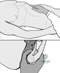
A 25-year-old professional musician undergoes surgical repair for a chronic soft tissue mallet finger with significant tendon retraction. The surgeon decides to perform a tendon-to-bone reattachment using drill holes and a pull-out suture technique. Post-operatively, the DIP joint is immobilized with a transarticular K-wire. The image below shows a typical K-wire placement for this procedure.
What is the primary purpose of this K-wire fixation in the immediate post-operative period?
Explanation
Correct Answer: C
The 'Internal Fixation (K-wire)' section states: 'After the tendon repair, transarticular K-wire fixation of the DIP joint is almost always performed to protect the repair and maintain the joint in the desired position during healing.' It further specifies: 'The DIP joint is gently placed in full extension or slight hyperextension (0-10 degrees).' The image clearly depicts a K-wire traversing the DIP joint, holding it in extension.
Option A is incorrect because the K-wire is not primarily for dynamic compression or bone healing in a soft tissue injury. Its role is static immobilization.
Option B is incorrect because the K-wire prevents active range of motion at the DIP joint, which is crucial to protect the healing tendon. Early active motion is contraindicated.
Option D is incorrect because while over-extension should be avoided to prevent volar plate injury, the primary purpose of the K-wire is not to prevent this specific injury during rehabilitation, but rather to immobilize the joint for tendon healing.
Option E is incorrect because the K-wire immobilizes the joint and tendon, which can actually contribute to stiffness if not managed properly in rehabilitation. It does not facilitate tendon gliding or prevent adhesions directly.
Question 7
A 30-year-old athlete undergoes surgical repair for a chronic soft tissue mallet finger. During the procedure, the surgeon identifies significant retraction of the proximal tendon stump and a very short distal stump. The image below shows a K-wire used for post-operative immobilization.
Which of the following surgical techniques is most appropriate for re-establishing the attachment of the terminal extensor tendon to the distal phalanx in this scenario?
Explanation
Correct Answer: B
The 'Detailed Surgical Approach / Technique' section, under 'Repair Technique,' describes 'Tendon-to-Bone Reattachment (Direct Repair)' as the most common technique for acute and subacute mallets, involving drill holes or suture anchors. For chronic injuries, it states: 'For chronic injuries with significant retraction and scar tissue, direct reattachment may lead to excessive tension. The tendon may be advanced distally and reattached to the distal phalanx as described above.' This directly addresses the scenario of significant retraction and a short distal stump, where advancement and reattachment to the bone are necessary.
Option A (Direct tendon-to-tendon repair) is typically used when a sufficient distal tendon stump remains, which is rare in Type I mallet injuries, especially chronic ones with a 'very short distal stump.' It's more common for lacerations.
Option C (Primary DIP joint arthrodesis) is a salvage procedure for severe, irreparable damage with significant functional impairment, not a primary repair technique for a chronic mallet finger, especially in an athlete.
Option D (Central slip tenotomy) would weaken PIP extension and is not a technique for repairing a mallet finger; it might be considered in severe swan neck deformities but not as a primary mallet repair.
Option E (Lateral band reconstruction without addressing the terminal tendon) is incomplete and would not restore DIP extension. Lateral band involvement may be addressed in chronic cases, but the primary issue is the terminal extensor tendon.
Question 8
A 48-year-old painter undergoes surgical repair of a chronic soft tissue mallet finger. Post-operatively, the DIP joint is immobilized with a K-wire as shown in the image. During the rehabilitation phase, which of the following is the MOST critical instruction for the patient during the initial 6-week immobilization period?
Explanation
Correct Answer: C
The 'Post-Operative Rehabilitation Protocols' section, under 'Phase 1: Immobilization (Weeks 0-6),' explicitly states for DIP Joint Immobilization: 'Crucial Principle: Absolutely no active or passive DIP joint flexion is permitted. The patient must be meticulously educated on this, particularly for activities of daily living.' This is the most critical instruction to protect the healing tendon repair.
Option A is incorrect because active DIP flexion would directly stress and likely rupture the healing tendon.
Option B is incorrect because passive DIP extension is already maintained by the K-wire/splint, and passive flexion is contraindicated.
Option D is incorrect because light resistive exercises for DIP extension are typically introduced much later, in Phase 3 (Weeks 12+), after the tendon has had significant time to heal.
Option E is incorrect because K-wires should only be removed by a medical professional, typically at 6 weeks post-operatively, not by the patient at home due to discomfort, as this could lead to complications like infection or re-injury.
Question 9
A 70-year-old retired teacher presents with a persistent 25-degree extensor lag after 10 weeks of continuous splinting for an acute soft tissue mallet finger. She is now developing a mild, flexible hyperextension of her PIP joint. The surgeon is considering surgical intervention. Which of the following complications is most commonly associated with unsatisfactory outcomes after soft tissue mallet finger repair, and what is a potential salvage strategy for a severe, irreparable case?
Explanation
Correct Answer: C
The 'Complications & Management' section identifies 'Extensor Lag / Re-rupture' as the 'Most common (5-20%), higher in chronic cases' and 'the most common reason for unsatisfactory outcomes.' For salvage strategies, it states: 'For persistent, functionally significant lag, revision surgery may be considered. Options include repeat direct repair, tendon advancement (if proximal tissue allows), tenodesis (using a portion of the lateral band or a small palmaris longus graft), or, in cases of severe, irreparable damage with significant functional impairment, DIP joint arthrodesis (fusion) in a functional position (typically 10-15 degrees of flexion).'
Option A (Infection) is a complication, but its incidence is low (1-5%), and aggressive physical therapy is not the primary salvage for infection.
Option B (Stiffness / Loss of Motion) is common, but early active DIP flexion is contraindicated and would worsen the outcome, not salvage it.
Option D (Nail Deformity) is often cosmetic and not typically salvaged with nerve release/neurolysis, which is for nerve irritation/neuroma.
Option E (Skin Necrosis) is low incidence (1-3%), and prolonged splinting is not a salvage for skin necrosis; it requires wound care, debridement, or skin grafting.
Question 10
A 35-year-old construction worker presents with an acute open mallet finger injury to his small finger, sustained from a laceration over the DIP joint. On examination, there is a clear disruption of the extensor tendon, and he has a significant extensor lag. Which of the following statements best reflects the consensus on managing this specific type of mallet finger injury?
Explanation
Correct Answer: B
The 'Indications & Contraindications' section, under 'Operative Indications,' lists 'Open Mallet Injuries: Lacerations over the DIP joint with extensor tendon disruption, which require surgical debridement and primary repair to prevent infection and facilitate healing.' The 'Summary of Key Literature / Guidelines' further reinforces this: 'Acute open mallet finger lacerations require surgical debridement and primary repair, irrespective of the degree of lag, to prevent infection and restore tendon integrity.'
Option A is incorrect because open injuries carry a high risk of infection and require surgical intervention, unlike most closed injuries.
Option C is incorrect because while swan neck deformity is a potential secondary complication, the immediate priority for an open injury is infection prevention and primary tendon repair.
Option D is incorrect because MRI is generally not indicated for routine mallet finger, and for an acute open injury, surgical exploration and repair are more urgent than advanced imaging.
Option E is incorrect because DIP joint arthrodesis is a salvage procedure for severe, irreparable cases, not the preferred initial treatment for an acute open mallet finger, especially in a young, active patient.
Question 11
A 35-year-old right-hand dominant male presents with acute right thumb pain and instability after a skiing accident. He fell while gripping a ski pole, which forced his thumb into violent abduction and hyperextension. He reported an immediate 'pop' and profound weakness in pinch grip. Which of the following statements best describes the biomechanical sequence of ligamentous failure in this classic injury pattern?

Explanation
Correct Answer: C
The case explicitly states, 'When the metacarpophalangeal joint is in extension during the traumatic event, the accessory ulnar collateral ligament and volar plate bear the initial stress. However, as the joint is forced into flexion and abduction, the proper ulnar collateral ligament becomes the primary restraint and subsequently fails.' This accurately describes the sequential failure of the ulnar collateral ligament complex in a Skier's Thumb injury.
Option A is incorrect because the proper UCL is the primary restraint in flexion, not extension, and the accessory UCL bears initial stress in extension.
Option B is incorrect because while the volar plate contributes to stability in extension, it is not the primary restraint in both positions, nor does it typically fail before the entire UCL complex in this specific mechanism.
Option D is incorrect because the adductor pollicis aponeurosis is a muscular aponeurosis, not a primary ligamentous restraint, and its interposition (Stener lesion) occurs after the UCL rupture, not as a primary failure leading to it.
Option E is incorrect because a forced abduction injury primarily stresses the ulnar collateral ligament, not the radial collateral ligament, which resists varus forces.
Question 12
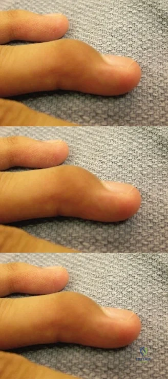
During the clinical examination of the patient, a distinct, firm, highly tender, pea-sized mass was appreciated proximally and ulnarly to the metacarpophalangeal joint line. This finding is pathognomonic for which of the following conditions?

Explanation
Correct Answer: C
The case explicitly states, 'During palpation of the ulnar joint line, a distinct, firm, highly tender, pea-sized mass was appreciated proximally and ulnarly to the metacarpophalangeal joint line. This palpable mass is the pathognomonic clinical hallmark of a Stener lesion. It represents the avulsed, retracted distal end of the ulnar collateral ligament that has displaced superficial to the proximal edge of the adductor pollicis aponeurosis.' This interposition prevents healing and necessitates surgical intervention.
Option A is incorrect because a partial tear would not typically present with a palpable, retracted mass, and would likely have a firm endpoint on stress testing.
Option B is incorrect because while an avulsion fracture can occur, the description of a 'pea-sized mass' refers to soft tissue (the ligament stump), and radiographs would be needed to confirm a bony avulsion, which were negative in this case.
Option D is incorrect because a ganglion cyst is typically a chronic, non-traumatic finding, and while it can be firm, it would not be acutely tender in this context or pathognomonic for an acute ligamentous injury.
Option E is incorrect because a flexor pollicis longus tendon rupture would present with an inability to flex the IP joint and a different palpable mass location, not specifically at the MCP joint ulnar aspect.
Question 13
Following a local anesthetic block, valgus stress testing was performed on the patient's right thumb metacarpophalangeal joint. In 30 degrees of flexion, the injured thumb demonstrated approximately 40 degrees of valgus angulation with a distinctly soft endpoint, compared to 10 degrees with a firm endpoint on the contralateral uninjured thumb. What is the most accurate interpretation of these findings?

Explanation
Correct Answer: C
The case states, 'The absolute angulation exceeding 35 degrees, combined with a side-to-side difference greater than 15 degrees, provided definitive clinical evidence of a complete, full-thickness rupture of the proper ulnar collateral ligament.' The finding of 40 degrees of valgus angulation in 30 degrees of flexion, with a side-to-side difference of 30 degrees (40 vs 10), and a soft endpoint, unequivocally indicates a complete rupture. Testing in 30 degrees of flexion specifically isolates the proper ulnar collateral ligament.
Options A and B are incorrect because partial sprains (Grade I or II) would typically present with less laxity (often <30 degrees) and, crucially, a firm endpoint, indicating some intact ligamentous fibers.
Option D is incorrect because an isolated accessory UCL injury would primarily manifest as instability in extension, not gross instability in 30 degrees of flexion where the proper UCL is the primary restraint.
Option E is incorrect because a volar plate avulsion primarily causes hyperextension instability, and while it can be part of a complex injury, the gross valgus instability in flexion points directly to UCL failure.
Question 14
The case highlights the importance of specific techniques during valgus stress testing of the thumb metacarpophalangeal joint. Which of the following statements represents a critical 'pearl' for accurate assessment of ulnar collateral ligament integrity?

Explanation
Correct Answer: C
The case explicitly states under 'Clinical Pearls and Pitfalls': 'Local Anesthesia is Mandatory: Attempting to grade laxity in an acutely injured, unanesthetized thumb is highly unreliable due to involuntary muscle guarding by the adductor pollicis. A local intra-articular or digital block is essential for an accurate physical examination.' This is a crucial step for accurate diagnosis.
Option A is incorrect because while testing in extension is part of the assessment, the '30-Degree Flexion Rule' is critical for isolating the proper UCL, which is often the primary injury. Testing only in extension may yield a false negative if the volar plate and accessory ligament are intact.
Option B is incorrect because the case states, 'in the modern clinical setting, stress radiography is largely contraindicated when a Stener lesion is suspected clinically' due to the risk of iatrogenic Stener lesion formation or conversion of a partial to a complete tear.
Option D is incorrect because the case advises, 'Palpation Precedes Stress: Always palpate the ulnar joint line for a Stener lesion before applying valgus stress. Forceful stress testing of a non-displaced complete tear can iatrogenically displace the ligament superficial to the aponeurosis, converting a potentially non-operative injury into an operative one.'
Option E is incorrect because the interphalangeal joint's motion does not significantly confound MCP joint stress testing, and its immobilization is not a standard or critical step for this specific assessment.
Question 15
Magnetic Resonance Imaging (MRI) was performed to definitively map the pathoanatomy. The MRI findings were described as a 'yo-yo on a string sign' on coronal T2-weighted fat-suppressed images. Which of the following best describes this specific MRI finding?

Explanation
Correct Answer: C
The case explicitly describes the MRI findings: 'Crucially, the MRI demonstrated the classic yo yo on a string sign. The torn, retracted ulnar collateral ligament stump appeared as a balled-up, nodular mass of low signal intensity (the 'yo-yo') resting superficial and proximal to the low-signal band of the adductor pollicis aponeurosis (the 'string'). This confirmed the presence of a Stener lesion.' This is the pathognomonic MRI sign for a Stener lesion.
Option A is incorrect because plain radiographs already ruled out a bony avulsion, and the 'yo-yo on a string' sign specifically refers to soft tissue interposition.
Option B is incorrect because this describes a partial sprain (Grade I or II), not a complete rupture with retraction and aponeurotic interposition.
Option D is incorrect because this describes a radial collateral ligament injury, which is on the opposite side of the joint and has a different mechanism and clinical presentation.
Option E is incorrect because while an intra-articular hematoma is expected with acute trauma, it does not represent the 'yo-yo on a string' sign, which specifically identifies the displaced ligament stump and aponeurosis.
Question 16
Given the patient's clinical presentation and MRI findings, which of the following is the most appropriate management strategy?

Explanation
Correct Answer: C
The case clearly states, 'In this patient's case, the clinical examination demonstrating gross valgus instability lacking a firm endpoint, combined with the palpable mass and definitive MRI evidence of a Stener lesion, served as absolute indications for operative intervention.' The Stener lesion creates a mechanical barrier to healing, making non-operative management futile. Open surgical repair with suture anchor fixation is the standard of care for acute, complete UCL ruptures with a Stener lesion.
Option A is incorrect because non-operative management is strictly reserved for partial tears or non-displaced bony avulsions, not for complete ruptures with a Stener lesion where spontaneous healing is impossible.
Option B is incorrect because the diagnosis is highly probable clinically and definitively confirmed by MRI, making a second opinion for diagnosis unnecessary and delaying definitive treatment.
Option D is incorrect because percutaneous pinning is typically used for fractures or dislocations requiring temporary stabilization, not for direct ligamentous repair in the presence of a Stener lesion which requires open reduction.
Option E is incorrect because corticosteroid injections are contraindicated in acute ligamentous injuries, especially ruptures, as they can impair healing and increase the risk of further damage. They are also not a definitive treatment for mechanical instability.
Question 17
During the surgical repair of the ulnar collateral ligament, the surgeon plans a lazy-S incision over the ulnar aspect of the thumb metacarpophalangeal joint. Which of the following structures is at highest risk of iatrogenic injury during the initial skin and subcutaneous dissection, and requires meticulous protection?

Explanation
Correct Answer: C
The case explicitly highlights this surgical pitfall: 'Radial Sensory Nerve Injury: The dorsal sensory branches of the radial nerve are highly variable and course directly through the surgical field. Meticulous blunt dissection and gentle retraction are critical. A postoperative neuroma in this location is often debilitating.' These branches are superficial and easily injured during the initial approach to the ulnar aspect of the thumb MCP joint.
Option A is incorrect because the flexor pollicis longus tendon is on the volar aspect of the thumb and is not typically encountered or at risk with an ulnar approach to the MCP joint.
Option B is incorrect because the radial artery is more proximally located in the wrist (anatomic snuffbox) and not directly in the superficial surgical field for a thumb MCP ulnar collateral ligament repair.
Option D is incorrect because the ulnar nerve is located on the ulnar side of the hand and wrist, but its sensory branches to the thumb are not typically in the immediate superficial field of a thumb MCP ulnar approach.
Option E is incorrect because the extensor pollicis brevis tendon is on the dorsal-radial aspect of the thumb and while it is part of the extensor mechanism, the dorsal sensory radial nerve branches are more superficial and at higher risk during the initial skin incision.
Question 18
Following successful surgical repair of the ulnar collateral ligament, the patient enters the acute immobilization phase (0-2 weeks). Which of the following instructions is most critical for the patient during this initial postoperative period?

Explanation
Correct Answer: B
The case outlines the acute immobilization phase: 'The patient is instructed to maintain strict elevation of the operative extremity above the level of the heart to mitigate swelling. Active range of motion of the shoulder, elbow, and non-involved digits is strongly encouraged to prevent stiffness and promote venous return. The interphalangeal joint of the involved thumb must be actively flexed and extended multiple times daily to prevent tethering of the flexor pollicis longus tendon.'
Option A is incorrect because active abduction would place direct stress on the healing UCL repair and is strictly contraindicated in the early phase.
Option C is incorrect because passive valgus stress testing would risk disrupting the fresh repair and is not part of the early rehabilitation protocol.
Option D is incorrect because the splint is typically removed by a therapist at the 2-week mark, and strict immobilization is crucial for protection during the initial healing phase.
Option E is incorrect because progressive strengthening exercises are initiated much later, typically at 6 weeks postoperatively, after initial ligament-to-bone healing has occurred.
Question 19
A 28-year-old rock climber presents with chronic ulnar-sided thumb pain and instability, 6 months after a fall where he sustained a similar mechanism of injury to the case patient. Clinical examination reveals gross valgus laxity of the MCP joint with a soft endpoint, and MRI confirms a complete UCL rupture with significant retraction and scarring. Based on the case's discussion of surgical decision-making, what is the most likely surgical approach for this patient?

Explanation
Correct Answer: C
The case discusses the classification of UCL injuries by chronicity: 'Acute injuries (typically <3 weeks old) are amenable to direct primary repair. Chronic injuries (>3-4 weeks old) often present with retracted, scarred, and attenuated ligamentous tissue that is inadequate for primary repair, necessitating complex ligamentous reconstruction using autograft (e.g., palmaris longus or plantaris tendon).' A 6-month-old injury with significant retraction and scarring falls squarely into the chronic category, requiring reconstruction.
Option A is incorrect because direct primary repair is reserved for acute injuries where the ligament tissue is healthy and can be reapproximated to its footprint. In chronic cases, the tissue is often too attenuated or scarred.
Option B is incorrect because non-operative management is ineffective for complete ruptures, especially chronic ones with gross instability, and would lead to persistent pain and dysfunction.
Option D is incorrect because percutaneous pinning is a temporary fixation method and does not address the underlying ligamentous deficiency in chronic instability.
Option E is incorrect because while arthrodesis provides stability, it sacrifices motion and is typically reserved as a salvage procedure for severe, painful, degenerative arthritis or failed reconstructions, not as a primary treatment for chronic instability in an otherwise healthy joint.
Question 20
A surgeon is performing an open repair of a Skier's Thumb. After incising the adductor aponeurosis and exposing the joint, the torn ulnar collateral ligament stump is mobilized. The surgeon then uses a small curette and burr to decorticate the bony footprint at the volar-ulnar aspect of the proximal phalanx base. What is the primary purpose of this crucial step?

Explanation
Correct Answer: C
The case describes this step: 'Using a small curette and a fine surgical burr, the cortical bone at the footprint was decorticated to expose bleeding cancellous bone. This crucial step stimulates the local inflammatory cascade and provides marrow-derived osteoprogenitor cells to enhance ligament-to-bone healing.' This is a fundamental principle in orthopedic surgery to promote robust soft tissue-to-bone healing.
Option A is incorrect because while anchor placement is important, decortication's primary role is biological, not purely mechanical for anchor purchase, which is achieved by proper anchor design and insertion technique.
Option B is incorrect because debridement of frayed ligament edges is done on the ligament itself, not by decorticating the bone. Decortication is about preparing the bone for healing, not removing tissue.
Option D is incorrect because decortication does not primarily decompress the joint space; that is achieved by proper reduction and irrigation. It also doesn't directly prevent stiffness.
Option E is incorrect because suture anchors are used for fixation, not direct suture passage through the bone in this context. Decortication is not for easier suture passage.
Question 21
A 24-year-old competitive skier falls while holding his ski pole and sustains an acute injury to his right thumb. On examination, there is localized tenderness over the ulnar aspect of the thumb metacarpophalangeal (MCP) joint. Valgus stress testing reveals 40 degrees of laxity when the MCP joint is flexed to 30 degrees, but only 5 degrees of laxity when the MCP joint is in full extension. Which of the following is the most accurate interpretation of these physical examination findings?
Explanation
Question 22
A 31-year-old male presents with a painful, swollen thumb 3 days after a fall onto an outstretched hand. Clinical examination raises suspicion for a complete ulnar collateral ligament (UCL) tear. MRI demonstrates a complete rupture of the UCL with the torn end retracted and resting superficial to a distinct anatomical structure, preventing anatomical reduction. Which structure is interposing between the torn UCL and its insertion site in this classic lesion?
Explanation
Question 23
A 25-year-old man presents to the emergency department after being struck on the tip of his right long finger by a baseball.
Assuming the provided radiograph demonstrates a dorsal avulsion fracture of the distal phalanx involving 60% of the articular surface with associated volar subluxation of the remaining distal phalanx, what is the most appropriate definitive management?
Explanation
Question 24
A 10-year-old boy falls while snowboarding and complains of severe pain at the ulnar aspect of his thumb MCP joint. Valgus stress testing reveals significant laxity compared to the contralateral side without a definitive endpoint. Radiographs show a widened physis at the ulnar base of the proximal phalanx. What is the most likely diagnosis in this pediatric patient?
Explanation
Question 25
During the surgical repair of an acute, retracted ulnar collateral ligament (UCL) tear of the thumb (Skier's thumb), the surgeon makes a lazy-S incision over the ulnar aspect of the MCP joint. Which of the following nerve branches is at greatest risk of injury during this surgical exposure?
Explanation
Question 26
A 45-year-old woman is undergoing conservative treatment for a soft tissue mallet finger of her index finger. She was instructed to wear an extension splint continuously for 8 weeks. At her 4-week follow-up, she admits she removed the splint for 5 minutes to wash her hand, during which her fingertip dropped into flexion. What is the most appropriate next step in her management?
Explanation
Question 27
A 62-year-old farmer presents with chronic instability and pain in his right thumb MCP joint, sustained from an injury 10 years ago. He has a positive pinch grip test with significant weakness. Radiographs demonstrate severe joint space narrowing, subchondral sclerosis, and osteophyte formation at the MCP joint. What is the most appropriate surgical treatment?
Explanation
Question 28
A 35-year-old musician presents with a soft-tissue mallet finger of the little finger. You decide to treat him conservatively with a custom thermoplastic splint. To optimize healing and prevent complications, what is the ideal position for splinting the affected digit?
Explanation
Question 29
A 29-year-old professional athlete is diagnosed with an acute thumb UCL tear. An MRI is obtained to evaluate for a Stener lesion. What classic MRI appearance is highly specific for the presence of a Stener lesion?
Explanation
Question 30
When performing an open repair of an acute ulnar collateral ligament tear of the thumb with a suture anchor, exact anatomic placement of the anchor is critical. Where is the normal anatomic insertion site of the proper UCL on the proximal phalanx?
Explanation
Question 31
A 30-year-old construction worker presents with a bony mallet finger of his middle finger involving 40% of the articular surface. Lateral radiographs show the fracture is slightly displaced, but the distal phalanx remains perfectly congruous with the head of the middle phalanx without any volar subluxation. What is the most appropriate management?
Explanation
Question 32
A surgeon is utilizing the Ishiguro technique (extension block pinning) for a displaced bony mallet finger with volar subluxation. Which of the following describes the correct sequence and placement of the pins?
Explanation
Question 33
A 50-year-old woman presents with a chronic soft tissue mallet deformity of her ring finger of 6 months duration. She has a 40-degree extensor lag at the DIP joint and a 20-degree hyperextension deformity at the PIP joint. Which of the following describes the fundamental pathophysiology of the secondary PIP joint deformity seen in this patient?
Explanation
Question 34
What is the most frequently encountered complication of conservative management (continuous extension splinting) for soft tissue mallet finger injuries?
Explanation
Question 35
A patient is undergoing surgical repair of an acute UCL tear of the thumb. During the procedure, the surgeon inadvertently places the suture anchor at the insertion site too dorsally on the base of the proximal phalanx. What specific postoperative biomechanical deficit is most likely to occur as a result of this technical error?
Explanation
Question 36
A 33-year-old male presents with acute valgus instability of the thumb MCP joint. Clinical examination and stress radiographs confirm a diagnosis of a complete UCL rupture. According to established guidelines, what degree of absolute valgus laxity in 30 degrees of MCP flexion is generally considered an absolute indication for surgical intervention?
Explanation
Question 37
A 55-year-old patient presents with a 5-week-old soft tissue mallet injury to the small finger. He has not received any prior treatment and has a 40-degree extensor lag. According to current literature, what is the most appropriate initial management for this delayed presentation?
Explanation
Question 38
In a patient presenting with an acute thumb injury, differentiating between a Skier's thumb (UCL injury) and a radial collateral ligament (RCL) injury is important. Which of the following clinical presentations or mechanisms is highly characteristic of an RCL injury rather than a UCL injury?
Explanation
Question 39
A 40-year-old man presents with a chronic, rigid mallet deformity of his index finger 2 years after an untreated injury. He complains of pain and difficulty performing fine motor tasks. Radiographs demonstrate advanced osteoarthritis of the DIP joint with joint space narrowing and osteophytes. What is the most reliable surgical option for this patient?
Explanation
Question 40
A 22-year-old athlete sustains a bony avulsion of the thumb UCL. Radiographs demonstrate a displaced bone fragment at the volar-ulnar base of the proximal phalanx that measures approximately 30% of the articular surface. What is the preferred surgical approach for this specific injury?
Explanation
Question 41
A 25-year-old skier falls while holding his ski pole and presents with ulnar-sided thumb pain. On examination, there is lack of a firm endpoint when a valgus stress is applied to the thumb metacarpophalangeal (MCP) joint in 30 degrees of flexion. Which of the following anatomical structures must be interposed to create a Stener lesion?
Explanation
Question 42
When examining a suspected ulnar collateral ligament (UCL) injury of the thumb, valgus stress testing is performed in both full extension and 30 degrees of flexion. Laxity detected exclusively in 30 degrees of flexion indicates an isolated tear of which structure?
Explanation
Question 43
A 45-year-old male sustains a soft tissue mallet finger injury to his right index finger. He is treated with a strict continuous DIP joint extension splint. During his 4-week follow-up, he admits the splint slipped off for 10 minutes while showering, causing the finger to flex. What is the most appropriate next step in management?
Explanation
Question 44
Which of the following is considered an absolute indication for operative intervention in a bony mallet finger injury?
Explanation
Question 45
A patient with an untreated chronic mallet finger presents with a new secondary deformity consisting of PIP joint hyperextension and DIP joint flexion. What is the primary biomechanical cause of this secondary PIP hyperextension?
Explanation
Question 46
A 12-year-old boy presents with a crush injury to his right long finger tip. Examination reveals a laceration through the nail bed, the nail plate is avulsed proximally, and the DIP joint is flexed. Radiographs show a widened distal phalanx physis. What is the most critical step in management?
Explanation
Question 47
During surgical repair of an acute Skier's thumb, the surgeon utilizes a dorsal-ulnar approach to the thumb MCP joint. Which of the following neurologic structures is at the highest risk of iatrogenic injury during this approach?
Explanation
Question 48
A 30-year-old laborer presents with an 8-week-old soft tissue mallet finger of the ring finger. He has had no previous treatment. What is the recommended initial management?
Explanation
Question 49
A patient presents with a thumb UCL injury. MRI confirms a Stener lesion. During surgical exploration and repair, where will the distal stump of the torn UCL be found?
Explanation
Question 50
A 28-year-old patient presents with pain and a supination deformity of the thumb following a fall onto an outstretched hand. Stress testing of the thumb MCP joint demonstrates laxity with radial stress. Which ligament is injured?
Explanation
Question 51
A 35-year-old male requires surgical intervention for a chronic thumb UCL injury that occurred 6 months ago. Intraoperatively, the native UCL tissue is found to be deficient and cannot be primarily repaired. What is the most appropriate surgical technique?
Explanation
Question 52
A 50-year-old patient undergoes extension block pinning (Ishiguro technique) for a bony mallet finger with volar subluxation.
Which of the following describes the correct pin configuration for this technique?
Explanation
Question 53
Which structure anatomically links the PIP and DIP joints, contributing to the coordinated extension of the DIP joint when the PIP joint extends?
Explanation
Question 54
A 22-year-old college athlete sustains a 'jammed finger'. Radiographs reveal a bony avulsion fracture of the dorsal base of the distal phalanx involving 10% of the articular surface with no joint subluxation. What is the most appropriate management?
Explanation
Question 55
What is the most common complication associated with non-operative extension splinting of a mallet finger?
Explanation
Question 56
In the Doyle classification of mallet finger injuries, a Type IVC injury is defined by which of the following characteristics?
Explanation
Question 57
A patient with a diagnosed thumb UCL injury is deemed appropriate for non-operative management. What is the standard conservative protocol?
Explanation
Question 58
A 65-year-old woman presents with a painful, stiff DIP joint secondary to an untreated chronic mallet finger sustained 10 years ago. Radiographs show severe osteoarthritis of the DIP joint. What is the most reliable definitive treatment?
Explanation
Question 59
When evaluating a patient with a 'Gamekeeper's thumb,' how does the pathophysiology traditionally differ from a 'Skier's thumb'?
Explanation
Question 60
During the physical examination of a suspected mallet finger, what clinical finding most accurately distinguishes a terminal tendon rupture from a central slip rupture (boutonniere deformity)?
Explanation
Question 61
A 28-year-old male skier presents after falling with his thumb caught in a ski pole strap. Examination reveals point tenderness over the ulnar aspect of the thumb metacarpophalangeal (MCP) joint and a palpable mass. A Stener lesion is suspected. Which of the following describes the anatomic basis of a Stener lesion?
Explanation
Question 62
In the evaluation of a suspected Skier's thumb, valgus stress testing is performed to assess the integrity of the ulnar collateral ligament (UCL) complex. Which of the following statements regarding the biomechanical function of the thumb MCP joint ligaments is correct?
Explanation
Question 63
A 10-year-old boy presents with a crush injury to his right long finger tip after it was caught in a door. Clinically, he has a flexed posture of the DIP joint and a laceration through the nail bed with the nail plate avulsed proximally. Radiographs show a widening of the distal phalanx physis. What is the most appropriate definitive management?
Explanation
Question 64
A 32-year-old woman presents 5 weeks after sustaining a closed soft-tissue mallet finger injury to her small finger. She has not received any prior treatment and currently has a 45-degree extensor lag. Which of the following is the most appropriate initial management?
Explanation
Question 65
A 25-year-old female presents after a skiing accident with pain and swelling at the ulnar base of her right thumb. On examination, valgus stress testing reveals 40 degrees of laxity with the metacarpophalangeal (MCP) joint in 30 degrees of flexion, compared to 10 degrees on the contralateral thumb. There is no palpable end-point. MRI confirms a complete ligament rupture. During surgical repair, the torn proximal stump of the ligament is most likely to be found superficial to which of the following structures?
Explanation
Question 66
When evaluating a patient for a suspected acute injury to the thumb metacarpophalangeal (MCP) joint, physical examination includes valgus stress testing in different joint positions. Testing the MCP joint in 30 degrees of flexion primarily isolates which of the following stabilizing structures?
Explanation
Question 67
A 35-year-old male sustains a bony mallet finger to his dominant right long finger while playing baseball. Radiographs demonstrate a dorsal avulsion fracture of the distal phalanx involving 40% of the articular surface. Which of the following radiographic findings serves as an absolute indication for surgical intervention rather than non-operative splinting?
Explanation
Question 68
A 10-year-old boy presents to the emergency department after 'jamming' his right middle finger while catching a football. He exhibits a flexed posture at the distal interphalangeal (DIP) joint and cannot actively extend it. Examination reveals swelling and bleeding from beneath the proximal nail fold. Radiographs show a widened distal phalanx physis. What is the most appropriate management?
Explanation
Question 69
A 45-year-old female presents with a 6-month history of a symptomatic chronic soft-tissue mallet finger of her left index finger. She has developed a swan neck deformity with a 35-degree extensor lag at the DIP joint and PIP joint hyperextension. Both joints remain fully passively correctable, and there is no radiographic evidence of osteoarthritis. Which of the following surgical interventions is most appropriate?
Explanation
Question 70
A 50-year-old male presents to your clinic 5 weeks after sustaining a closed soft-tissue mallet injury to his right small finger. He has not sought prior medical attention and currently has a 40-degree extensor lag at the DIP joint. Radiographs are negative for fractures. What is the recommended initial management for this delayed presentation?
Explanation
Question 71
During the surgical approach for repairing a completely ruptured ulnar collateral ligament of the thumb with an associated Stener lesion, an incision is made over the ulnar aspect of the thumb MCP joint. Which of the following neurologic structures is at greatest risk of iatrogenic injury during this approach?
Explanation
Question 72
A patient with a soft-tissue mallet finger has been undergoing continuous extension splinting. At the 4-week mark, the patient admits to removing the splint to wash his hands, during which the DIP joint fell into full flexion. What is the most appropriate next step in management?
Explanation
Question 73
A 32-year-old mechanic sustains a closed soft-tissue mallet finger. He is compliant with 8 weeks of continuous extension splinting, followed by a weaning protocol. At his final follow-up, he notes a firm, painless bump over the dorsal aspect of the DIP joint but has full active extension. What is the most likely etiology of this dorsal prominence?
Explanation
Question 74
A 28-year-old male presents with a thumb injury.
Assuming the radiograph demonstrates a bony mallet finger with a large dorsal fragment (45% of the articular surface) and volar subluxation of the distal phalanx, what is the most widely accepted surgical technique for this specific pattern?
Explanation
Question 75
A 22-year-old skier presents after a fall with a painful thumb. Radiographs demonstrate a 3 mm displaced avulsion fracture of the ulnar base of the thumb proximal phalanx. The MCP joint is subluxated radially. What is the most appropriate management?
Explanation
Question 76
A 62-year-old male presents with a chronic mallet finger of his right index finger sustained 5 years ago. He complains of significant pain, difficulty with pinch grip, and a 45-degree extensor lag. Radiographs reveal complete loss of joint space, subchondral sclerosis, and osteophytes at the DIP joint. What is the definitive treatment of choice?
Explanation
Question 77
Which of the following complications is most frequently encountered during the non-operative management of a soft-tissue mallet finger with strict continuous splinting?
Explanation
Question 78
The terminal extensor tendon, which ruptures in a mallet finger injury, is formed anatomically by the convergence of which of the following structures over the middle phalanx?
Explanation
Question 79
A 40-year-old gamekeeper presents with a history of chronic pain and weakness in his right thumb. He has noticeable laxity of the thumb MCP joint on examination. Radiographs show no degenerative changes. He desires surgical intervention to improve his grip and pinch strength. In the setting of chronic UCL instability without arthrosis, what is the most appropriate surgical option?
Explanation
Question 80
In the pathogenesis of a swan neck deformity resulting from an untreated mallet finger, the initial flexed posture of the DIP joint leads to a biomechanical cascade. Which of the following best describes the primary anatomic change at the PIP joint?
Explanation
Question 81
A 14-year-old boy falls while snowboarding and complains of thumb pain. Examination reveals significant swelling over the ulnar aspect of the thumb MCP joint and laxity with valgus stress testing. Radiographs show a displaced fracture through the epiphysis and exiting the articular surface of the ulnar base of the proximal phalanx. The proper UCL is functionally attached to this fragment. What is the most likely diagnosis?
Explanation
Question 82
While diagnosing a suspected complete tear of the proper ulnar collateral ligament of the thumb, stress radiography is sometimes utilized. According to established criteria, a complete rupture is strongly suspected if there is an absolute valgus opening greater than what threshold?
Explanation
Question 83
A patient presents with an acute hyperextension injury to the thumb MCP joint. Which structure provides the primary static restraint to valgus stress when the thumb MCP joint is in 0 degrees of extension?
Explanation
Question 84
A 55-year-old rheumatoid arthritis patient presents with bilateral spontaneous mallet finger deformities. There is no history of acute trauma. Radiographs reveal diffuse osteopenia and erosive changes at the radiocarpal and distal radioulnar joints. What is the most likely pathophysiologic mechanism for these specific mallet deformities?
Explanation
None
