Adult Acquired Flatfoot Reconstruction: FDL Transfer & Medial Displacement Calcaneal Osteotomy Masterclass
Key Takeaway
This masterclass details the surgical management of adult acquired flatfoot deformity with flexor digitorum longus (FDL) transfer and medial displacement calcaneal osteotomy. We cover comprehensive anatomy, meticulous preoperative planning, and a granular, step-by-step intraoperative execution, emphasizing critical pearls, potential pitfalls, and robust postoperative rehabilitation strategies for optimal patient outcomes.
Welcome, colleagues, to this definitive masterclass on the reconstructive management of adult acquired flatfoot deformity (AAFD), increasingly referred to in contemporary literature as Progressive Collapsing Foot Deformity (PCFD). Today, we are focusing our attention on a ubiquitous yet biomechanically complex pathology: Stage II Posterior Tibial Tendon Dysfunction (PTTD). This chapter will exhaustively detail the comprehensive reconstructive approach utilizing a Flexor Digitorum Longus (FDL) tendon transfer augmented by a Medial Displacement Calcaneal Osteotomy (MDCO). This specific combination of procedures represents the gold standard motion-sparing technique designed to restore the medial longitudinal arch, re-establish functional hindfoot stability, and halt the progression of degenerative collapse.
Comprehensive Introduction and Patho-Epidemiology
The evolution of our understanding regarding adult acquired flatfoot deformity has shifted from viewing it as an isolated tendinopathy to recognizing it as a global ligamentous and tendinous failure of the medial column. The posterior tibial tendon (PTT) acts as the primary dynamic stabilizer of the medial longitudinal arch. As it undergoes progressive tearing, attenuation, and eventual failure, the foot gradually collapses into a predictable planovalgus configuration. This pathological cascade is overwhelmingly degenerative rather than acute or inflammatory. Histological analysis of excised PTT specimens routinely demonstrates an absence of inflammatory cells, revealing instead a replacement of normal linear collagen bundles with amorphous scar tissue, mucinous degeneration, and fibroblast hypercellularity.
This degenerative process most frequently occurs within a well-documented hypovascular zone located approximately 1 to 1.5 centimeters distal to the medial malleolus, just proximal to the tendon's broad insertion onto the navicular tuberosity. The presence of an accessory navicular ossicle (Type II) can act as a local mechanical stressor and a site of painful synchondrosis, further predisposing the tendon to early degeneration and functional incompetence. Epidemiologically, this condition predominantly affects women in their fifth to seventh decades of life, often presenting with a high body mass index and medical comorbidities such as hypertension or seronegative arthropathies, though it can undoubtedly occur in athletic or younger populations subjected to repetitive microtrauma.
The natural history of PTTD dictates a progressive collapse of the arch initiated by the dysfunction of the posterior tibialis. The widely accepted Johnson and Strom classification, later modified by Myerson, delineates this progression. Stage I represents tenosynovitis with pain and swelling but no architectural collapse; the length of the tendon remains intact. Stage II, the primary focus of our operative intervention today, is characterized by a flexible planovalgus deformity. The tendon is elongated or ruptured, resulting in a clinically apparent loss of the medial arch, hindfoot valgus, and forefoot abduction. Stage III indicates a rigid, fixed deformity where the subtalar and talonavicular joints are no longer passively correctable, necessitating arthrodesis rather than joint-sparing reconstruction. Finally, Stage IV involves secondary failure of the deltoid ligament complex, leading to tibiotalar tilt and ankle joint arthritis.
Detailed Surgical Anatomy and Biomechanics
A profound, three-dimensional understanding of the hindfoot and midfoot anatomy is the absolute prerequisite for executing this reconstruction safely and effectively. The posterior tibial tendon originates from the posterior surfaces of the tibia, fibula, and the intervening interosseous membrane. It courses distally, passing immediately posterior to the medial malleolus within a dedicated fibro-osseous tunnel secured by the flexor retinaculum. Distal to the malleolus, it fans out to insert primarily onto the navicular tuberosity, with robust secondary plantar slips attaching to the cuneiforms, the cuboid, and the bases of the second through fourth metatarsals. Biomechanically, it is the strongest invertor of the foot and works synergistically with the Achilles tendon to lock the transverse tarsal joints during the terminal stance phase of gait, converting the foot into a rigid lever for push-off.
The Flexor Digitorum Longus (FDL) tendon serves as our premier workhorse for tendon transfer in this pathology. Originating from the posterior tibia, it passes behind the medial malleolus, situated anterior to the posterior tibial neurovascular bundle and superficial to the sustentaculum tali. Distally, it splits into four individual slips inserting into the distal phalanges of the lesser toes. Crucially, in the plantar midfoot, the FDL tendon crosses superficial to the Flexor Hallucis Longus (FHL) tendon at the Master Knot of Henry. This anatomical decussation is the exact site where we will harvest the FDL, often requiring careful dissection to separate the fibrous interconnections between the two tendons to maximize FDL excursion. The FDL is chosen because its vector of pull closely mimics the PTT, it fires in the same phase of gait, and its sacrifice results in minimal functional deficit to the lesser digits.
Navigating the neurovascular anatomy demands meticulous surgical technique. The posterior tibial neurovascular bundle—comprising the posterior tibial artery, accompanying venae comitantes, and the tibial nerve—lies immediately posterior to the FDL tendon and anterior to the FHL deep within the tarsal tunnel. Extreme caution is paramount during the medial dissection, particularly when exposing the sustentaculum tali and harvesting the FDL. Laterally, the sural nerve is highly vulnerable during the approach for the calcaneal osteotomy. It courses subcutaneously along the posterolateral aspect of the ankle and foot. Meticulous, sharp dissection through the subcutaneous layers, strictly avoiding aggressive blunt retractor placement, is required to prevent debilitating postoperative sural neuritis.
Biomechanically, the Medial Displacement Calcaneal Osteotomy (MDCO) is the linchpin of this reconstructive pairing. Tendon transfer alone in the presence of a structurally uncorrected planovalgus foot is doomed to stretch out and fail. The MDCO physically translates the posterior tuberosity of the calcaneus medially, typically by 10 millimeters. This medial shift fundamentally alters the hindfoot biomechanics by moving the insertion vector of the Achilles tendon medial to the subtalar joint axis. Consequently, the Achilles is converted from a deforming evertor force into a corrective invertor force, inherently stabilizing the hindfoot and profoundly unburdening the newly transferred FDL tendon.
Exhaustive Indications and Contraindications
Patient selection is the ultimate determinant of surgical success. The classic indication for an FDL transfer combined with an MDCO is a symptomatic, flexible Stage II adult acquired flatfoot deformity that has failed exhaustive conservative management. Conservative measures should include at least three to six months of immobilization in a controlled ankle motion (CAM) boot, followed by custom articulated ankle-foot orthoses (AFOs), aggressive physical therapy focusing on eccentric strengthening, and judicious use of non-steroidal anti-inflammatory medications. When these modalities fail to relieve pain or halt deformity progression, surgical intervention is warranted.
The flexibility of the deformity is the critical determining factor. The surgeon must be able to passively correct the hindfoot valgus to neutral and reduce the talonavicular joint. If the subtalar joint is rigidly contracted and cannot be brought out of valgus, a joint-sparing procedure is contraindicated, and the patient must be counseled toward a subtalar or triple arthrodesis. Furthermore, the presence of significant degenerative joint disease within the subtalar, talonavicular, or calcaneocuboid joints precludes this motion-sparing approach. Fusing an arthritic joint is necessary for reliable pain relief; attempting to preserve motion in a globally arthritic hindfoot will invariably lead to chronic postoperative pain and patient dissatisfaction.
Relative contraindications must also be carefully weighed. Morbid obesity (BMI > 40) places immense mechanical stress on the reconstructed soft tissues and the calcaneal osteotomy site. While not an absolute contraindication, these patients are at a statistically higher risk for attenuation of the transfer, loss of correction, and hardware failure. In such scenarios, a primary fusion may offer a more durable, albeit stiffer, construct. Severe peripheral neuropathy, uncontrolled diabetes with poor glycemic control (HbA1c > 8.0%), and active smoking are profound risk factors for wound dehiscence, deep infection, and nonunion, requiring extensive preoperative optimization.
| Parameter | Indications for FDL Transfer + MDCO | Contraindications (Absolute & Relative) |
|---|---|---|
| Pathology Stage | Stage II PTTD (Johnson & Strom) | Stage III (Rigid), Stage IV (Ankle Valgus) |
| Deformity Flexibility | Fully passively correctable hindfoot/midfoot | Fixed, rigid hindfoot valgus or forefoot varus |
| Joint Status | Preserved cartilage, no significant arthritis | Advanced subtalar or talonavicular osteoarthritis |
| Patient Factors | Failed 3-6 months conservative therapy | Morbid Obesity (Relative), Active Smoker (Relative) |
| Tendon Status | Elongated, attenuated, or ruptured PTT | Intact PTT with isolated tenosynovitis (Stage I) |
Pre-Operative Planning, Templating, and Patient Positioning
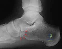
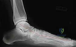
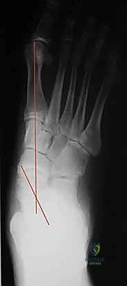
Meticulous preoperative planning forms the blueprint for intraoperative execution. Standardized, weight-bearing radiographs of the foot and ankle are non-negotiable. The anterior-posterior (AP) view is utilized to assess the talonavicular coverage angle, quantifying the degree of forefoot abduction. The lateral weight-bearing view is critical for evaluating the talo-first metatarsal angle (Meary's angle), which should normally be zero degrees. In PTTD, a plantar sag at the talonavicular or naviculocuneiform joint results in a break in this line. Additionally, the calcaneal pitch is measured to assess the global loss of the medial longitudinal arch. A standing ankle mortise view is mandatory to rule out deltoid ligament incompetence and talar tilt, which would upgrade the diagnosis to Stage IV.
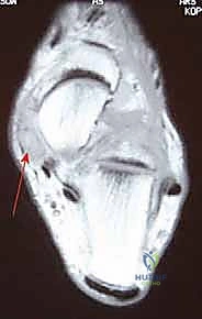
Advanced imaging, specifically Magnetic Resonance Imaging (MRI), is highly valuable, though the diagnosis remains primarily clinical and radiographic. MRI provides exceptional detail regarding the structural integrity of the PTT, identifying interstitial tearing, tenosynovitis, fluid within the tendon sheath, and complete ruptures. Furthermore, MRI allows for the evaluation of the spring ligament complex (superomedial and inferior calcaneonavicular ligaments), which is frequently attenuated in advanced Stage II disease and may require concomitant imbrication or reconstruction. MRI also helps rule out subtle subchondral cysts or early arthritic changes not readily apparent on plain radiographs.
Clinical assessment of the Achilles tendon and gastrocnemius-soleus complex is imperative. The Silfverskiöld test is performed to differentiate between isolated gastrocnemius tightness and a combined Achilles contracture. With the arch collapsed, the functional length of the Achilles is often shortened. Correcting the arch intraoperatively will unmask this tightness, potentially restricting ankle dorsiflexion and placing undue stress on the midfoot reconstruction. If the ankle cannot be dorsiflexed past neutral with the knee extended (but can with the knee flexed), a gastrocnemius recession (Strayer procedure) is indicated. If dorsiflexion is restricted with both knee flexion and extension, a percutaneous Achilles tendon lengthening (tendo-Achilles lengthening, TAL) may be required.
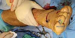
For patient positioning, the patient is placed supine on a radiolucent operating table. A substantial gel bump is placed under the ipsilateral hip to internally rotate the operative leg. This brings the posterolateral aspect of the heel into direct view, optimizing the approach for the initial calcaneal osteotomy. A well-padded pneumatic tourniquet is applied to the proximal thigh to ensure a bloodless surgical field, which is critical for identifying delicate neurovascular structures. Once the lateral osteotomy is completed and provisionally or definitively fixed, the hip bump is removed, allowing the leg to externally rotate naturally, thereby providing excellent exposure to the medial aspect of the foot and ankle for the tendon transfer phase.
Step-by-Step Surgical Approach and Fixation Technique
Part 1: The Lateral Approach and Medial Displacement Calcaneal Osteotomy
The procedure universally begins with the calcaneal osteotomy. Correcting the bony architecture first provides the mechanical foundation necessary to protect the subsequent soft tissue reconstruction.
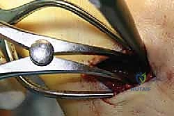
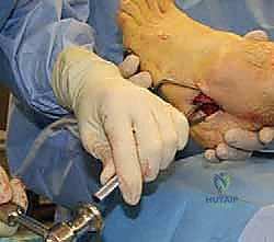
We initiate an oblique incision, approximately 4 to 5 centimeters in length, over the lateral aspect of the calcaneal tuberosity. This incision is positioned posterior to the peroneal tendons and the sural nerve, angled parallel to the relaxed skin tension lines to optimize wound healing. Dissection through the subcutaneous tissue must be meticulous and sharp. The sural nerve is the primary structure at risk here; it courses from proximal-posterior to distal-anterior in the subcutaneous fat. We utilize blunt dissection with a hemostat only when necessary to gently mobilize and protect the nerve, strictly avoiding rigorous retraction that could induce a traction neuropraxia.
Upon reaching the periosteum of the lateral calcaneal wall, a longitudinal incision is made. Using a Cobb elevator, we raise full-thickness subperiosteal flaps superiorly and inferiorly. Superiorly, the dissection extends to the dorsal aspect of the tuberosity, anterior to the Achilles insertion. Inferiorly, we dissect down to the plantar aspect of the calcaneus, taking great care to stay anterior to the origin of the plantar fascia to avoid iatrogenic plantar fasciitis. Hohmann retractors are placed dorsally and plantarly to protect the soft tissues and expose the entire lateral face of the calcaneus.
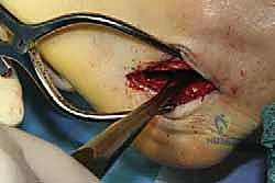
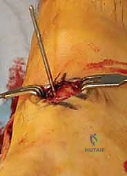
Using a microsagittal saw with a narrow blade, the osteotomy is performed in-line with the skin incision, angled approximately 45 degrees to the plantar surface of the foot. The cut is made from lateral to medial. As the saw blade approaches the medial cortex, the surgeon must rely on tactile feedback, feeling the change in resistance to avoid plunging into the medial neurovascular bundle. We often leave a millimeter of the medial cortex intact and complete the osteotomy with a broad osteotome, utilizing a gentle prying motion to cleanly fracture the remaining bone.
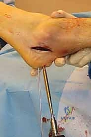
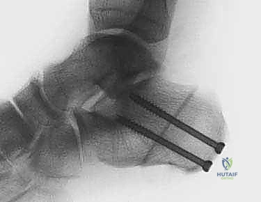
Once the osteotomy is mobile, a lamina spreader or specialized calcaneal displacement tool is inserted into the osteotomy site to stretch the medial periosteum. The posterior tuberosity fragment is then physically translated medially, typically by 10 millimeters, or approximately 50% of the width of the calcaneus. The displacement is provisionally held with a stout Kirschner wire driven from the posterior heel, across the osteotomy, and into the anterior calcaneal body. Axial and lateral fluoroscopic views are obtained to confirm the magnitude of displacement and ensure the osteotomy has not hinged into varus or valgus. Definitive fixation is achieved utilizing one or two large fragment (6.5mm or 7.0mm) partially threaded cannulated screws, inserted either percutaneously from the posterior heel or through the superior aspect of the lateral incision.
Part 2: Medial Dissection and Posterior Tibial Tendon Debridement
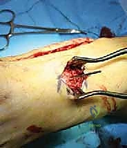
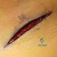
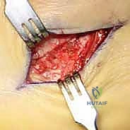
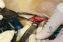
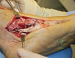
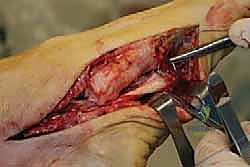
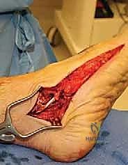
With the bony foundation corrected, the hip bump is removed, and attention is turned to the medial aspect of the foot. A curvilinear incision is made starting 2 centimeters proximal to the tip of the medial malleolus, coursing distally along the path of the PTT, and extending to the medial cuneiform. The subcutaneous tissues are carefully divided, taking care to coagulate crossing venous branches. The flexor retinaculum is identified and longitudinally incised to open the first fibro-osseous compartment, exposing the diseased posterior tibial tendon.

The PTT is systematically inspected. In Stage II disease, the tendon is typically grossly thickened, hyperemic, and demonstrates longitudinal interstitial tearing with profound loss of tension. The diseased portion of the tendon, usually extending from the medial malleolus to its insertion on the navicular, is sharply excised. We retain a small, healthy stump of the distal insertion at the navicular to serve as a biological marker and potential tissue for imbrication later. The tendon sheath is thoroughly debrided of inflammatory synovium.
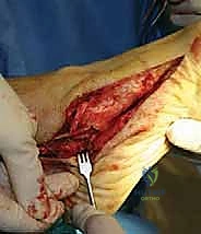
Part 3: FDL Harvest and Navicular Preparation
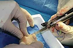
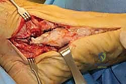
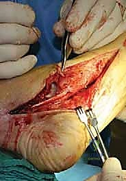
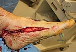
Immediately plantar and lateral to the excised PTT lies the Flexor Digitorum Longus tendon. The FDL is identified by its smaller caliber and its excursion when traction is applied, noting flexion of the lesser toes. The dissection is carried distally into the plantar midfoot, tracing the FDL toward the Master Knot of Henry. Retracting the abductor hallucis muscle plantarly aids in this exposure. The fibrous interconnections between the FDL and the deeper FHL tendon at the Master Knot are carefully sharply divided. The FDL is then transected as distally as possible, maximizing the length of the graft. A heavy, non-absorbable locking whipstitch (e.g., #2 FiberWire) is immediately placed into the distal end of the harvested FDL tendon.
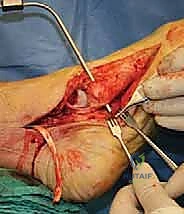
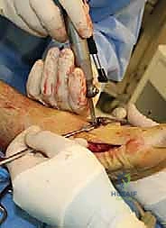
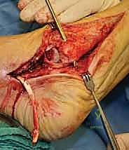
Attention is then directed to the navicular. The periosteum over the navicular tuberosity is elevated. Using a cannulated drill system over a guidewire, a bone tunnel is created through the navicular. The trajectory of this tunnel is critical; it is typically drilled from dorsal-superior to plantar-inferior, aiming slightly lateral to match the natural pull vector of the transferred tendon. The diameter of the drill hole is matched to the diameter of the harvested FDL tendon, usually requiring a 5.0mm to 6.0mm reamer. The edges of the tunnel are chamfered to prevent abrasive wear on the tendon graft.
Part 4: Tendon Transfer, Tensioning, and Fixation
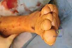
The whipstitched FDL tendon is passed through the navicular bone tunnel from plantar to dorsal. This is a critical juncture where the assistant's role becomes paramount. The assistant must hold the foot in maximum plantarflexion and maximum inversion. This position fully reduces the talonavicular joint, recreates the medial longitudinal arch, and removes all tension from the medial soft tissues.

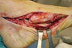
With the foot held rigidly in this corrected position, maximal tension is applied to the FDL graft pulling dorsally. Fixation is achieved using a bioabsorbable or PEEK interference screw (typically 5.5mm to 6.25mm) inserted into the dorsal aspect of the navicular tunnel, securely wedging the tendon against the bone. Alternatively, a biotenodesis screw system can be utilized. Once the screw is seated, the tension is tested. The foot should rest naturally in a neutral to slightly inverted position.
Following tendon fixation, the spring ligament complex is routinely inspected. If attenuated, it is imbricated using heavy absorbable sutures to further support the talar head. The remaining stump of the native PTT can also be sutured
Clinical & Radiographic Imaging Archive