ABOS Part I & OITE Orthopaedic Deformity Correction Exam Review | Paley Principles & FAN | Part 22009
Key Takeaway
This comprehensive review prepares orthopaedic surgeons for ABOS Part I and OITE exams, focusing on deformity correction. It covers Paley's principles, osteotomy planning, mechanical axis analysis (MAD, CORA, mLDFA, MPTA), Fixator Assisted Nailing (FAN), and Poller screw techniques through advanced multiple-choice questions for optimal exam readiness.
ABOS Part I & OITE Orthopaedic Deformity Correction Exam Review | Paley Principles & FAN | Part 22009
Comprehensive 100-Question Exam
00:00
Start Quiz
Question 1
A 32-year-old male presents with chronic right knee pain and a progressive varus deformity. Standing long-leg radiographs reveal a Mechanical Axis Deviation (MAD) of 25mm medial to the center of the knee. Further analysis shows a Mechanical Lateral Distal Femoral Angle (mLDFA) of 88° and a Medial Proximal Tibial Angle (MPTA) of 75°. The Joint Line Convergence Angle (JLCA) is 1°. Based on Paley's principles, which of the following statements best describes the location and nature of this patient's deformity?
Explanation
Correct Answer: C
The patient presents with a significant varus deformity, indicated by the MAD of 25mm medial to the center of the knee (normal is 8-10mm medial). To localize the deformity, we use the joint orientation angles. The mLDFA is 88°, which is within the normal range (85-90°, average 88°), indicating no significant deformity in the distal femur. However, the MPTA is 75°, which is significantly outside the normal range (85-90°, average 87°). An abnormal MPTA points directly to a tibial-based deformity, specifically in the proximal tibia. The JLCA of 1° is normal (0-2°), ruling out significant intra-articular pathology as the primary cause of the angular deformity.
Option A is incorrect because the mLDFA is normal, ruling out a primary femoral deformity.
Option B is incorrect because the JLCA is normal, suggesting the primary issue is extra-articular bony malalignment, not intra-articular pathology or laxity.
Option D is incorrect because the mLDFA is normal, localizing the deformity solely to the tibia.
Option E is incorrect because a MAD of 25mm medial is significantly abnormal, indicating a clear varus deformity.
Question 2
A 55-year-old patient presents with a severe tibial varus deformity, as shown in the preoperative planning radiograph below. The surgeon has meticulously drawn the mechanical axes of the proximal and distal tibial segments, identifying their intersection point. Which of the following statements accurately describes the significance of this intersection point in Paley's methodology?

Explanation
Correct Answer: C
The image on the left (a) in the provided figure demonstrates the identification of the Center of Rotation of Angulation (CORA). The CORA is the geometric point where the proximal and distal mechanical (or anatomic) axes of a deformed bone intersect. According to Paley's principles, identifying the CORA is the single most important step in preoperative planning as it dictates the ideal location for the osteotomy and governs the mechanical behavior of the entire limb during correction. Placing the osteotomy and the hinge of correction (ACA) at the CORA (Rule One) allows for pure angulation correction without translation.
Option A is incorrect. The nail entry point is typically suprapatellar or parapatellar for the tibia, not necessarily at the CORA.
Option B is incorrect. The mechanical axis of the entire limb is drawn from the femoral head to the ankle mortise, and its restoration is the goal, but the intersection of the segmental axes defines the CORA.
Option D is incorrect. While severe deformity can lead to cartilage wear, the CORA is a geometric construct for bony deformity, not a direct indicator of cartilage pathology.
Option E is incorrect. Poller screws are placed strategically around the nail in the metaphysis to prevent toggle, which is related to the corrected alignment, not the CORA itself.
Question 3
A 40-year-old patient requires correction of a distal femoral valgus deformity. Preoperative planning reveals the CORA is located precisely at the articular surface of the lateral femoral condyle. To achieve perfect mechanical axis restoration while minimizing surgical morbidity and preserving the joint, the surgeon plans an osteotomy 5 cm proximal to the CORA, with the hinge of correction maintained at the CORA. According to Paley's osteotomy rules, what is the expected biomechanical result of this approach?
Explanation
Correct Answer: C
This scenario describes Paley's Rule Two: Correction with Obligatory Translation. The geometric rule states that the hinge of correction (ACA) is placed at the CORA, but the actual bone cut (osteotomy) is performed at a different level (in this case, 5 cm proximal to the CORA). This is often necessary when the CORA is in an inaccessible or undesirable location, such as within the joint line. The biomechanical result is that the overall mechanical axis of the limb is perfectly restored, but a translation (sliding) of the bone fragments at the osteotomy site is an unavoidable geometric consequence. This translation must be anticipated and accommodated by the chosen hardware.
Option A is incorrect. Pure angulation with no translation occurs only when both the osteotomy and the hinge are exactly at the CORA (Rule One).
Option B is incorrect. Creation of a new, secondary translational deformity (a 'dog-leg') occurs when both the osteotomy and the hinge are at a level different from the CORA (Rule Three), which is generally to be avoided.
Option D is incorrect. If planned correctly according to Rule Two, the angular deformity is fully corrected, and the mechanical axis is restored.
Option E is incorrect. While an opening wedge osteotomy can lengthen the limb, the primary outcome described here is angular correction with translation, not necessarily lengthening, and certainly not without angular correction.
Question 4
A 28-year-old patient with a large, multiplanar angular deformity of the tibia requires correction while preserving limb length and maximizing bony contact for rapid healing. The surgeon opts for a focal dome osteotomy, as depicted in the intraoperative fluoroscopy image below. What is the primary advantage of this specific osteotomy design in this clinical scenario?

Explanation
Correct Answer: B
The focal dome osteotomy is an advanced technique characterized by a cylindrical, semi-circular cut made along the arc of a circle centered at the CORA. As seen in the image, the cut surfaces are curved, allowing the bone fragments to slide and rotate against each other during correction while maintaining maximum, continuous cortical contact. This unique geometric property provides extraordinary intrinsic stability and promotes rapid osseous union, often without the need for bone graft. It is particularly advantageous for large angular corrections where preserving limb length and maximizing bony contact are critical.
Option A is incorrect. The focal dome osteotomy is designed to correct angular deformity without altering limb length, unlike an opening wedge osteotomy which lengthens.
Option C is incorrect. Focal dome osteotomies are technically demanding and often require specialized guides and drill bits, making them more complex than simple transverse cuts.
Option D is incorrect. The focal dome osteotomy preserves limb length, it does not inherently shorten it. Closing wedge osteotomies shorten the limb.
Option E is incorrect. While it provides intrinsic stability, fixation (internal or external) is still required to hold the correction and allow for healing.
Question 5
A 60-year-old patient with severe bilateral genu varum is undergoing surgical correction. The surgeon plans to use Fixator Assisted Nailing (FAN) for the tibial deformities. Which of the following statements best describes the primary advantage of the FAN technique over traditional methods using only circular external fixators for such complex deformities?
Explanation
Correct Answer: C
Fixator Assisted Nailing (FAN) is a hybrid technique that combines the multi-planar precision of external fixation with the biological and mechanical superiority of intramedullary nailing. The external fixator is applied temporarily in the operating room to achieve acute, millimeter-accurate correction of the deformity. Once corrected, an intramedullary nail is inserted and locked, and the external fixator is removed before the patient leaves the operating room. This allows the patient to benefit from the stability of internal fixation, early mobilization, and avoids the prolonged complications associated with long-term external frame wear.
Option A is incorrect. FAN relies on intramedullary nailing as the definitive internal fixation.
Option B is incorrect. FAN achieves acute correction in the operating room. Gradual correction is characteristic of long-term external fixator use.
Option D is incorrect. While FAN can be adapted for lengthening, its primary strength lies in precise angular and translational deformity correction.
Option E is incorrect. By removing the external fixator immediately, FAN significantly reduces the risk of long-term pin tract infections and other frame-related complications compared to prolonged external fixation.
Question 6
During a Fixator Assisted Nailing (FAN) procedure for a proximal tibial varus deformity, the surgeon is preparing to place the half-pins for the monolateral external fixator. To ensure an unimpeded path for the intramedullary reamers and the definitive nail, what is the ideal strategic placement for these pins in the proximal and distal fragments?
Explanation
Correct Answer: C
The case explicitly states the ideal strategy for tibial FAN pin placement: 'place two half-pins posteriorly in the proximal fragment and two posteriorly in the distal fragment. This posterior placement leaves the entire anterior-to-posterior and medial-to-lateral trajectory of the medullary canal completely free and unimpeded.' This ensures that the guide wire, reamers, and intramedullary nail can be inserted without obstruction from the external fixator pins.
Option A is incorrect as anterior placement would obstruct the typical anterior entry point and reaming path for a tibial nail.
Option B is incorrect as medial/lateral placement could obstruct the nail's path, especially if the nail is placed centrally or slightly off-center.
Option D is incorrect as placing pins directly through the medullary canal would make intramedullary nailing impossible.
Option E is incorrect. While eccentric placement is key, using only a single pin per fragment would provide insufficient stability for acute correction and holding the alignment for nailing.
Question 7
A 48-year-old patient presents with a significant genu varum deformity and a concomitant 1.5 cm leg length discrepancy (LLD) in the affected limb. The deformity is localized to the proximal tibia. The surgeon's primary goal is to correct the varus and simultaneously address the LLD. Which osteotomy design is most appropriate for this patient?
Explanation
Correct Answer: C
The patient has a varus deformity and a concomitant leg length discrepancy (LLD) where the affected limb is shorter. An opening wedge osteotomy is performed by making a single transverse or oblique cut and opening a gap on the concave side of the deformity. A key advantage of this technique is that it lengthens the limb, which is highly beneficial for patients with a pre-existing LLD. While it requires bone graft and has a longer consolidation time, it directly addresses both the angular deformity and the length discrepancy.
Option A is incorrect. A closing wedge osteotomy inherently shortens the limb, which would exacerbate the existing LLD.
Option B is incorrect. A focal dome osteotomy corrects angular deformity without altering limb length, so it would not address the LLD.
Option D is incorrect. A transverse osteotomy with acute shortening would worsen the LLD.
Option E is incorrect. While bi-level osteotomies exist, the deformity is localized to the proximal tibia, and the question asks for the most appropriate osteotomy design for the given goals, not necessarily the number of osteotomies. An opening wedge at the tibia can address both issues.
Question 8
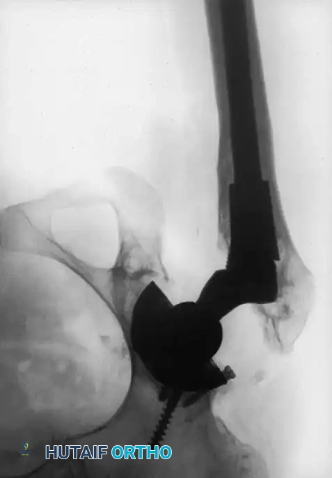
Following a Fixator Assisted Nailing (FAN) procedure for a proximal tibial varus deformity, the external fixator is removed, and the intramedullary nail is locked. The surgeon then considers the need for interference (Poller) screws. The close-up fluoroscopic image below shows two interference screws strategically placed on the medial side of the nail in the proximal tibia. What is the primary biomechanical purpose of these screws in this specific context?

Explanation
Correct Answer: C
The image clearly shows two interference (Poller) screws placed on the medial side of the intramedullary nail in the proximal tibia. The case describes the 'bell-clapper effect,' where the nail can toggle or slide within the wide metaphyseal canal, leading to loss of correction. For a varus deformity, the bone tends to settle back into varus, meaning the nail would drift medially. Therefore, placing Poller screws on the medial side of the nail physically blocks this medial drift, creating a rigid channel that forces the nail to maintain the limb's newly corrected alignment and preventing recurrence of the varus deformity.
Option A is incorrect. While they contribute to overall stability, their primary role in the metaphysis is to prevent translation/toggle, not specifically rotational stability in the diaphysis (which is handled by locking screws).
Option B is incorrect. Proximal/distal migration is prevented by the proximal and distal locking screws of the nail.
Option D is incorrect. Poller screws do not primarily compress the osteotomy site; their role is to control nail position within the canal.
Option E is incorrect. While Poller screws can be placed before nailing to guide reaming and nail insertion, their primary biomechanical purpose after nail insertion and locking is to block unwanted translation and maintain correction.
Question 9
A 35-year-old patient undergoes Fixator Assisted Nailing (FAN) for a valgus deformity of the distal femur. The deformity has been acutely corrected, and the intramedullary nail is in place. To prevent the 'bell-clapper effect' and maintain the corrected alignment, the surgeon plans to insert interference (Poller) screws. According to the golden rules for Poller screw placement, where should these screws be strategically positioned?
Explanation
Correct Answer: B
The golden rule for interference screw placement is to 'Place on the Concave Side of the Deformity' and 'Place in the Acute Angle'. For a valgus deformity, the concavity is on the lateral side of the limb. Therefore, to block the nail from drifting laterally and prevent the bone from settling back into a valgus position, the blocking screws must be placed on the lateral side of the nail. They are placed in the acute angle formed by the nail and the bone's axis to effectively narrow the canal and prevent toggle.
Option A is incorrect. Medial placement would be for a varus deformity, not valgus.
Option C and D are incorrect. While Poller screws can be placed in the sagittal plane, the primary rule for angular deformity correction is based on the coronal plane concavity/convexity. Anterior/posterior placement would address sagittal plane instability, but for a valgus deformity (coronal plane), lateral placement is key.
Option E is incorrect. Poller screws are placed around the nail, typically in the metaphyseal region where the canal is wide, not specifically proximal or distal to the locking screws, which have a different function.
Question 10
The multi-panel image below illustrates the journey of a patient undergoing Fixator Assisted Nailing (FAN) for bilateral bowleg deformities. Panel (g) shows the postoperative clinical appearance, and panel (e) shows the postoperative standing long-leg radiograph. What critical aspect of deformity correction is best demonstrated by the comparison of the preoperative state (f) and the postoperative results (e and g)?

Explanation
Correct Answer: C
The multi-panel image, particularly comparing the preoperative varus deformity (f) with the postoperative aligned limbs (g) and the perfectly restored mechanical axis on the standing long-leg radiograph (e), highlights the power of combining Paley's principles with the FAN technique. The case emphasizes that FAN achieves 'a beautifully restored mechanical axis' and 'dramatic cosmetic and functional improvement' through acute correction. This demonstrates the ability to reliably restore normal biomechanics and improve patient quality of life.
Option A is incorrect. FAN achieves acute correction, not gradual lengthening over months. Gradual lengthening is characteristic of long-term external fixator use.
Option B is incorrect. FAN's advantage is precisely to avoid prolonged external fixation by using it temporarily and then removing it.
Option D is incorrect. While interference screws are important, their primary role is to prevent toggle and maintain alignment in the metaphysis, not provide rotational stability throughout the diaphysis (which is the role of the nail's locking screws).
Option E is incorrect. While Rule Two involves obligatory translation, the goal is always to restore the mechanical axis, not to create a 'controlled translational deformity' as an end goal in itself, unless addressing a pre-existing translational deformity. The overall outcome shown is perfect alignment, not a deliberate residual translational deformity.
Question 11
A 38-year-old male presents with progressive knee pain and a noticeable bowing deformity of his left lower extremity. A full-length weight-bearing radiograph is obtained, and the surgeon begins the Paley method of deformity analysis. The initial step involves drawing a line from the center of the femoral head to the center of the talar dome. This line is observed to pass 25 mm medial to the center of the knee joint. The surgeon then identifies the intersection point of the proximal and distal mechanical axes of the deformed bone segment, as shown in the diagram below.
Which of the following statements accurately describes the initial findings and their significance in this patient's case?

Explanation
Correct Answer: B
The patient has a varus deformity with a Mechanical Axis Deviation (MAD) of 25 mm medial, which is the primary biomechanical driver of premature osteoarthritis. The text defines MAD as the perpendicular distance from the mechanical axis line to the center of the knee joint. A line passing medial to the knee center indicates a varus deformity, and a deviation of 25 mm is significant. This chronic maldistribution of force is explicitly stated as the primary biomechanical driver of premature osteoarthritis, ligamentous instability, and functional decline.
Incorrect Options:
- A: A MAD of 25 mm medial indicates a varus deformity, not valgus. Valgus would be a lateral deviation.
- C: The identified intersection point is indeed the CORA, which dictates the apex of the deformity and guides the osteotomy. However, the MAD, not the CORA, quantifies the magnitude of the deformity and its impact on the weight-bearing axis. The CORA tells you where the deformity originates, not its magnitude.
- D: In a neutrally aligned limb, the mechanical axis should pass slightly medial to the exact center of the knee, typically bisecting the medial tibial spine, but a 25 mm medial deviation is well outside the normal range and indicates a significant varus deformity, not normal alignment.
- E: The CORA dictates the rules of the osteotomy (angulation and translation), not the normal joint orientation angles. These angles (mLDFA, MPTA) are target values for correction, not determined by the CORA itself.
Question 12
A 55-year-old female presents with a severe post-traumatic varus malunion of the distal femur. Preoperative planning reveals a CORA located 5 cm proximal to the knee joint line, within the diaphyseal bone. The surgeon plans a distal femoral osteotomy. Given the CORA's location, the surgeon decides to perform the osteotomy directly at the CORA. Which of Paley's Osteotomy Rules applies to this scenario, and what is the expected outcome regarding correction?

Explanation
Correct Answer: C
Rule 1 applies; a pure angular correction (hinging) at the osteotomy site will perfectly realign the mechanical axis without translation. The case explicitly states that the osteotomy is performed 'directly at the level of the CORA.' According to the text and diagram (specifically panel A of the image), Paley's Osteotomy Rule One states: 'When the osteotomy is performed directly at the level of the CORA, a pure angular correction (hinging) will perfectly realign the mechanical axis. No translation is required.'
Incorrect Options:
- A: Rule 2 applies when the osteotomy is different from the CORA, requiring both angulation and translation. This is contrary to the scenario described.
- B: Rule 3 describes a common pitfall when the osteotomy is different from the CORA and angulation occurs without translation, leading to malalignment. This is not applicable when the osteotomy is at the CORA.
- D: Rule 1 explicitly states 'No translation is required' when the osteotomy is at the CORA. The mLDFA is a target angle, but its achievement through a Rule 1 osteotomy does not require translation.
- E: Rule 2 applies when the osteotomy is away from the CORA. While it's true that osteotomies are often performed away from the CORA for fixation reasons, the scenario here specifies the osteotomy is at the CORA.
Question 13
A 28-year-old male presents with a severe congenital tibial varus deformity. Preoperative planning identifies the CORA located 2 cm distal to the knee joint line, making a direct osteotomy at this level challenging for stable internal fixation. The surgeon decides to perform a high tibial osteotomy 5 cm distal to the joint line, in the metaphyseal bone. During the procedure, after performing the osteotomy, the surgeon only performs an angular correction (hinging) at the osteotomy site without any translation. Postoperative radiographs show correction of the local bone angle but persistent overall mechanical axis malalignment. Which of Paley's Osteotomy Rules was violated, and what is the resulting deformity?
Explanation
Correct Answer: B
Rule 2 was violated; the surgeon failed to perform the mandatory translation required when the osteotomy is away from the CORA, resulting in a 'dog-leg' deformity. The scenario describes an osteotomy performed 'away from the CORA' (5 cm distal to the joint line vs. CORA at 2 cm distal) where 'only an angular correction (hinging) at the osteotomy site without any translation' was performed. The text explicitly states under 'Osteotomy Rule Three: The Common Pitfall' (which is a violation of Rule Two's requirements): 'If the osteotomy is performed at a level different from the CORA, and the angulation occurs around the osteotomy site itself (not the CORA) without translation, a secondary translational deformity is created, and the mechanical axis remains malaligned.' This results in a 'dog-leg' deformity, as illustrated in panel C of the provided image.
Incorrect Options:
- A: While performing the osteotomy at the CORA (Rule 1) would be ideal for pure angulation, the clinical scenario often necessitates moving the osteotomy away for fixation. The error here is not moving away from the CORA, but failing to translate once away.
- C: Rule 3 describes the consequence of violating Rule 2, not a correct application. The persistent malalignment is a direct result of the surgical error, not necessarily a misidentified CORA.
- D: While an external fixator can assist with translation, the core violation is the failure to understand and execute the biomechanical principle of translation itself, regardless of the tool used.
- E: The osteotome twist technique is for executing the osteotomy and translation, but the fundamental error was the decision to only angulate without translation when the osteotomy was away from the CORA.
Question 14
A 42-year-old male is undergoing a high tibial osteotomy for a varus deformity. Preoperative planning indicates a target Medial Proximal Tibial Angle (MPTA) of 87°. During the intraoperative fluoroscopic validation, the surgeon measures the MPTA as 82°. Which of the following statements best describes the significance of the MPTA and the necessary intraoperative adjustment?
Explanation
Correct Answer: B
An MPTA of 82° indicates a residual varus deformity, and the osteotomy needs to be opened further medially. The text states the normal range for MPTA is 85° to 90°, with an average target value of 87°. An MPTA of 82° is less than the normal range, meaning the medial side of the proximal tibia is angled too acutely downwards, indicating persistent varus. To correct this, the osteotomy needs to be opened further medially to increase the MPTA towards the target of 87°.
Incorrect Options:
- A: An MPTA of 82° is less than the target 87°, indicating varus, not overcorrection into valgus. Overcorrection into valgus would result in an MPTA greater than 90°.
- C: The MPTA is explicitly stated as 'The primary target in high tibial osteotomies,' while the mLDFA is crucial for distal femoral osteotomies.
- D: An MPTA of 82° is outside the normal range of 85° to 90°, requiring adjustment.
- E: The MPTA defines the relationship of the knee joint line to the mechanical axis of the tibia, not the femur. The mLDFA defines this relationship for the femur.
Question 15
A 60-year-old patient with severe genu varum is scheduled for a high tibial osteotomy. Preoperative planning using a full-length weight-bearing radiograph identifies the CORA 1 cm distal to the joint line. Due to concerns about bone quality and the need for stable internal fixation, the surgeon plans the osteotomy 4 cm distal to the joint line. To ensure a perfect correction, the surgeon utilizes the goniometer method to calculate the required translation. Which of the following steps is crucial for accurately determining the amount of translation needed?
Explanation
Correct Answer: C
The crucial step for accurately determining the amount of translation needed is placing the goniometer's pivot point directly over the CORA, simulating the correction, and then measuring the perpendicular distance from the center of the bone at the planned osteotomy site to the new, corrected distal axis line. The text describes the goniometer method: '1. Identify the CORA... 2. Plan the Osteotomy Level... 3. Position the Goniometer: Place the center pivot of the goniometer directly over the CORA. ... 5. Simulate the Correction: Rotate the goniometer arm representing the distal axis until the desired correction is achieved... 6. Measure the Translation: Observe exactly where this new, corrected axis line crosses your planned osteotomy level. Measure the perpendicular distance from the center of the bone at the osteotomy site to this new line. This distance, in millimeters, is the exact amount of translation required intraoperatively.'
Incorrect Options:
- A: The goniometer's pivot point must be over the CORA, not the osteotomy level, to accurately simulate the correction around the true apex of the deformity.
- B: Translation is measured at the osteotomy site, not at the CORA. The CORA is the pivot, the osteotomy site is where the translation occurs.
- D: While calculations are involved, this specific formula is not described as the goniometer method for determining translation. The goniometer method is a visual and direct measurement technique.
- E: The goniometer method is for calculating translation for the current deformity, not for confirming mLDFA after the osteotomy, which is an intraoperative fluoroscopic step.
Question 16
During a complex tibial osteotomy, the surgeon is utilizing the multiple drill hole and osteotome twist technique. After creating a series of bicortical drill holes, the surgeon inserts a wide, flat osteotome into the center of the drill hole pattern. Which of the following actions is the most appropriate next step to complete the osteotomy and achieve the planned translation, and what is its primary biomechanical advantage?

Explanation
Correct Answer: C
Twist the osteotome with slow, controlled rotational force to connect the cancellous bone bridges and lever the distal fragment into the exact translated position, preserving osteocytes. The text explicitly describes this: 'Insert a wider, flat osteotome into the center of the drill hole pattern. Now, instead of hammering aggressively, twist the osteotome. This rotational force will connect the remaining cancellous bone bridges and complete the osteotomy with a gentle, controlled crack.' It further states: 'The twisting motion is not just for breaking the bone; it is the mechanical engine for translation. By twisting the osteotome handle (e.g., counterclockwise), the broad blade acts as a cam, levering and pushing the distal fragment into the exact translated position calculated preoperatively.' The technique also 'drastically minimizes heat generation compared to a high-speed oscillating power saw, thereby preserving the vital osteocytes at the bone ends that are critical for rapid callus formation and healing.'
Incorrect Options:
- A: Aggressive hammering is discouraged; the text advises 'instead of hammering aggressively, twist the osteotome.' This can lead to uncontrolled fracture and comminution.
- B: The multiple drill hole and osteotome twist technique is specifically chosen to minimize heat generation compared to a high-speed oscillating saw, which can cause thermal necrosis.
- D: The technique involves a single wider osteotome for the central twist, not multiple narrow osteotomes hammered simultaneously.
- E: Translation is achieved after the osteotomy is complete or during the final separation via the twist, not by applying linear pressure before the cut is fully made.
Question 17
A 30-year-old patient is undergoing a complex proximal tibial osteotomy for a severe varus deformity. The surgeon is meticulously performing the osteotomy using the multiple drill hole technique. Which of the following surgical pearls is most critical to prevent a devastating complication during this specific procedure?
Explanation
Correct Answer: D
Protecting the common peroneal nerve and the posterior neurovascular bundle with blunt retractors is most critical to prevent a devastating complication during a proximal tibial osteotomy. The text specifically highlights this under 'Surgical Pearls for Flawless Osteotomy Execution': 'Protect Neurovascular Structures: Be acutely aware of nearby structures, particularly the common peroneal nerve during proximal tibial osteotomies. Use blunt retractors to protect the posterior neurovascular bundle.' Damage to these structures can lead to permanent neurological deficits or vascular compromise, which are devastating complications.
Incorrect Options:
- A, B, C, E: While all these are crucial surgical pearls mentioned in the text for a successful osteotomy and good bone healing, they primarily relate to the biomechanics of the cut and bone viability. Neurovascular injury, however, represents a more immediate and potentially devastating complication in terms of patient function and limb viability, especially in the context of a proximal tibial osteotomy where the common peroneal nerve is highly vulnerable.
Question 18
A 48-year-old patient with a severe bilateral varus deformity of the tibiae is undergoing Fixator-Assisted Nailing (FAN) for correction. After meticulous preoperative planning, strategic pin insertion, and osteotomy, the external fixator is locked, holding the limb in perfect corrected alignment. The surgeon then proceeds with reaming the intramedullary canal and inserting the intramedullary nail. What is the primary rationale for using a temporary external fixator in this scenario?
Explanation
Correct Answer: C
The primary rationale for using a temporary external fixator in this scenario is to rigidly hold the limb in the perfectly corrected position, preventing displacement during the forces of intramedullary reaming and nail insertion. The text states: 'The physical forces involved in sequentially reaming an intramedullary canal and hammering a titanium nail can easily displace a perfectly corrected but unsecured osteotomy. A temporary external fixator acts as a rigid, unyielding external scaffold, locking the limb in the perfectly corrected position while the internal work is done.'
Incorrect Options:
- A: The external fixator in FAN is temporary and removed once the internal nail is locked, not for definitive, long-term fixation.
- B: While some external fixators are used for gradual correction, in FAN, the correction is achieved acutely and then held rigidly while the nail is inserted.
- D: The FAN technique aims for rigid stability during internal fixation, not micromotion, which would risk losing the acute correction.
- E: While Poller screws are often inserted with the fixator in place, the primary rationale for the fixator itself is to maintain alignment during the reaming and nailing process, not solely for Poller screw insertion.
Question 19
During a Fixator-Assisted Nailing (FAN) procedure for a proximal tibial varus deformity, the surgeon considers inserting interference (Poller) screws. Which of the following statements accurately describes the purpose and optimal timing for inserting these screws in this context?
Explanation
Correct Answer: B
Poller screws are used to artificially narrow the medullary canal, forcing the nail into the optimal trajectory and preventing translation, and are typically inserted before removing the external fixator. The text states: 'For added mechanical stability, interference screws (often called blocking or Poller screws) may be inserted to artificially narrow the medullary canal. This forces the nail into the optimal trajectory and prevents the nail from translating within the wide metaphyseal bone.' It also notes: 'Note: These interference screws are typically inserted before removing the external fixator to ensure the alignment isn't lost during screw placement.'
Incorrect Options:
- A: Poller screws are for augmenting stability and guiding the nail, not for primary fixation or replacing the nail.
- C: The text specifically states: 'This step is vastly more important for proximal tibial FAN than for distal femoral FAN due to the funnel shape of the proximal tibia.'
- D: Poller screws are typically inserted before removing the external fixator to maintain alignment during their placement.
- E: Poller screws are for rigid internal fixation, not for creating a hinge point for gradual correction.
Question 20
A 35-year-old patient is undergoing a distal femoral osteotomy for a valgus deformity. The surgeon is aiming to restore normal joint orientation angles. Which of the following angles is the primary target for this specific correction, and what is its average target value?
Explanation
Correct Answer: B
The primary target for a distal femoral osteotomy is the Mechanical Lateral Distal Femoral Angle (mLDFA), with an average target value of 87°. The table in the text lists 'mLDFA' as 'Crucial for distal femoral osteotomies' with an 'Average Target Value' of '87°'.
Incorrect Options:
- A: MPTA is the primary target for high tibial osteotomies, not distal femoral.
- C: mLDTA is essential for supramalleolar corrections, not distal femoral.
- D: JLCA measures ligamentous laxity or cartilage loss, not a primary target for bone correction itself, though it's an important assessment.
- E: LPFA defines the relationship of the hip joint to the proximal femoral axis, not the distal femur.
Question 21
A surgeon is performing a high tibial osteotomy. After creating the osteotomy, the surgeon notices that the bone does not yield easily to the osteotome twist, despite having made multiple drill holes. What is the most appropriate next step, according to the surgical pearls, and why?
Explanation
Correct Answer: C
The most appropriate next step is to remove the osteotome and use the drill to connect any missed spots, as there is likely a remaining cortical bone bridge. The text's 'Surgical Pearls for Flawless Osteotomy Execution' explicitly states: 'Don't Force It: If the bone doesn't yield easily to the twist, there is a remaining cortical bone bridge. Remove the osteotome and use the drill to connect any missed spots.'
Incorrect Options:
- A: Aggressive hammering is discouraged and can lead to uncontrolled fracture or comminution.
- B: Switching to a high-speed saw defeats the purpose of the multiple drill hole technique, which is to minimize thermal necrosis.
- D: While twisting is key, the pearl 'Don't Force It' indicates that excessive force is counterproductive and suggests a missed bone bridge rather than simply needing more force.
- E: Abandoning the osteotomy level without first ensuring the current attempt is complete is premature and may not be necessary if a simple bone bridge is the issue.
Question 22
A patient requires correction of a diaphyseal tibial deformity. Preoperative planning identifies the Center of Rotation of Angulation (CORA). The surgeon performs the osteotomy exactly at the CORA and places the hinge axis at the CORA. According to Paley's osteotomy rules, what is the expected biomechanical outcome?
Explanation
Question 23
A surgeon is planning a distal femoral osteotomy for a severe valgus deformity. The CORA is located at the joint line. To preserve the joint capsule, the surgeon places the osteotomy 4 cm proximal to the CORA, but carefully sets the hinge axis of the external fixator exactly on the CORA. What will be the result of this correction?
Explanation
Question 24
A junior resident plans a proximal tibial osteotomy for a varus deformity. By mistake, both the osteotomy and the center of the hinge are placed 5 cm proximal to the true CORA. According to Paley's principles, what is the geometric consequence of this technical error?
Explanation
Question 25
An 18-year-old male presents with bilateral genu varum. Standing long-leg radiographs reveal a Mechanical Axis Deviation (MAD) of 35 mm medial to the center of the knee. The mechanical Lateral Distal Femoral Angle (mLDFA) is measured at 87 degrees, and the Medial Proximal Tibial Angle (MPTA) is measured at 76 degrees. The Joint Line Convergence Angle (JLCA) is 1 degree. Where is the primary source of the deformity?
Explanation
Question 26
During Fixator-Assisted Nailing (FAN) for a distal tibial valgus deformity, the surgeon intends to use a blocking (Poller) screw to prevent the intramedullary nail from following the path of least resistance into the deformed metaphysis. To effectively guide the nail and correct the valgus, where should the blocking screw be placed in the distal segment?
Explanation
Question 27
A 10-year-old girl with a congenital femoral deficiency has a current limb length discrepancy of 3 cm. The surgeon wishes to calculate her expected discrepancy at skeletal maturity using the Paley Multiplier Method. Which of the following parameters is strictly required for this specific calculation?
Explanation
Question 28
When evaluating sagittal plane deformities of the lower extremity using Paley's joint orientation angles, what is the normal expected Posterior Distal Femoral Angle (PDFA) referenced from the mechanical axis?
Explanation
Question 29
A 55-year-old female with knee pain has a medial Mechanical Axis Deviation (MAD). Her mLDFA is 88 degrees and MPTA is 87 degrees. However, her Joint Line Convergence Angle (JLCA) is 6 degrees, opening laterally. What is the most likely etiology of her medial MAD?
Explanation
Question 30
What is the primary mechanical advantage of the Taylor Spatial Frame (TSF) over traditional Ilizarov circular frames when managing complex diaphyseal deformities?
Explanation
Question 31
Which of the following describes the optimal rate and rhythm for distraction osteogenesis in a healthy adult undergoing tibial lengthening, as established by Ilizarov?
Explanation
Question 32
A 25-year-old male is undergoing 5 cm of tibial lengthening via distraction osteogenesis. At week four, he develops an equinus contracture of the ankle despite aggressive physical therapy. What is the most appropriate next step in management to prevent long-term morbidity?
Explanation
Question 33
When planning an opening wedge osteotomy to correct a valgus deformity of the proximal tibia using Paley principles, where must the physical hinge or axis of rotation be placed to avoid unintentional translation?
Explanation
Question 34
In the context of distraction osteogenesis and limb lengthening, how is the Bone Healing Index (BHI) defined?
Explanation
Question 35
A 40-year-old male presents with post-traumatic deformity. Standing radiographs show a lateral Mechanical Axis Deviation (MAD). Measurement of the mechanical Lateral Distal Femoral Angle (mLDFA) is 80 degrees, while the MPTA is 87 degrees. Based on Paley's normal parameters, what is the primary deformity?
Explanation
Question 36
During the surgical approach for distraction osteogenesis, the surgeon performs a low-energy corticotomy rather than a standard osteotomy with a power saw. What is the primary biological rationale for this technique according to Ilizarov principles?
Explanation
Question 37
When performing Fixator-Assisted Plating (FAP) for a complex distal femoral deformity, what is the primary role of the temporary external fixator?
Explanation
Question 38
A patient with a severe procurvatum deformity of the tibia is treated with a closing wedge osteotomy. To achieve angular correction without introducing secondary translation, where must the geometric hinge axis be placed?
Explanation
Question 39
You plan to lengthen the femur of a 30-year-old using a motorized intramedullary lengthening nail. The patient has a 4 cm length discrepancy but a perfectly normal mechanical axis. To minimize the risk of inducing a new angular deformity during lengthening, where is the optimal location for the osteotomy?
Explanation
Question 40
When constructing an Ilizarov circular frame for a tibial deformity, the surgeon inserts two tensioned transfixion wires on a single ring. To maximize the frame's stiffness against axial loading and torsional forces, what should the crossing angle between the two wires ideally be?
Explanation
Question 41
A patient undergoing tibial lengthening is evaluated at the 3-week post-operative mark. Radiographs show early premature consolidation of the regenerate bone. The patient admits they have only been distracting 0.25 mm per day. What is the most appropriate next step in management?
Explanation
Question 42
A surgeon is planning to correct a diaphyseal tibial deformity. The Center of Rotation of Angulation (CORA) is identified in the mid-diaphysis. According to Paley's Osteotomy Rule 1, if both the osteotomy and the Axis of Correction of Angulation (ACA) are placed exactly at the CORA, what will be the resulting alignment of the bone segments?
Explanation
Question 43
During preoperative planning for a distal femoral valgus deformity, the CORA is located intra-articularly. To preserve the joint capsule and hardware placement, the surgeon performs the osteotomy 4 cm proximal to the CORA but places the ACA at the CORA hinge. According to Paley's Osteotomy Rule 2, what is the expected outcome?
Explanation
Question 44
A patient undergoes a corrective osteotomy for a tibial varus deformity. The surgeon inadvertently places both the osteotomy and the ACA 3 cm distal to the true CORA. According to Paley's Osteotomy Rule 3, what biomechanical consequence will occur?
Explanation
Question 45
When utilizing the Fixator-Assisted Nailing (FAN) technique for correcting a complex lower extremity deformity, what is the most critical step regarding the placement of the temporary half-pins?
Explanation
Question 46
A 45-year-old patient has a mechanical axis deviation (MAD) of 35 mm lateral to the knee center. The Mechanical Lateral Distal Femoral Angle (mLDFA) is 81 degrees, and the Medial Proximal Tibial Angle (MPTA) is 87 degrees. The Joint Line Convergence Angle (JLCA) is 1 degree. Where is the primary source of the deformity?
Explanation
Question 47
You are treating a proximal third tibia fracture with a planned intramedullary nail and want to prevent the common valgus/procurvatum malalignment. Using the blocking (Poller) screw principle described in deformity correction, where should the blocking screws be placed in the proximal segment relative to the desired nail path?
Explanation
Question 48
Which of the following is the correct sequential order of steps when performing Fixator-Assisted Nailing (FAN) for deformity correction?
Explanation
Question 49
A patient presents with a multi-apical tibial deformity demonstrating two distinct CORAs. If the surgeon decides to correct the entire deformity using a single osteotomy at the proximal CORA, what compensatory action must be incorporated to achieve a collinear mechanical axis?
Explanation
Question 50
In assessing lower extremity alignment, a Joint Line Convergence Angle (JLCA) of 6 degrees (opening laterally) is measured on a standing AP radiograph. What does this abnormal JLCA most likely represent in the context of varus deformity?
Explanation
Question 51
A patient presents with a diaphyseal femoral deformity measuring 15 degrees of varus on the AP radiograph and 20 degrees of apex anterior bowing (procurvatum) on the lateral radiograph. Using the principles of oblique plane deformity, what is the approximate true magnitude of this deformity?
Explanation
Question 52
During preoperative planning for a sagittal plane tibial deformity, the Posterior Proximal Tibial Angle (PPTA) is measured. The normal anatomic PPTA is approximately 81 degrees. If a patient has a PPTA of 95 degrees, what type of deformity is present?
Explanation
Question 53
A pure translation deformity of the tibial diaphysis is noted on radiographs, with zero degrees of angular deformity. Where is the Center of Rotation of Angulation (CORA) located in this specific scenario?
Explanation
Question 54
When performing a closing wedge osteotomy at the CORA for a varus deformity of the tibia, where should the Axis of Correction of Angulation (ACA) be positioned to prevent any iatrogenic translation?
Explanation
Question 55
You are treating a patient with a severe distal tibial deformity using a circular external fixator. The surgeon plans a neutral wedge (dome) osteotomy. Where is the ACA located in a perfectly executed neutral wedge osteotomy?
Explanation
Question 56
In the context of the Fixator-Assisted Nailing (FAN) technique, which of the following is an absolute contraindication to utilizing this method for deformity correction?
Explanation
Question 57
A surgeon is evaluating a patient with a significant diaphyseal femur deformity. The angle between the anatomic axis and the mechanical axis of the normal femur (Anatomic-Mechanical Angle, AMA) is typically:
Explanation
Question 58
A patient is undergoing Fixator-Assisted Plating (FAP) instead of FAN for a distal femoral valgus deformity. What is the primary biomechanical advantage of FAP over FAN in a very distal peri-articular deformity?
Explanation
Question 59
Using Paley's malalignment test, you drop a plumb line from the center of the femoral head to the center of the ankle. The line passes 20 mm medial to the center of the knee joint. The MPTA is 80 degrees, and the mLDFA is 88 degrees. Which of the following describes the deformity?
Explanation
Question 60
During a FAN procedure for a proximal tibial deformity, the surgeon plans to use a Poller screw to correct a procurvatum tendency. To properly narrow the canal and push the nail posteriorly, where should the Poller screw be placed in the proximal fragment?
Explanation
Question 61
When calculating the magnitude of translation generated by applying Paley's Rule 2 (ACA at CORA, osteotomy distant from CORA), the amount of translation is mathematically dependent on which two factors?
Explanation
Question 62
A surgeon is planning to correct a diaphyseal tibial deformity. The mechanical axes of the proximal and distal segments intersect at the Center of Rotation of Angulation (CORA). The surgeon decides to perform the osteotomy 4 cm distal to the CORA but places the axis of correction (hinge) exactly at the CORA. According to Paley's osteotomy rules, what is the expected biomechanical outcome?
Explanation
Question 63
During a Fixator-Assisted Nailing (FAN) procedure for a proximal third tibia fracture with a valgus deformity, the surgeon plans to use Poller (blocking) screws to maintain alignment during reaming and nail insertion. Where is the most biomechanically advantageous position to place the blocking screw in the proximal segment?
Explanation
Question 64
A 45-year-old patient presents with medial knee pain. Standing long-leg radiographs reveal a Mechanical Axis Deviation (MAD) of 35 mm medial to the midline. The Mechanical Proximal Tibial Angle (MPTA) is 87 degrees, the mechanical Lateral Distal Femoral Angle (mLDFA) is 96 degrees, and the Joint Line Convergence Angle (JLCA) is 1 degree. Where is the primary source of the patient's deformity?
Explanation
Question 65
According to Paley's Osteotomy Rule 3, what occurs when both the osteotomy and the axis of correction (hinge) are located away from the Center of Rotation of Angulation (CORA)?
Explanation
Question 66
In deformity planning for a patient with severe osteoarthritis and a varus knee, the Joint Line Convergence Angle (JLCA) is measured at 6 degrees (opening laterally). What does this abnormal JLCA primarily indicate in this clinical scenario?
Explanation
Question 67
When using a Taylor Spatial Frame (TSF) for correcting a complex, multi-planar deformity, what represents the primary biomechanical advantage of its 'virtual hinge' over classic Ilizarov frames?
Explanation
Question 68
A 28-year-old male is undergoing treatment for a 5 cm post-traumatic tibial leg length discrepancy using the Lengthening Over a Nail (LON) technique. What is the primary advantage of LON compared to classic distraction osteogenesis using solely a circular external fixator?
Explanation
Question 69
A surgeon is evaluating a patient for a sagittal plane deformity of the proximal tibia prior to an osteotomy. What is the normal accepted range for the Posterior Proximal Tibial Angle (PPTA)?
Explanation
Question 70
A surgeon plans an acute valgus-producing high tibial osteotomy to correct a severe proximal tibial varus deformity. During the acute correction into valgus, which neurovascular structure is at the greatest risk of stretch injury?
Explanation
Question 71
During Fixator-Assisted Nailing (FAN) of a distal third femur fracture, the gastrocnemius muscle routinely pulls the distal segment into recurvatum (apex posterior angulation). To prevent this deformity during reaming and nailing, a Poller (blocking) screw should be strategically placed:
Explanation
Question 72
A 12-year-old girl has a leg length discrepancy. Using the Paley Multiplier Method to predict her leg length discrepancy at maturity, the surgeon multiplies her current discrepancy by an age-specific multiplier. What physiological event marks the stabilization of the multiplier at 1.0 (indicating skeletal maturity) for females in this system?
Explanation
Question 73
When calculating parameters for femoral deformity correction, what is the normal average Anatomic-Mechanical Angle (AMA) of the femur, and how do the axes relate?
Explanation
Question 74
A patient presents with a Mechanical Axis Deviation (MAD) of only 5 mm, but radiographs reveal a distal femoral valgus deformity (mLDFA of 80 degrees) and a proximal tibial varus deformity (MPTA of 80 degrees). This scenario best illustrates which of the following concepts?
Explanation
Question 75
A surgeon utilizes Fixator-Assisted Plating (FAP) for a complex distal tibia deformity. What is the primary procedural rationale for choosing FAP over traditional free-hand plating or pure external fixation?
Explanation
Question 76
If a surgeon attempts to correct a multi-apical diaphyseal deformity of the tibia by performing a single osteotomy at the intersection point of the proximal and distal mechanical axes (the apparent single CORA), what is the inevitable geometric result?
Explanation
Question 77
A patient undergoes a corticotomy and distraction osteogenesis of the tibia. What is the standard accepted rate of distraction to optimize bone regenerate formation while preventing premature consolidation?
Explanation
Question 78
You are assessing a standing long-leg AP radiograph.
The mechanical axis line passes 20 mm lateral to the center of the knee. The MPTA is 95 degrees, and the mLDFA is 87 degrees. What is the diagnosis?
Explanation
Question 79
A patient undergoes correction of a diaphyseal tibial malunion. The preoperative plan localizes the Center of Rotation of Angulation (CORA) at the mid-diaphysis. The surgeon performs the osteotomy precisely at the CORA and places the hinge of the external fixator at this exact same location. According to Paley's osteotomy rules, what is the expected biomechanical outcome of this correction?
Explanation
Question 80
A 25-year-old female presents with a distal femoral valgus deformity. Preoperative templating identifies the CORA precisely at the knee joint line. To preserve the joint capsule and secure adequate fixation, the surgeon performs a metaphyseal osteotomy 4 cm proximal to the joint line, but places the hinge of the corrective device exactly at the CORA. Which of the following describes the resulting alignment?
Explanation
Question 81
During the correction of a mid-diaphyseal femoral varus deformity, the surgeon performs the osteotomy 5 cm proximal to the true CORA. Inadvertently, the hinge of the external fixator is placed at the level of the osteotomy rather than at the CORA. What is the resulting mechanical consequence of this technical error?
Explanation
Question 82
A surgeon utilizes the Fixator-Assisted Nailing (FAN) technique to correct a severe distal tibial varus deformity. To guide the intramedullary nail and prevent recurrent deformity, blocking (Poller) screws are planned. Where must the blocking screws be placed relative to the medullary canal to achieve optimal correction?
Explanation
Question 83
A 45-year-old male presents with bilateral knee pain. Standing full-length radiographs reveal a mechanical axis deviation (MAD) of 35 mm medial to the center of the knee joint bilaterally. Radiographic measurements show a mechanical lateral distal femoral angle (mLDFA) of 87 degrees, a medial proximal tibial angle (MPTA) of 72 degrees, and a joint line convergence angle (JLCA) of 1 degree. What is the primary anatomic source of this patient's malalignment?
Explanation
Question 84
A patient with severe medial compartment osteoarthritis presents with a clinically apparent varus alignment. Radiographic analysis yields an mLDFA of 88 degrees, an MPTA of 86 degrees, and a mechanical axis deviation of 25 mm medial. The joint line convergence angle (JLCA) is 7 degrees, opening laterally. What is the most likely cause of the medial mechanical axis deviation?
Explanation
Question 85
The Fixator-Assisted Nailing (FAN) technique combines external fixation principles with intramedullary nailing. Which of the following best describes the critical sequence of steps in the FAN technique?
Explanation
Question 86
When analyzing a complex diaphyseal deformity, the surgeon draws the proximal and distal anatomical axes, but they do not intersect at a single apex. Instead, there is an intercalary mid-diaphyseal segment that does not align with either the proximal or distal axis. This radiographic finding is pathognomonic for:
Explanation
Question 87
During distraction osteogenesis utilizing an Ilizarov circular frame, a specific protocol must be followed to optimize the bone healing index and prevent premature consolidation. What is the standard recommended rate and rhythm of distraction for a healthy adult?
Explanation
Question 88
Evaluation of sagittal plane alignment is crucial prior to proximal tibial osteotomy to avoid inadvertently altering knee kinematics. What is the approximate normal value for the posterior proximal tibial angle (PPTA)?
Explanation
Question 89
A surgeon plans a medial opening wedge high tibial osteotomy for varus gonarthrosis. Based on Paley's principles, how will a pure opening wedge osteotomy at the proximal tibia affect the patient's leg length?
Explanation
Question 90
A focal dome osteotomy is advantageous in complex deformity corrections because of its unique biomechanical properties. Which of the following is a key advantage of utilizing a focal dome osteotomy when the osteotomy site is displaced from the CORA?
Explanation
Question 91
A 21-year-old female undergoes an acute medial closing wedge osteotomy for a severe valgus proximal tibial deformity. Immediately postoperatively, she exhibits a profound foot drop and inability to extend her great toe. Which peripheral nerve is at the highest risk during this specific deformity correction?
Explanation
Question 92
The "Lengthening Over a Nail" (LON) technique is frequently utilized in modern limb lengthening. What is the primary clinical advantage of LON compared to classic distraction osteogenesis using solely a circular external fixator?
Explanation
Question 93
When executing a malalignment test on a standing full-length radiograph, the mechanical lateral distal femoral angle (mLDFA) is precisely measured. Which of the following correctly defines the mLDFA?
Explanation
Question 94
Following a corticotomy for distraction osteogenesis, a latent period is mandatory before initiating distraction. According to standard Ilizarov principles, what is the optimal duration of this latent period in a healthy adult?
Explanation
Question 95
When using anatomical axes for preoperative templating of a distal femoral deformity, the surgeon must account for the native anatomical-mechanical angle (AMA) of the femur. In a normal adult, what is the typical magnitude of the AMA?
Explanation
Question 96
The Reverse Planning Method (RPM) is a powerful technique utilized for complex, multiapical lower extremity deformities. Which of the following principles forms the foundation of the RPM?
Explanation
None
