ABOS Part I & AAOS OITE Pelvic Ring Injury Review: Diagnosis, Management, Complications | Part 21567
Key Takeaway
This module provides a comprehensive review of pelvic ring injuries for the ABOS Part I and AAOS OITE examinations. It covers critical aspects including diagnosis, classification (Tile, Young & Burgess), management strategies, surgical techniques (iliosacral screws, spino-pelvic fixation), and potential complications like hemorrhage and nerve injury, through 22 advanced multiple-choice questions.
ABOS Part I & AAOS OITE Pelvic Ring Injury Review: Diagnosis, Management, Complications | Part 21567
Comprehensive 100-Question Exam
00:00
Start Quiz
Question 1
A 38-year-old male presents to your clinic 6 months after sustaining a high-energy pelvic ring injury (Young & Burgess Vertical Shear type) that was initially treated with anterior plating of the pubic symphysis and percutaneous iliosacral screw fixation. He complains of persistent, activity-limiting pain in his left sacroiliac region and a noticeable limp. Radiographs reveal a 1.5 cm vertical displacement of the left hemipelvis compared to his initial post-operative films, despite intact hardware. Which of the following best describes the patient's current condition?

Explanation
Correct Answer: C
The patient's presentation of persistent, activity-limiting pain, a noticeable limp, and radiographic evidence of progressive vertical displacement (1.5 cm) 6 months after initial fixation, despite intact hardware, is the classic definition of residual pelvic instability. The case explicitly states that residual instability refers to 'the continued or recurrent mechanical incompetence of the pelvic ring after initial treatment, either surgical or non-operative,' often masked by temporary fixation or insufficient evaluation, leading to chronic pain and functional impairment. The progressive displacement is a key indicator of ongoing mechanical incompetence.
Option A (Acute pelvic instability requiring immediate re-stabilization) is incorrect because 'acute' refers to the immediate post-injury phase. This patient is 6 months post-injury, and the instability has developed or persisted chronically.
Option B (A stable malunion of the posterior pelvic ring) is incorrect. While there is malalignment, the key feature is that it is 'progressive' and 'symptomatic,' indicating ongoing instability rather than a stable, albeit malaligned, union. A stable malunion would typically not show progressive displacement.
Option D (Expected post-traumatic pain, manageable with conservative measures) is incorrect. While some post-traumatic pain is expected, the severity (activity-limiting), the functional impairment (limp), and especially the objective radiographic evidence of progressive displacement indicate a mechanical problem beyond typical post-traumatic pain that would likely not be resolved with conservative measures alone, as per the 'Indications for Intervention' section.
Option E (Hardware failure without associated instability) is incorrect. The case states 'despite intact hardware,' implying the hardware itself has not failed (e.g., fractured or pulled out). Even if hardware failure were present, the primary issue is the resulting instability, not just the hardware failure in isolation.
Question 2
A 25-year-old female sustains a high-energy pelvic injury. Initial AP pelvis radiographs show significant widening of the pubic symphysis and a subtle widening of the left sacroiliac joint. Inlet and outlet views are difficult to interpret due to patient guarding. A CT scan with 3D reconstructions is performed. Which of the following posterior ligamentous structures is considered the primary stabilizer preventing distraction of the SI joint and is most critical for resisting vertical shear forces?

Explanation
Correct Answer: D
The case content, under 'Anatomy of the Pelvic Ring' and 'Posterior Pelvic Ring,' explicitly states: 'The interosseous ligaments are the primary stabilizers, preventing distraction of the SI joint.' It further elaborates that the posterior SI ligaments (which include the interosseous) are 'extremely strong' and 'predominantly resisted by the posterior SI ligaments and the mechanical interlock of the sacrum within the iliac wings' for shear stability.
Option A (Anterior Sacroiliac Ligaments) is incorrect. The case states these are 'thinner' and 'less critical for stability than posterior ligaments.'
Option B (Sacrospinous Ligaments) is incorrect. While they 'resist external rotation and vertical shear forces,' they are not described as the 'primary stabilizers preventing distraction of the SI joint' in the same way the interosseous ligaments are.
Option C (Iliolumbar Ligaments) is incorrect. These ligaments 'connect the transverse processes of L4 and L5 to the iliac crest' and 'contribute to lumbopelvic stability and transfer lumbar forces to the pelvis,' but they are not the primary stabilizers of the SI joint itself against distraction or vertical shear.
Option E (Long Posterior Sacroiliac Ligaments) is incorrect. While they are part of the strong posterior ligamentous complex and 'resist external rotation and vertical sheer forces,' the text specifically identifies the 'interosseous ligaments' as the 'primary stabilizers, preventing distraction of the SI joint.'
Question 3
A 42-year-old male presents with chronic left-sided pelvic pain and instability following a motor vehicle collision 1 year prior. Initial radiographs were interpreted as a stable lateral compression injury, but his symptoms have worsened. Current AP pelvis radiographs show subtle widening of the left SI joint. A follow-up CT scan reveals an avulsion of the iliac cortical rim adjacent to the SI joint. This specific radiographic finding is pathognomonic for which of the following?

Explanation
Correct Answer: C
The case content, under 'Biomechanics of Pelvic Stability' and 'Radiographic insights,' explicitly states: 'The "crescent sign" (avulsion of the iliac cortical rim) is pathognomonic for severe posterior ligamentous injury.' This sign indicates a significant disruption of the strong posterior ligamentous complex, which is crucial for pelvic stability.
Option A (Anterior sacroiliac ligamentous injury) is incorrect. While anterior SI ligaments can be injured, the crescent sign specifically points to the posterior complex due to the strong attachments to the iliac rim.
Option B (Sacrotuberous ligament avulsion) is incorrect. The sacrotuberous ligament connects the sacrum/PSIS to the ischial tuberosity. While its avulsion indicates significant injury, the 'crescent sign' specifically refers to the iliac cortical rim avulsion, which is more directly associated with the posterior SI ligaments.
Option D (Iliolumbar ligamentous sprain) is incorrect. Iliolumbar ligaments connect the lumbar spine to the iliac crest and are not directly associated with the 'crescent sign' at the SI joint.
Option E (Pubic symphysis instability) is incorrect. Pubic symphysis instability is an anterior ring injury and would manifest as symphyseal widening, not an iliac cortical rim avulsion.
Question 4
A 55-year-old male presents with persistent pain and difficulty ambulating 9 months after sustaining a pelvic ring injury. His initial injury was characterized by a widely diastased pubic symphysis and partial disruption of the posterior sacroiliac ligaments, without significant vertical displacement. According to the Tile classification, this initial injury pattern is best described as:

Explanation
Correct Answer: B
The case content, under 'Summary of Key Literature / Guidelines' and 'Tile Classification,' describes: 'Type B: Rotationally unstable, vertically stable (e.g., "open book," lateral compression). Disruption of anterior and partial posterior ligaments.' The patient's initial injury, with a 'widely diastased pubic symphysis' (anterior disruption) and 'partial disruption of the posterior sacroiliac ligaments, without significant vertical displacement,' perfectly matches the description of a Tile Type B injury.
Option A (Type A: Stable) is incorrect. Type A injuries are stable and involve minimal disruption, such as isolated rami fractures or avulsions. This patient's injury involves significant anterior and partial posterior disruption, making it unstable.
Option C (Type C: Rotationally and vertically unstable) is incorrect. Type C injuries involve complete disruption of both anterior and posterior ligaments, leading to vertical instability. The patient's injury explicitly states 'without significant vertical displacement,' ruling out Type C.
Option D (Young & Burgess Vertical Shear) is incorrect. While Young & Burgess is a valid classification, the question specifically asks for the Tile classification. A Vertical Shear injury would correspond to a Tile Type C due to its vertical instability.
Option E (AO/OTA Type 61-C3) is incorrect. While AO/OTA is a comprehensive classification, the question specifically asks for the Tile classification. Furthermore, a C3 injury in AO/OTA would imply a highly unstable, complex injury, likely with vertical instability, which is not described for this patient's initial injury.
Question 5
A 60-year-old female, 18 months post-pelvic ring injury, presents with chronic, severe, and activity-limiting pain in her right SI joint region. She has a significant waddling gait, requires a walker for ambulation, and reports difficulty with all activities of daily living. Radiographs show a persistent 1.2 cm diastasis of the right SI joint and a 0.8 cm vertical migration of the right hemipelvis, which has progressed from 0.5 cm at 6 months post-op. She has undergone extensive physical therapy and multiple SI joint injections without sustained relief. Which of the following is the most compelling indication for surgical intervention in this patient?

Explanation
Correct Answer: E
The case content, under 'Indications for Intervention,' lists several operative indications. This patient presents with multiple, clear indications:
- Persistent Pain: 'Unremitting or activity-limiting pain localized to the pelvis or lumbosacral region, not responsive to conservative management.' The patient has 'chronic, severe, and activity-limiting pain' refractory to 'extensive physical therapy and multiple SI joint injections.'
- Progressive Deformity: 'Radiographic evidence of ongoing displacement, widening of the pubic symphysis, or vertical migration of the hemipelvis (e.g., >1 cm displacement).' The patient has 'persistent 1.2 cm diastasis of the right SI joint and a 0.8 cm vertical migration of the right hemipelvis, which has progressed from 0.5 cm.'
- Functional Impairment: 'Significant gait disturbance (e.g., waddling gait, limb length discrepancy), difficulty with weight-bearing, or inability to perform activities of daily living due to instability.' The patient has a 'significant waddling gait, requires a walker for ambulation, and reports difficulty with all activities of daily living.'
While 'Risk of future degenerative changes' is a valid long-term concern for untreated instability, the immediate and most compelling reasons for surgery are the severe pain, functional impairment, and progressive radiographic deformity. Since all listed options (A, B, C) are strong, independent indications for surgery, and the patient exhibits all of them, 'All of the above' is the most accurate answer.
Question 6
A 48-year-old male is scheduled for revision surgery for a symptomatic sacral non-union and persistent vertical instability following a complex pelvic fracture. The surgical plan involves spino-pelvic fixation using S2 Alar-Iliac (S2AI) screws. Which of the following imaging modalities is most critical for detailed pre-operative planning of screw trajectories, understanding complex fracture morphology, and identifying the relationship of bone fragments to neural structures?

Explanation
Correct Answer: C
The case content, under 'Pre-Operative Planning & Patient Positioning' and 'Detailed Radiographic Assessment,' highlights the importance of various imaging modalities. For complex fracture morphology, sacral foramina, relationship to neural structures, and planning screw trajectories, it states: 'Computed Tomography (CT) Scan: ...Axial Slices: Detailed visualization of fracture patterns, sacral foramina, SI joint pathology, and relationship of bone fragments to neural structures. Essential for identifying missed sacral fractures... 3D Reconstructions: Invaluable for understanding complex fracture morphology, planning screw trajectories (especially for SI screws), and simulating reduction maneuvers. This allows for precise measurement of any residual displacement and angulation.'
Option A (Anteroposterior (AP) Pelvis Radiographs) and Option B (Inlet and Outlet Pelvis Radiographs) are important for initial assessment of overall alignment and gross displacement, but they lack the detailed 3D information needed for complex screw trajectory planning and precise assessment of neural structures.
Option D (Magnetic Resonance Imaging (MRI)) is best for evaluating soft tissue injuries (ligamentous integrity), neurological assessment (nerve root compression), and occult fractures. While useful for soft tissue, it is not the primary modality for detailed bony morphology and screw trajectory planning in the same way CT is.
Option E (Dynamic Stress Radiographs) are useful for unmasking subtle instability not apparent on static views, particularly for dynamic components of residual instability, but they do not provide the detailed anatomical information for surgical planning of screw placement.
Question 7
During percutaneous iliosacral screw placement for a residual SI joint diastasis in a 30-year-old male, the surgeon notes a sudden increase in resistance during K-wire insertion into the S1 body, followed by a brief twitching of the patient's great toe. Which of the following neurological structures is most likely at risk of iatrogenic injury in this scenario?

Explanation
Correct Answer: C
The case content, under 'Complications & Management' and 'Neurological Injury,' explicitly warns about nerve injuries during iliosacral screw placement: 'For lumbosacral plexus/sciatic nerve (often with SI screw malposition or direct trauma).' The S1 nerve root is particularly vulnerable during S1 iliosacral screw placement, as it exits the S1 foramen. A 'twitching of the great toe' is a classic sign of S1 nerve root irritation or impingement, as the S1 nerve root contributes to plantarflexion and sensation in the foot, including the great toe.
Option A (Femoral nerve) and Option B (Obturator nerve) are typically at risk with anterior approaches or acetabular fixation, not directly with posterior iliosacral screw placement into S1.
Option D (Superior gluteal nerve) is at risk during open posterior approaches, particularly with extensive dissection around the greater sciatic notch, but less directly from a percutaneous S1 iliosacral screw trajectory unless the screw is significantly malpositioned laterally and superiorly.
Option E (Pudendal nerve) is located more inferiorly and medially in the pelvis and is not typically at direct risk during S1 iliosacral screw placement.
Question 8
A 72-year-old female presents with severe chronic low back and pelvic pain, inability to sit upright, and significant functional disability following a fall 2 years ago. Imaging reveals a high transverse sacral fracture (Denis zone III) with significant kyphotic deformity and complete disruption between the lumbar spine and the pelvis. This condition, described as lumbopelvic dissociation, is best managed surgically with which of the following fixation techniques?

Explanation
Correct Answer: C
The case content, under 'Surgical Anatomy & Biomechanics' and 'Patterns of Instability,' defines 'Lumbopelvic Dissociation' as 'A severe form of instability involving a complete disruption between the lumbar spine and the pelvis, typically through a transverse sacral fracture (Denis zone III).' Under 'Detailed Surgical Approach / Technique' and 'Sacral Non-union/Malunion,' it states that 'Spino-Pelvic Fixation' is indicated 'For high transverse sacral fractures (Denis zone III) or lumbopelvic dissociation (e.g., U-type, H-type sacral fractures). This involves connecting the lumbar spine (L4/L5) to the ilium... S2 Alar-Iliac (S2AI) screws are utilized. Rods connect the lumbar screws to the iliac/S2AI screws, creating a stable lumbopelvic construct.'
Option A (Anterior plating of the pubic symphysis) is incorrect. This addresses anterior ring instability and would not stabilize a lumbopelvic dissociation.
Option B (Percutaneous iliosacral screw fixation) is typically used for SI joint diastasis or sacral ala fractures, but it is insufficient for the severe instability and complete disruption seen in lumbopelvic dissociation.
Option D (SI joint plating) is used for SI joint disruption but, like iliosacral screws, does not provide the necessary stability to bridge the lumbar spine to the pelvis for lumbopelvic dissociation.
Option E (External fixation) is generally used for acute temporary stabilization or for specific anterior ring injuries, but it is rarely a definitive solution for chronic, severe lumbopelvic dissociation.
Question 9
A 40-year-old male undergoes surgical stabilization of residual pelvic instability involving a symptomatic pubic symphysis non-union and a vertically unstable sacral fracture. The surgeon performs dual plating of the pubic symphysis and two well-placed iliosacral screws. In the immediate post-operative phase, what is the most appropriate weight-bearing protocol for this patient, assuming stable fixation and good bone quality?

Explanation
Correct Answer: C
The case content, under 'Post-Operative Rehabilitation Protocols' and 'Immediate Post-Operative Phase,' states: 'Stable Fixation (e.g., bicortical plating of symphysis, two well-placed iliosacral screws): Protected weight-bearing (toe-touch or partial WB, typically 25-50% body weight) on the affected side using crutches or a walker.' The patient's fixation (dual plating of symphysis and two iliosacral screws) is described as 'stable fixation and good bone quality,' fitting this description.
Option A (Non-weight bearing (NWB) on both lower extremities) is generally too restrictive for stable fixation and would hinder early mobilization.
Option B (Non-weight bearing (NWB) on the affected side) is indicated for 'Less Stable Fixation (e.g., unilateral fixation, poor bone quality, spino-pelvic fusion for highly unstable injuries),' which is not the case here.
Option D (Full weight-bearing (FWB) as tolerated) is typically reserved for the intermediate rehabilitation phase (8-12 weeks) after radiographic healing is evident, not immediately post-op.
Option E (Continuous bed rest for 6 weeks) is an outdated and harmful protocol, increasing the risk of complications like DVT, pressure ulcers, and muscle atrophy. Early mobilization is a key principle of modern fracture management.
Question 10
A 50-year-old male, 1 year after surgical fixation of a vertically unstable pelvic injury, presents with recurrent pain and instability. Radiographs reveal a fractured iliosacral screw and loss of reduction of the SI joint. He is otherwise medically optimized for surgery. Which of the following is the most appropriate management strategy for this patient?

Explanation
Correct Answer: C
The case content, under 'Complications & Management' and 'Hardware Failure / Loosening,' states: 'Management: Symptomatic hardware failure necessitates revision surgery. Removal of failed hardware, debridement, re-reduction, and re-fixation with stronger constructs, larger diameter screws, or different fixation points. Bone grafting for associated non-unions.' This patient has symptomatic hardware failure (fractured screw) leading to loss of reduction and recurrent instability, making revision surgery the most appropriate course of action.
Option A (Multidisciplinary pain management and continued physical therapy) and Option B (Observation with activity modification and serial radiographs) are conservative approaches that are indicated for asymptomatic malunions or stable residual deformities. They are inappropriate for symptomatic hardware failure with loss of reduction and recurrent instability, as this indicates a mechanical problem requiring surgical correction.
Option D (Hardware removal only, followed by non-weight bearing) is incorrect. Simply removing the failed hardware without addressing the underlying instability and loss of reduction would leave the patient with an unstable pelvis, likely worsening their symptoms and functional outcome. Hardware removal is typically considered only if it is symptomatic and the underlying fracture is completely healed and stable.
Option E (Consideration of amputation in extremis) is an extreme measure reserved for non-reconstructible anatomy in rare, highly comminuted chronic cases, as mentioned under 'Absolute Contraindications.' This is not indicated for a fractured iliosacral screw with loss of SI joint reduction, which is a reconstructible problem.
Question 11
A 28-year-old male presents with chronic pain and instability following an 'open book' pelvic injury sustained in a motorcycle accident 1 year prior. Initial treatment involved a pelvic binder and limited weight-bearing. Current imaging reveals a persistent 2.5 cm pubic symphysis diastasis and a partially healed sacral ala fracture with some rotational malalignment. Based on the case's discussion of patterns of instability, this injury primarily represents:

Explanation
Correct Answer: C
The case content, under 'Biomechanics of Pelvic Stability' and 'Patterns of Instability,' describes: 'Rotational Instability: Disruption of the anterior ring (pubic symphysis or rami) combined with partial disruption of the posterior ring ligaments (e.g., external rotation injuries like "open book").' The patient's initial injury was an 'open book' type, and current imaging shows 'persistent 2.5 cm pubic symphysis diastasis' (anterior ring disruption) and a 'partially healed sacral ala fracture with some rotational malalignment' (partial posterior ring disruption), without explicit mention of significant vertical displacement, making it primarily a rotational instability.
Option A (Vertical Instability) is incorrect. Vertical instability involves 'Disruption of both anterior and posterior ring structures, including the strong posterior SI ligaments and often a sacral fracture or SI joint dislocation. This is the most severe form.' While there is a sacral fracture, the primary characteristic described is rotational malalignment and symphyseal diastasis, typical of rotational instability, not severe vertical displacement.
Option B (Lumbopelvic Dissociation) is incorrect. This is a severe form of instability involving a complete disruption between the lumbar spine and the pelvis, typically through a transverse sacral fracture (Denis zone III), which is not described here.
Option D (Combined Mechanical Instability (Young & Burgess)) is a classification type, but the question asks what the injury 'primarily represents' in terms of instability pattern. While an 'open book' injury could be classified as APC II or III (Young & Burgess), the underlying mechanical instability is rotational.
Option E (Stable Avulsion Fracture) is incorrect. A 2.5 cm pubic symphysis diastasis and a sacral fracture clearly indicate an unstable injury, not a stable avulsion.
Question 12
A 32-year-old male presents to the emergency department after a high-speed motor vehicle collision. He is hemodynamically unstable, with a blood pressure of 80/40 mmHg and a heart rate of 130 bpm. Physical examination reveals gross instability of the pelvis, significant perineal ecchymosis, and a suspected open fracture of the pubic symphysis. Initial resuscitation with crystalloids and blood products is underway, and a pelvic binder has been applied. Despite these measures, his blood pressure remains low. A FAST exam is negative for intra-abdominal fluid. Given the patient's persistent hemodynamic instability and the nature of his injury, which of the following is the MOST appropriate next step in management?
Explanation
Correct Answer: C
The patient presents with persistent hemodynamic instability despite initial resuscitation and pelvic binder application, strongly suggesting ongoing hemorrhage from the pelvic fracture. The negative FAST exam makes a significant intra-abdominal source less likely. In this scenario, the priority is to control pelvic hemorrhage. Emergent angiography with embolization is indicated for suspected arterial bleeding (often identified by CT angiography, though not yet performed here, persistent instability points to it), while preperitoneal pelvic packing is highly effective for venous bleeding, which accounts for 80-90% of significant pelvic hemorrhage. Both are critical damage control interventions aimed at stabilizing the patient before definitive fixation.
Option A (Immediate definitive internal fixation) is incorrect. Definitive fixation is typically delayed until the patient is hemodynamically stable and physiologically optimized, often following damage control procedures like embolization or packing. Attempting definitive fixation in an unstable patient increases morbidity and mortality.
Option B (Diagnostic peritoneal lavage (DPL)) is incorrect. While DPL can detect intra-abdominal hemorrhage, the negative FAST exam makes it less urgent, and the primary concern is clearly pelvic hemorrhage given the gross pelvic instability and persistent shock. Furthermore, DPL is less commonly used now with the widespread availability of FAST and CT scans.
Option D (Transfer to the operating room for exploratory laparotomy) is incorrect. Exploratory laparotomy is indicated for intra-abdominal hemorrhage, especially if the FAST is positive or there are signs of peritonitis. With a negative FAST and clear signs of pelvic instability, the hemorrhage is most likely retroperitoneal from the pelvis, which is not directly addressed by a standard laparotomy. Preperitoneal packing is a more targeted approach for pelvic hemorrhage.
Option E (Application of a traction table for reduction of vertical displacement) is incorrect. While a traction table can aid in reduction, it is a step towards definitive fixation and does not directly address life-threatening hemorrhage. Hemodynamic stabilization takes precedence over fracture reduction in this emergent setting.
Question 13
A 55-year-old female sustains a pelvic fracture after a fall from a height. She is hemodynamically stable. Initial radiographs reveal a Young-Burgess Vertical Shear (VS) injury. Which of the following statements regarding this fracture pattern is most accurate?
Explanation
Correct Answer: C
A Young-Burgess Vertical Shear (VS) injury is a high-energy injury resulting from an axial load (e.g., fall from height landing on feet). It is characterized by complete disruption of both the anterior and posterior pelvic rings, leading to significant vertical displacement. This pattern is highly unstable, both rotationally and vertically, and carries a very high morbidity and mortality.
Option A (It is typically managed non-operatively with bed rest and pain control) is incorrect. VS injuries are inherently unstable and always require operative stabilization due to complete disruption of both anterior and posterior rings.
Option B (It is characterized by pubic symphysis diastasis less than 2.5 cm with intact posterior ligaments) is incorrect. This description aligns with a Young-Burgess Anteroposterior Compression Type I (APC I) injury, which is rotationally stable.
Option D (It is a rotationally unstable but vertically stable injury, often referred to as an 'open book' fracture) is incorrect. This describes a Young-Burgess Anteroposterior Compression Type II (APC II) injury. VS injuries are both rotationally and vertically unstable.
Option E (The primary source of hemorrhage is usually arterial, originating from the superior gluteal artery) is incorrect. While arterial bleeding (e.g., from the superior gluteal artery) can occur, the vast majority (80-90%) of significant pelvic hemorrhage, especially in high-energy unstable fractures like VS, is venous in origin, primarily from the presacral venous plexus and internal iliac veins.
Question 14
A 28-year-old male presents with a Tile Type B2 pelvic fracture (lateral compression with ipsilateral posterior injury) after a motorcycle accident. He is hemodynamically stable. During the secondary survey, a rectal exam reveals gross blood, and a urethral injury is suspected due to blood at the meatus. Which of the following imaging studies is most appropriate to evaluate for a urethral injury in this patient?
Explanation
Correct Answer: C
The presence of blood at the meatus and gross blood on rectal exam in a male with a pelvic fracture strongly suggests a urethral injury. A retrograde urethrogram (RUG) is the gold standard for evaluating suspected urethral trauma. It involves injecting contrast into the urethra to visualize its integrity and identify any extravasation, strictures, or ruptures.
Option A (CT cystogram) is incorrect. A CT cystogram evaluates the bladder for rupture by filling it with contrast. While bladder injury can coexist with urethral injury, the RUG specifically targets the urethra, which is the primary concern with blood at the meatus.
Option B (Intravenous pyelogram (IVP)) is incorrect. An IVP evaluates the kidneys and ureters for injury by observing the excretion of intravenous contrast. It does not directly visualize the urethra or bladder and is not the appropriate study for suspected urethral trauma.
Option D (MRI of the pelvis) is incorrect. MRI provides excellent soft tissue detail but is not typically the initial or most appropriate study for acute urethral trauma due to its cost, time, and limited availability in the acute trauma setting. It is also less effective than RUG for visualizing urethral integrity.
Option E (Repeat AP pelvis radiograph) is incorrect. A repeat AP pelvis radiograph would provide information about the bony pelvis but not about the integrity of the urethra. The initial radiographs have already classified the fracture.
Question 15
A 40-year-old male with a Young-Burgess APC III pelvic fracture is undergoing definitive fixation. The surgeon plans to perform percutaneous iliosacral screw fixation for the posterior ring instability. Which of the following nerve roots is at the highest risk of iatrogenic injury during S1 iliosacral screw placement if the trajectory is too anterior or caudal?
Explanation
Correct Answer: C
During percutaneous S1 iliosacral screw placement, the L5 nerve root is at the highest risk of iatrogenic injury. The L5 nerve root exits the sacrum through the L5-S1 foramen, which is located immediately anterior and slightly caudal to the typical entry point and trajectory for an S1 iliosacral screw. If the screw trajectory is too anterior, too caudal, or penetrates the anterior cortex of the sacrum, it can directly impinge upon or injure the L5 nerve root, leading to foot drop or other neurological deficits.
Option A (Femoral nerve) is incorrect. The femoral nerve is located more anteriorly in the thigh and is not typically at risk during posterior iliosacral screw placement.
Option B (Obturator nerve) is incorrect. The obturator nerve passes through the obturator foramen and is not directly in the path of an S1 iliosacral screw.
Option D (Sciatic nerve) is incorrect. The sciatic nerve exits the pelvis via the greater sciatic foramen, which is lateral and inferior to the S1 screw trajectory. While it can be injured in very complex sacral fractures or with extremely aberrant screw placement, the L5 nerve root is more directly vulnerable with standard S1 screw placement.
Option E (Pudendal nerve) is incorrect. The pudendal nerve exits the pelvis through the greater sciatic foramen, curves around the ischial spine, and re-enters through the lesser sciatic foramen. It is not typically at risk during S1 iliosacral screw placement.
Question 16
A 60-year-old male presents with a stable, minimally displaced pubic rami fracture (Young-Burgess APC I) after a low-energy fall. He is hemodynamically stable and has no associated injuries. Which of the following is the most appropriate initial management strategy?
Explanation
Correct Answer: C
A Young-Burgess APC I injury (pubic symphysis diastasis < 2.5 cm, intact posterior ligaments) is considered rotationally stable. Isolated, minimally displaced pubic rami fractures, especially in a hemodynamically stable patient without associated injuries, are typically managed non-operatively. The focus is on pain control, early mobilization (weight-bearing as tolerated), and physical therapy to prevent complications like DVT and pneumonia, and to restore function.
Option A (Emergent external fixation of the pelvis) is incorrect. External fixation is reserved for hemodynamically unstable patients or mechanically unstable fractures (e.g., APC II/III, VS) to provide temporary stability and aid in hemorrhage control. It is not indicated for stable APC I injuries.
Option B (Percutaneous iliosacral screw fixation) is incorrect. Iliosacral screw fixation addresses posterior pelvic ring instability (e.g., SI joint disruption, sacral fractures). An APC I injury has an intact posterior ring, so this intervention is not necessary.
Option D (Open reduction and internal fixation of the pubic rami) is incorrect. ORIF of isolated pubic rami fractures is rarely indicated, especially if they are minimally displaced and stable. Non-operative management is the standard.
Option E (Preperitoneal pelvic packing) is incorrect. Preperitoneal packing is a damage control procedure for life-threatening pelvic hemorrhage in hemodynamically unstable patients. It is not indicated for a stable, minimally displaced fracture in a hemodynamically stable patient.
Question 17
A 48-year-old male sustains a severe pelvic fracture in a crush injury. He is hemodynamically unstable and requires massive transfusion. After initial resuscitation and application of a pelvic binder, a CT scan of the pelvis with contrast is performed, which shows a large retroperitoneal hematoma and an active arterial blush originating from a branch of the internal iliac artery. Which of the following hemorrhage control strategies is most specifically indicated at this point?
Explanation
Correct Answer: C
The CT scan identifying an 'active arterial blush' from a branch of the internal iliac artery is a direct indication for emergent angiography and embolization. Angiography allows for precise localization of the bleeding vessel, and embolization can effectively occlude the arterial source, which is crucial for controlling arterial hemorrhage. While other interventions may be part of the overall management, angiography and embolization specifically target the identified arterial bleed.
Option A (Immediate preperitoneal pelvic packing) is incorrect as the primary, most effective intervention for an identified arterial blush. While packing is effective for venous bleeding and can be used in conjunction with embolization or if embolization fails, it is not the most specific or primary treatment for an identified arterial source.
Option B (External fixation of the pelvis) is incorrect as the primary, most effective intervention for an identified arterial blush. External fixation provides mechanical stability and helps tamponade venous bleeding by reducing pelvic volume, but it does not directly control an active arterial extravasation.
Option D (Damage control laparotomy) is incorrect. A standard laparotomy primarily addresses intra-abdominal injuries. While preperitoneal packing can be performed via a suprapubic incision, a full laparotomy is not the most targeted approach for retroperitoneal arterial bleeding from a pelvic fracture, especially when angiography is available and indicated.
Option E (Application of a second pelvic binder) is incorrect. A single, properly applied pelvic binder is sufficient for external compression. Applying a second binder is unlikely to provide additional benefit and may cause skin complications.
Question 18
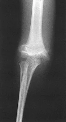
A 35-year-old male presents with a complex pelvic fracture. As part of the initial imaging workup, the following radiographs are obtained:
Based on the provided image, which view is depicted, and what specific information does it primarily provide regarding pelvic fracture assessment?
Explanation
Correct Answer: C
The image provided is an Outlet view of the pelvis. This view is characterized by the projection of the pubic symphysis over the sacrum, allowing for clear visualization of the vertical alignment of the hemipelvis. It is primarily used to assess for superior or inferior vertical migration (displacement) of the hemipelvis and to evaluate the sacral lordosis or angulation.
Option A (AP Pelvis view) is incorrect. An AP pelvis view shows the entire pelvic ring, symphysis, and SI joints without significant superimposition, but it is not the view shown. It is the initial mandatory view.
Option B (Inlet view) is incorrect. An inlet view projects the posterior structures (sacrum, SI joints) without superimposition, useful for assessing sacral kyphosis/angulation and anterior-posterior displacement of the posterior ring. The image does not show the characteristic appearance of an inlet view where the pelvic brim is clearly visualized.
Option D (Judet view (Obturator Oblique)) is incorrect. Judet views are specific for acetabular fractures. The obturator oblique view visualizes the anterior column and posterior wall of the acetabulum. The image is a pelvic ring view, not an acetabular view.
Option E (Judet view (Iliac Oblique)) is incorrect. The iliac oblique view visualizes the posterior column and anterior wall of the acetabulum. Again, the image is a pelvic ring view.
Question 19
A 72-year-old female with multiple comorbidities sustains a Tile Type B1 pelvic fracture (APC II, 'open book' injury) after a fall. She is hemodynamically stable after initial resuscitation. The pubic symphysis diastasis measures 3.5 cm. Given her age and comorbidities, the trauma team is considering the timing of definitive fixation. According to current guidelines, what is generally considered the 'golden window' for definitive fixation of mechanically unstable pelvic fractures once the patient is physiologically stable?
Explanation
Correct Answer: C
The case study explicitly states: 'The "golden window" for definitive fixation is generally considered within 5-7 days.' This timing allows for initial resuscitation, stabilization of associated injuries, and optimization of the patient's physiological status, while still providing the benefits of early definitive stabilization, such as reduced complication rates and improved outcomes compared to prolonged external fixation.
Option A (Within 12 hours of injury) is incorrect. While emergent interventions for hemorrhage control (e.g., external fixation, embolization, packing) may occur within hours, definitive internal fixation is typically not performed this early unless the patient is fully stable and there are no other pressing issues. The focus in the immediate hours is on damage control.
Option B (Within 24-48 hours of injury) is incorrect. While some urgent fixations may occur in this timeframe, the 'golden window' is generally considered slightly longer to allow for full physiological optimization, especially in polytrauma patients or those with comorbidities.
Option D (After 2-3 weeks, once soft tissue swelling has resolved) is incorrect. Delaying definitive fixation beyond the 'golden window' can lead to increased difficulty in reduction, higher rates of malunion, and potentially increased morbidity, especially for unstable fractures.
Option E (Only if non-operative management fails after 6 weeks) is incorrect. A Tile Type B1 (APC II) fracture with 3.5 cm symphysis diastasis is rotationally unstable and requires operative stabilization. Non-operative management is not appropriate for this degree of instability, and waiting 6 weeks would lead to malunion and poor outcomes.
Question 20
A 22-year-old male presents with a Young-Burgess APC II pelvic fracture. He is hemodynamically stable. The pubic symphysis diastasis is 3.0 cm. The posterior sacroiliac ligaments are intact. Which of the following statements best describes the stability of this fracture pattern?
Explanation
Correct Answer: C
A Young-Burgess Anteroposterior Compression Type II (APC II) injury is characterized by pubic symphysis diastasis greater than 2.5 cm and disruption of the anterior sacroiliac ligaments, but with intact posterior sacroiliac ligaments. This pattern makes the pelvic ring rotationally unstable (often described as an 'open book' injury) but vertically stable because the strong posterior ligaments remain intact, preventing vertical displacement.
Option A (Rotationally and vertically unstable) is incorrect. This describes an APC III or Vertical Shear (VS) injury, where both anterior and posterior ligamentous complexes are disrupted.
Option B (Rotationally stable and vertically stable) is incorrect. This describes an APC I or LC I injury, which are considered stable pelvic fractures.
Option D (Vertically unstable but rotationally stable) is incorrect. This combination is not typical for a primary pelvic ring injury pattern. Vertical instability almost always implies rotational instability due to posterior ring disruption.
Option E (Stable to all forces due to intact posterior arch) is incorrect. While the posterior arch (posterior SI ligaments) is intact, the anterior disruption (symphysis diastasis > 2.5 cm and anterior SI ligament disruption) makes the pelvis rotationally unstable.
Question 21
A 50-year-old male with a complex pelvic fracture is undergoing definitive posterior fixation. The surgeon plans to use percutaneous iliosacral screws. To ensure accurate placement and avoid neurovascular injury, which combination of fluoroscopic views is essential for verifying guide wire and screw trajectory?
Explanation
Correct Answer: B
The case study explicitly states that percutaneous iliosacral screw placement is performed under fluoroscopic guidance using 'inlet, outlet, lateral sacral views, +/- Judet views for SI joint'. These three views (inlet, outlet, and lateral sacral) are crucial for confirming the correct trajectory of the guide wire and screw, ensuring it remains within the safe corridor of the sacral ala and body, avoiding the sacral foramina and neurovascular structures. The inlet view assesses anterior-posterior displacement and sacral kyphosis, the outlet view assesses vertical displacement and sacral lordosis, and the lateral sacral view confirms the depth and trajectory within the sacral body.
Option A (AP pelvis and lateral hip views) is incorrect. While an AP pelvis is a standard initial view, a lateral hip view is not specific for iliosacral screw placement. The image provided is an outlet view, which is one of the critical views.
Option C (Judet views (iliac oblique and obturator oblique)) is incorrect. Judet views are primarily used for acetabular fractures and are not the primary views for iliosacral screw placement, although they can be helpful for assessing the SI joint itself.
Option D (Frog-leg lateral and cross-table lateral views) is incorrect. These are typically hip views and are not used for iliosacral screw placement.
Option E (CT scan with 3D reconstruction only) is incorrect. While a pre-operative CT scan with 3D reconstruction is invaluable for planning, intra-operative fluoroscopy is essential for real-time guidance during screw insertion. Relying solely on a pre-operative CT without intra-operative imaging is unsafe.
Question 22
A 38-year-old male undergoes definitive internal fixation for a Tile Type C pelvic fracture. Post-operatively, he develops severe pain, swelling, and tenderness in the gluteal region, accompanied by neurological deficits in the sciatic nerve distribution. His intracompartmental pressures are elevated. Which of the following early complications is most likely occurring?
Explanation
Correct Answer: D
The patient's symptoms of severe pain, swelling, tenderness, neurological deficits (sciatic nerve distribution), and elevated intracompartmental pressures in the gluteal region are classic signs of compartment syndrome. While rare in the gluteal compartment, it can occur after high-energy pelvic trauma, especially with high-volume fluid resuscitation and prolonged immobilization. Urgent fasciotomy is indicated for this life-threatening condition.
Option A (Deep venous thrombosis (DVT)) is incorrect. While DVT is a common complication of pelvic fractures, its symptoms typically include leg swelling, pain, and tenderness, but not elevated intracompartmental pressures or acute neurological deficits in the sciatic nerve distribution.
Option B (Heterotopic ossification (HO)) is incorrect. HO is a late complication involving ectopic bone formation in soft tissues, usually presenting weeks to months after injury with stiffness and pain, not acute compartment syndrome.
Option C (Pin site infection) is incorrect. Pin site infections are associated with external fixation and present with localized redness, warmth, drainage, and pain around the pins, not diffuse gluteal swelling, neurological deficits, and elevated intracompartmental pressures.
Option E (Malunion) is incorrect. Malunion is a late complication where the fracture heals in an unacceptable position, leading to chronic pain and dysfunction, not an acute post-operative emergency like compartment syndrome.
Question 23
A 45-year-old male is brought to the trauma bay following a high-speed motorcycle collision. He is hemodynamically unstable despite receiving 2 units of uncrossmatched blood. A pelvic binder is applied, and an AP pelvis radiograph demonstrates an anteroposterior compression (APC) type III pelvic ring injury. If an arterial source of hemorrhage is present, which of the following vessels is most commonly injured in this specific fracture pattern?
Explanation
Question 24
A 28-year-old male sustains an unstable sacral fracture with extension into the central sacral canal (Denis Zone III). Which of the following neurologic deficits is most specifically associated with fractures involving this zone?
Explanation
Question 25
A 35-year-old female sustains a closed lateral compression type II (LC-II) pelvic ring injury. Physical examination reveals a fluctuant, ballotable soft-tissue swelling overlying her left greater trochanter. If open reduction and internal fixation is planned directly through this area, what is the most significant anticipated complication?
Explanation
Question 26
During the acute resuscitation of a patient with a mechanically unstable pelvic ring injury, the trauma team decides to place a circumferential pelvic binder. To optimally reduce pelvic volume and provide biomechanical stability, the binder must be centered over which anatomic landmark?
Explanation
Question 27
A 50-year-old male undergoes percutaneous iliosacral screw fixation for a completely displaced sacroiliac joint disruption. Preoperative pelvic radiographs reveal L5 transverse processes that articulate with the ilium and non-circular upper sacral neural foramina. These radiographic findings indicate an increased risk of which of the following during screw placement?
Explanation
Question 28
A 32-year-old male falls from a height of 30 feet, landing directly on his feet. Imaging confirms a U-shaped sacral fracture with severe displacement. Neurologic examination reveals profound bilateral lower extremity weakness and saddle anesthesia. Which of the following surgical constructs is most appropriate to restore pelvic stability and allow mobilization?
Explanation
Question 29
Which of the following pelvic radiograph views is most appropriate to evaluate for subtle cranial (vertical) displacement of the left hemipelvis in a suspected vertical shear injury?
Explanation
Question 30
A 24-year-old male sustains an open book pelvic fracture (APC II). On secondary survey, there is blood at the urethral meatus and the prostate is high-riding on digital rectal exam. Which of the following is the most appropriate next step in the management of his genitourinary system?
Explanation
Question 31
During an anterior intrapelvic (Stoppa) approach for acetabular/pelvic ring fixation, profuse bleeding occurs just posterior to the superior pubic ramus near the symphysis. This hemorrhage is most likely originating from an anastomosis between the external iliac system and which internal iliac branch?
Explanation
Question 32
A patient with an APC III pelvic ring injury undergoes successful open reduction and internal fixation of the pubic symphysis and percutaneous posterior fixation. Assuming no contraindications, what is the most appropriate timeline for initiating pharmacologic venous thromboembolism (VTE) prophylaxis?
Explanation
Question 33
When utilizing intraoperative fluoroscopy for the placement of an S1 iliosacral screw, the surgeon uses the inlet view to monitor the screw's trajectory. The anterior margin of the 'safe zone' on this specific view represents which anatomic structure?
Explanation
Question 34
A 41-year-old female complains of persistent dyspareunia and pelvic pain one year after undergoing anterior symphyseal plating and bilateral SI joint screw fixation for an APC II injury. Radiographs show a healed pelvic ring with intact hardware. What is the most likely cause of her dyspareunia?
Explanation
Question 35
A 75-year-old female sustains a fragility fracture of the pelvis (FFP) following a ground-level fall. Imaging reveals an undisplaced unilateral sacral alar fracture and an ipsilateral superior pubic ramus fracture (Lateral Compression type I equivalent). She experiences intractable pain and cannot mobilize out of bed after 4 days of optimal medical management. What is the most appropriate next step in management?
Explanation
Question 36
A crescent fracture of the ilium is most classically associated with which type of pelvic ring injury pattern in the Young-Burgess classification?
Explanation
Question 37
The image represents a high-energy pelvic ring disruption. In the acute trauma setting, the finding of Destot's sign is highly suggestive of this class of injury. What is Destot's sign?
Explanation
Question 38
Which of the following ligaments is considered the primary static stabilizer of the pelvic ring, providing the greatest resistance against vertical shear forces?
Explanation
Question 39
A 29-year-old male presents with an open pelvic fracture involving a severe perineal laceration. Examination reveals gross fecal contamination of the pelvic fracture site and absent anal sphincter tone. Immediate management, alongside aggressive debridement and skeletal stabilization, must include which of the following?
Explanation
Question 40
A 38-year-old male sustains a severe pelvic crush injury resulting in an LC-III fracture pattern (windswept pelvis). This injury pattern is characterized by which of the following combinations of forces?
Explanation
Question 41
A 35-year-old male sustains a high-energy pelvic ring injury. AP radiograph demonstrates 4 cm of pubic symphyseal widening and widening of the left sacroiliac joint. He is hemodynamically stable. An MRI is obtained which confirms disruption of the anterior sacroiliac, sacrotuberous, and sacrospinous ligaments. Which of the following ligamentous structures remains intact in an Anteroposterior Compression Type II (APC-II) injury but is disrupted in an APC-III injury?
Explanation
Question 42
A 45-year-old female presents to the trauma bay in hemorrhagic shock following a high-speed motor vehicle collision. Her pelvis is clinically unstable to compression. The trauma team decides to place a non-invasive commercial pelvic binder. To be maximally effective at reducing pelvic volume, the binder should be centered directly over which anatomical landmark?
Explanation
Question 43
A 28-year-old male presents with a pelvic ring injury.
The orthopedic surgeon requests standard trauma pelvic views. What is the correct radiographic beam projection required to obtain a standard pelvic inlet view to best assess anterior-posterior hemipelvic translation?
Explanation
Question 44
During the ilioinguinal approach for anterior pelvic ring fixation, significant brisk arterial bleeding is encountered posterior to the superior pubic ramus near the symphysis. This hemorrhage is most likely originating from an anastomosis between which of the following vessels?
Explanation
Question 45
A 22-year-old male sustains an APC-III pelvic fracture and arrives hypotensive. Fluid resuscitation and a pelvic binder are applied, but he remains persistently hypotensive with a blood pressure of 75/40 mmHg. FAST exam is negative. What is the most appropriate next step in the management of this patient?
Explanation
Question 46
Which of the following is a classic radiographic characteristic of a dysmorphic sacrum, which increases the risk of neurologic injury during percutaneous placement of an S1 iliosacral screw?
Explanation
Question 47
A patient with a vertical shear pelvic fracture undergoes closed reduction and percutaneous iliosacral screw fixation. During placement of an S1 screw, the drill breaches the anterior cortex of the sacral ala. Which of the following neurologic structures is at greatest immediate risk of injury?
Explanation
Question 48
A 55-year-old male presents with a pelvic ring injury following a crush mechanism. Radiographs demonstrate an impacted, stable fracture of the anterior sacral ala and fractures of the ipsilateral superior and inferior pubic rami. According to the Young-Burgess classification, which mechanism of injury is responsible for this pattern?
Explanation
Question 49
A 30-year-old male presents with a completely unstable pelvic ring injury and gross blood at the urethral meatus. A retrograde urethrogram demonstrates a posterior urethral disruption. What is the most appropriate initial urologic management before definitive pelvic ring fixation?
Explanation
Question 50
A patient with a severe lateral compression pelvic fracture is noted to have a large, fluctuant swelling over the left greater trochanteric region. Skin integrity is intact, but the skin feels highly mobile over the deep fascia. If left untreated, this specific lesion most strongly predisposes the patient to which of the following complications?
Explanation
Question 51
A 40-year-old female sustains a Denis Zone 3 sacral fracture in an equestrian accident. Which of the following neurologic deficits is most commonly associated with fractures occurring in this specific anatomic zone?
Explanation
Question 52
Which of the following posterior pelvic ring injuries is classically described in a Lateral Compression Type 2 (LC-2) injury according to the Young-Burgess classification?
Explanation
Question 53
A 24-year-old male is treated with an anterior subcutaneous pelvic internal fixator (INFIX) for an APC-II pelvic injury. Post-operatively, he complains of burning pain and numbness over the anterolateral aspect of his thigh. Injury to which of the following nerves is the most likely cause?
Explanation
Question 54
Which of the following veins is the most common anatomical source of massive retroperitoneal hemorrhage following a high-energy pelvic ring disruption?
Explanation
Question 55
A 32-year-old construction worker falls from a height, sustaining bilateral vertical transforaminal sacral fractures combined with a transverse fracture through the S2 body. This injury is clinically classified as a spinopelvic dissociation. Which of the following is the most appropriate surgical treatment to address this specific pathology?
Explanation
Question 56
An 18-year-old female sustains an unstable pelvic fracture. She is successfully stabilized in the ICU. The surgical team plans for internal fixation on hospital day 3. According to major orthopedic trauma guidelines, what is the optimal strategy for deep vein thrombosis (DVT) prophylaxis in this patient?
Explanation
Question 57
A 45-year-old male sustains an APC-III pelvic injury and undergoes urgent pelvic packing and application of an external fixator. Forty-eight hours later, he is brought back to the OR for definitive open reduction and internal fixation of the pubic symphysis. The symphysis is reduced and fixed with a multi-hole plate. What is the most common mode of failure for isolated anterior plate fixation in a completely unstable posterior ring injury?
Explanation
Question 58
A Day Type 1 crescent fracture involves the posterior iliac wing and extends into the sacroiliac joint. Based on the Day classification system, in which section of the sacroiliac joint does a Type 1 crescent fracture exit?
Explanation
Question 59
A trauma patient has an open pelvic ring fracture with a large laceration extending from the perineum into the anal sphincter. Fecal contamination is present. In addition to thorough debridement and pelvic stabilization, what is the most critical adjunctive procedure to minimize mortality from pelvic sepsis?
Explanation
Question 60
During radiographic evaluation of a pelvic ring injury, the 'outlet' view is best utilized to assess which of the following deformities?
Explanation
Question 61
What is the correct anatomical landmark for the optimal placement of a pelvic circumferential compression device (binder) to most effectively reduce pelvic volume in a hemodynamically unstable trauma patient?
Explanation
Question 62
A hemodynamically unstable patient with an Anteroposterior Compression Type III (APC-III) pelvic injury remains hypotensive despite initial fluid resuscitation, blood transfusion, and application of a pelvic binder. The FAST scan is negative. What is the next most appropriate step in management?
Explanation
Question 63
During the anterior intrapelvic (Stoppa) approach for a pelvic ring injury, massive hemorrhage occurs from a vessel located superior to the superior pubic ramus. This vessel is an anastomosis between which two vascular systems?
Explanation
Question 64
Which nerve root is most commonly injured in a vertical shear pelvic ring injury that involves a displaced transforaminal sacral fracture?
Explanation
Question 65
Which intraoperative fluoroscopic view is most critical to evaluate for anterior-posterior translation of the sacroiliac joint during percutaneous iliosacral screw fixation?
Explanation
Question 66
A male patient presents with an APC-II pelvic ring fracture and blood at the urethral meatus. A retrograde urethrogram confirms a posterior urethral injury. What is the most common anatomical site of urethral disruption in this setting?
Explanation
Question 67
When planning for percutaneous iliosacral screw fixation, which of the following is a classic radiographic hallmark of sacral dysmorphism?
Explanation
Question 68
A 65-year-old female sustains a Lateral Compression Type II (LC-2) pelvic ring injury. By the Young-Burgess classification, this injury involves a fracture of which of the following posterior structures?
Explanation
Question 69
A patient has a pelvic ring injury with a transverse fracture through the S2 foramina. According to the Denis classification, what zone does this represent, and what is the most likely neurologic deficit?
Explanation
Question 70
A patient with an APC-III pelvic ring injury develops a large, fluctuant fluid collection over the greater trochanter after a high-speed motorcycle crash. Aspiration yields serosanguinous fluid. What is the pathophysiologic mechanism of this lesion?
Explanation
Question 71
A 45-year-old male is treated for an APC-III pelvic ring injury. Follow-up radiographs reveal failure of the anterior symphyseal plate. What is the most common reason for failure of isolated anterior symphyseal plating in a completely unstable pelvic ring?
Explanation
Question 72
When is it generally considered safe and most efficacious to initiate pharmacologic deep vein thrombosis (DVT) prophylaxis in a patient with a surgically stabilized pelvic ring injury and no associated traumatic brain or solid organ injury?
Explanation
Question 73
A trauma patient is transferred from an outside hospital with a pelvic binder in place for 36 hours. What is the most immediate clinical complication specifically associated with prolonged continuous pelvic binder application?
Explanation
Question 74
A patient undergoes placement of a subcutaneous anterior pelvic internal fixator (INFIX) for an LC-1 pelvic ring injury. Postoperatively, the patient complains of numbness, tingling, and a burning sensation over the anterolateral aspect of the thigh. Which nerve is most likely affected by the implant?
Explanation
Question 75
In the Tile classification of pelvic ring injuries, a Tile Type C injury is primarily characterized by which of the following biomechanical features?
Explanation
Question 76
Which ligamentous structure is considered the strongest in the pelvis and provides the most significant resistance to vertical shear forces acting on the sacroiliac joint?
Explanation
Question 77
When placing an S1 iliosacral screw on a pelvic outlet fluoroscopic view, what is the most critical anatomical boundary that must be identified to avoid iatrogenic injury to the L5 nerve root?
Explanation
Question 78
A patient sustains an open pelvic ring injury with a large perineal wound and gross fecal contamination. Following acute hemorrhage control and temporary skeletal stabilization, what is the next most critical surgical step to reduce mortality?
Explanation
Question 79
Which of the following pelvic ring injury patterns is classically associated with the highest requirement for massive blood transfusion and the highest overall mortality rate?
Explanation
Question 80
In the surgical management of a completely unstable sacroiliac joint disruption, what is the primary biomechanical advantage of utilizing two iliosacral screws rather than a single screw?
Explanation
Question 81
A 35-year-old male is brought in hemodynamically unstable after a motorcycle crash. A pelvic binder is applied. Which of the following anatomic structures is responsible for the vast majority (approximately 80%) of hemorrhage in pelvic ring injuries?
Explanation
None
