Orthopedic Deformity Correction: ABOS Part I Board Exam Review MCQs & Clinical Cases | Part 22015
Key Takeaway
Orthopedic deformity correction involves surgical techniques like osteotomies to realign bone segments, guided by principles such as Paley's rules. These methods address conditions like varus/valgus deformities, using internal or external fixation. Accurate preoperative planning, including CORA identification and joint angle measurements, is crucial for restoring mechanical axis alignment and preventing iatrogenic deformities.
Orthopedic Deformity Correction: ABOS Part I Board Exam Review MCQs & Clinical Cases | Part 22015
Comprehensive 100-Question Exam
00:00
Start Quiz
Question 1
A 38-year-old male presents with progressive right knee pain and a varus deformity. Full-length, weight-bearing radiographs reveal a mechanical axis that passes 25mm medial to the center of the knee joint. The Mechanical Lateral Distal Femoral Angle (mLDFA) is measured at 88°, and the Medial Proximal Tibial Angle (MPTA) is 78°. The Center of Rotation of Angulation (CORA) is identified in the proximal tibia, 3 cm distal to the joint line. The surgeon plans a closing wedge osteotomy to correct the deformity. Based on Paley's principles, which of the following statements is TRUE regarding the patient's deformity and the planned correction?
Explanation
Correct Answer: D
The patient's MPTA is 78°. The normal range for the Medial Proximal Tibial Angle (MPTA) is 85-90°. An MPTA less than 85° indicates a varus deformity of the proximal tibia. Therefore, an MPTA of 78° confirms a significant proximal tibial varus deformity, which is consistent with the clinical presentation of varus knee pain and a mechanical axis passing medial to the knee.
Option A is incorrect: The mLDFA is 88°. The normal range for the Mechanical Lateral Distal Femoral Angle (mLDFA) is 85-90°. An mLDFA >90° indicates valgus, and <85° indicates varus. Since 88° falls within the normal range, there is no significant femoral varus or valgus deformity. The primary deformity is tibial.
Option B is incorrect: The goal of the osteotomy is to restore the MPTA to its normal range (85-90°), not to decrease it further. Decreasing it would exacerbate the varus deformity.
Option C is incorrect: A mechanical axis passing 25mm medial to the center of the knee joint indicates a varus alignment, not valgus. In varus, the mechanical axis falls medial to the knee, overloading the medial compartment. A lateral closing wedge osteotomy would correct a valgus deformity, not a varus deformity. A medial closing wedge osteotomy (or lateral opening wedge) would be used for varus correction.
Option E is incorrect: According to Paley's Rule One, when the osteotomy is performed exactly at the level of the CORA, pure angulation corrects the deformity without creating any translation. Since the CORA is identified in the proximal tibia and the osteotomy is planned at this level, only angulation will be required for correction, assuming it's a simple angular deformity.
Question 2
A 55-year-old female presents with severe medial compartment osteoarthritis of the left knee secondary to a long-standing varus deformity. Preoperative planning reveals a CORA located 5 cm distal to the knee joint line in the proximal tibia. Due to poor bone quality at the CORA from a previous trauma, the surgeon opts to perform the closing wedge osteotomy 8 cm distal to the knee joint line. Which of Paley's rules of osteotomy applies to this scenario, and what are its implications for the surgical technique?
Explanation
Correct Answer: B
The CORA is located 5 cm distal to the knee joint line, but the surgeon plans to perform the osteotomy 8 cm distal to the knee joint line. This means the osteotomy is being performed at a level different from the CORA. According to Paley's Rule Two, when the osteotomy is performed at a level different from the CORA, correction requires a combination of both angulation and translation. This typically necessitates a complete, through-and-through osteotomy, as the concave hinge cannot be reliably preserved if translation is required.
Option A is incorrect: Rule One applies when the osteotomy is performed exactly at the CORA, allowing for pure angulation and preservation of the concave cortex. This is not the case here.
Option C is incorrect: Rule Three describes the consequence of violating Rule Two by performing only angulation when translation is also required. While Rule Three is a warning relevant to this scenario if translation is omitted, Rule Two is the primary rule that dictates the required surgical approach (angulation + translation) when operating away from the CORA.
Option D is incorrect: This describes Rule One, which is not being followed in this scenario due to the surgeon's decision to operate away from the CORA.
Option E is incorrect: While an external fixator can be used for complex corrections, Rule Two itself does not mandate its use. It simply states that angulation and translation are required. Robust internal fixation can also achieve this. The choice of hardware is secondary to the biomechanical principle.
Question 3
During a closing wedge osteotomy for a tibial varus deformity, the surgeon meticulously makes two converging cuts with an oscillating saw. After removing the bone wedge, a gentle bending force is applied to close the osteotomy. The surgeon observes that the concave cortex, which was intentionally preserved, undergoes a controlled 'greenstick' fracture. What is the primary biomechanical advantage of this specific technique?
Explanation
Correct Answer: C
The case explicitly states that in a closing wedge osteotomy under Rule One, the concave cortex is carefully preserved and acts as a biological 'hinge.' When this hinge is gently cracked (plastically deformed) during closure, combined with its intact, thick periosteal sleeve, it provides exceptional rotational and translational stability. This stability is crucial as it allows the bone itself to share the mechanical load with the applied fixation, thereby reducing stress on the hardware and promoting healing.
Option A is incorrect: While some minor adjustments might be possible, the primary advantage is stability, not ease of adjustment. The angle is determined preoperatively.
Option B is incorrect: While preserving the periosteum does help maintain blood supply, the primary biomechanical advantage of the hinge itself is stability, not solely blood loss minimization.
Option D is incorrect: While the stability provided by the hinge reduces the mechanical stress on the hardware, it does not eliminate the need for internal fixation. The text mentions it reduces the risk of hardware fatigue and failure, implying hardware is still used.
Option E is incorrect: While bone healing is the ultimate goal, the 'greenstick' fracture of the concave cortex doesn't primarily increase surface area for callus formation. The stability it provides is what indirectly promotes healing by minimizing micromotion.
Question 4
A surgeon is performing a closing wedge osteotomy to correct a distal femoral valgus deformity. During the procedure, despite meticulous preoperative planning, the surgeon notices that as the osteotomy gap is closed, the distal segment of the femur inadvertently rotates into a flexion deformity. According to the case material, what is the most likely technical error leading to this complication?
Explanation
Correct Answer: C
The case explicitly states: 'The most frequent, frustrating, and functionally detrimental complication of a closing wedge osteotomy is the inadvertent creation of a new deformity in a completely different plane (for example, accidentally creating a flexion deformity while attempting to correct a varus deformity). This multiplanar error almost always results from a single, specific technical failure: the proximal and distal cuts of the bony wedge are not made perfectly parallel to each other in the desired plane of correction.' If the cuts are not parallel, they cannot close flush, forcing the bone into an unintended plane.
Option A is incorrect: Failing to account for the kerf leads to unintended overcorrection in the planned plane, not an out-of-plane deformity.
Option B is incorrect: Aggressive periosteal stripping on the concave side would compromise the hinge stability and blood supply, potentially leading to nonunion or hardware failure, but not directly an out-of-plane deformity during closure.
Option D is incorrect: The Krackow effect (micro-crushing from compression) also leads to slight overcorrection in the planned plane, not an out-of-plane deformity. Insufficient compression would lead to undercorrection or delayed union, not a new deformity in a different plane.
Option E is incorrect: Performing an osteotomy away from the CORA without translation (violating Rule Three) leads to a residual translation deformity and uncorrected MAD, but not necessarily an inadvertent rotation into a different plane during the closure of the wedge itself, assuming the cuts were parallel in the primary plane.
Question 5
A 28-year-old patient requires a complex multiplanar deformity correction of the tibia following a malunion. The surgeon plans to use an Ilizarov external fixator. According to the case, how can the external fixator be optimally utilized as a precision cutting guide for the closing wedge osteotomy?
Explanation
Correct Answer: B
The case describes the advanced technique: 'The external frame is applied strictly based on anatomical landmarks. The proximal rings are mounted perfectly parallel to the proximal joint line, and the distal rings are mounted parallel to the distal joint line. The mechanical hinge of the external fixator is then positioned to coincide exactly with the pre-planned CORA. Once the frame is rigidly secured to the limb, specialized cutting guides are attached directly to the rings. Because the rings are already perfectly aligned with the bone's true axes, these guides automatically orient the oscillating saw blade to resect the precise, mathematically determined wedge of bone needed.'
Option A is incorrect: While rings are parallel to axes, the key is aligning them to joint lines and positioning the hinge at the CORA for precision cutting guides.
Option C is incorrect: Rings are typically applied parallel to joint lines or bone axes, not perpendicular to the mechanical axis for cutting guides. Using the rings directly as guides without specialized attachments would be imprecise.
Option D is incorrect: While external fixators can distract, this is not their primary role as a cutting guide for a closing wedge osteotomy. Distraction is more common for opening wedge or lengthening procedures.
Option E is incorrect: The question specifically asks about using the fixator as a cutting guide, which implies its use before or during the osteotomy cuts, not just for post-osteotomy stabilization and adjustment.
Question 6
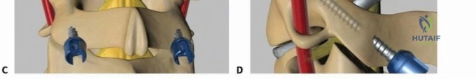
A 40-year-old male undergoes a closing wedge osteotomy for a severe tibial varus deformity. Preoperative planning indicates that the CORA is located intra-articularly, making a Rule One osteotomy impractical. The surgeon therefore plans to perform the osteotomy 4 cm distal to the CORA, requiring both angulation and translation. During the procedure, the surgeon performs the through-and-through osteotomy and then immediately closes the wedge to correct the angulation, before attempting to translate the distal segment. Based on Paley's principles and the provided image, what is the most likely outcome of this sequence?

Explanation
Correct Answer: C
The case explicitly details the 'Unforgiving Sequence: Order of Correction in Complex Cases' and illustrates it with the provided image. Path B (The Incorrect, 'Jammed' Method) shows that if the surgeon mistakenly first closes the wedge to correct the angulation, 'This premature action immediately creates massive friction and extreme compression between the two flat, raw osteotomy surfaces. Furthermore, the tightened concave periosteal sleeve acts as a tether, securely 'locking' the segments together. At this point, attempting to translate the bone is physically akin to trying to slide two pieces of rough sandpaper against each other while clamped in a heavy-duty vise. The required force is immense, risking iatrogenic fracture, severe soft tissue damage, and ultimately making the planned translation physically impossible. The deformity is left permanently 'jammed' in a poorly corrected, highly compromised position.'
Option A is incorrect: This contradicts the 'jammed' effect described in the text and image.
Option B is incorrect: The text clearly states this incorrect sequence leads to a 'poorly corrected, highly compromised position' and a residual deformity.
Option D is incorrect: For a Rule Two osteotomy requiring translation, a complete through-and-through osteotomy is typically performed, meaning the concave periosteal sleeve is not preserved as a hinge in the same way as a Rule One osteotomy. Even if some soft tissue remains, it acts as a tether, hindering translation.
Option E is incorrect: The scenario describes a situation where both angulation and translation are required (Rule Two). If translation is not achieved due to the incorrect sequence, the correction will not be pure angulation, and a residual translation deformity will persist.
Question 7
A 60-year-old patient undergoes a high tibial osteotomy for varus knee deformity. The surgeon plans a 10-degree closing wedge correction. To ensure sub-millimeter accuracy, the surgeon utilizes the parallel pin technique. After drilling two stout Steinmann pins, one proximal and one distal to the planned osteotomy site, they are oriented to form an angle of 10 degrees. The osteotomy is then performed. What is the expected observation if the wedge resection is perfectly accurate?
Explanation
Correct Answer: B
The case describes the parallel pin technique: 'Before any saw cuts are made, two stout Steinmann pins are drilled into the bone—one proximal and one distal to the planned osteotomy site. Using a sterile angle-measuring device (goniometer), the pins are oriented so the angle between them is exactly equal to the planned correction angle. The closing wedge osteotomy is then meticulously performed between the two pins. As the bony wedge is closed, the surgeon carefully observes the pins. A perfect, accurate correction has been achieved when the two previously angled pins become perfectly parallel to each other.' This technique visually confirms that the resected wedge precisely matches the planned angular correction.
Option A is incorrect: If the pins remained at a 10-degree angle after closure, it would mean no correction or an inaccurate correction was achieved.
Option C is incorrect: The goal is for the pins to become parallel, indicating the correction angle has been achieved and the segments are realigned.
Option D is incorrect: The parallel pin technique is for verifying angular accuracy, not primarily for assessing the need for compression. If the gap remains open, it suggests an under-resection or issues with closure, but the primary indicator of angular accuracy is the pins becoming parallel.
Option E is incorrect: If the pins converged beyond parallel, it would indicate an overcorrection, meaning the wedge removed was larger than planned.
Question 8
A 42-year-old male with a severe distal femoral recurvatum deformity (PDFA 108°) is scheduled for a closing wedge osteotomy. The CORA is identified at the anterior cortex and physis. The surgeon plans to perform a Rule One osteotomy, preserving the concave cortex as a hinge. Which of the following statements accurately reflects the biological and mechanical considerations for this approach?
Explanation
Correct Answer: C
The case states: 'If the concave cortex is left intact and utilized as the primary hinge, the osteotomy achieves such profound intrinsic stability that it can often be effectively stabilized with simple, low-profile hardware, such as surgical staples or a tension band wire construct, rather than massive, bulky locking plates.' In a recurvatum deformity, the anterior cortex is the concave side, and preserving it as a hinge provides this stability.
Option A is incorrect: The case explicitly states: 'For a Rule One osteotomy, surgical dissection on the concave side should be minimal to entirely non-existent. Preserving the periosteal sleeve is absolutely paramount for maintaining the local blood supply and ensuring the stability of the cortical hinge.'
Option B is incorrect: In a recurvatum deformity, the posterior side is the convex side where the wedge is removed. The anterior cortex is the concave side and is preserved as the hinge.
Option D is incorrect: The case mentions that in a Rule Two scenario (requiring translation), the half-wedge of bone that was resected can be preserved and used as vital autograft to fill the structural space created on the translated concave side. While not directly applicable to a pure Rule One osteotomy, it shows the resected bone can have utility.
Option E is incorrect: The primary risk of a closing wedge osteotomy is often the creation of an out-of-plane deformity if cuts are not parallel, or issues with hardware failure. Creating a varus deformity from an anterior hinge in a recurvatum correction is not the primary or expected risk; the hinge is designed to facilitate the planned correction.
Question 9
A 30-year-old patient presents with a complex post-traumatic deformity of the distal tibia, requiring a closing wedge osteotomy. Preoperative templating indicates a 15-degree correction. The surgeon is aware of potential intraoperative inaccuracies. Which of the following factors, if not accounted for, could lead to an unintended overcorrection of the deformity?
Explanation
Correct Answer: C
The case specifically highlights two factors that can lead to unintended overcorrection: 'Accounting for the Kerf: An oscillating saw blade does not simply magically part the bone; it physically vaporizes a strip of bone equal to the blade's thickness. ...When making two separate converging cuts for a closing wedge, the surgeon removes not only the planned wedge itself but also the width of two saw blade kerfs. ...This can easily translate to 1-2 degrees of unintended overcorrection.' And 'The Krackow Effect (The Biology of Compression): ...applying dynamic compression across an osteotomy site in soft, cancellous bone leads to the micro-crushing of the bony trabeculae. This physical impaction can slightly increase the final angular correction beyond what was geometrically resected with the saw.'
Option A is incorrect: Insufficient preservation of the concave hinge would lead to instability and potentially hardware failure, not necessarily overcorrection of the angle.
Option B is incorrect: Aggressive periosteal stripping on the convex side risks avascular necrosis, but not direct overcorrection of the angle.
Option D is incorrect: Performing an osteotomy away from the CORA without translation (Rule Three violation) leads to a residual translation deformity and uncorrected MAD, not an angular overcorrection.
Option E is incorrect: Non-parallel cuts lead to out-of-plane deformities, not necessarily an overcorrection in the intended plane.
Question 10
A 68-year-old patient with a long-standing genu varum deformity is scheduled for a high tibial osteotomy. Preoperative radiographs confirm a significant varus deformity with a Mechanical Lateral Distal Femoral Angle (mLDFA) of 87°, a Medial Proximal Tibial Angle (MPTA) of 75°, and a Joint Line Convergence Angle (JLCA) of 4°. The surgeon plans to correct the deformity to restore neutral alignment. Based on these findings, what is the most appropriate interpretation of the patient's deformity?
Explanation
Correct Answer: C
The Joint Line Convergence Angle (JLCA) is measured at 4°. The normal range for JLCA is 0-2°. The case states: '>2° strongly suggests joint space narrowing or ligamentous instability.' A JLCA of 4° is significantly elevated, indicating a substantial intra-articular component to the deformity, likely due to medial compartment cartilage loss or medial collateral ligament laxity, which contributes to the overall varus alignment.
Option A is incorrect: The mLDFA is 87°, which is within the normal range of 85-90°. This indicates no significant deformity in the distal femur. The MPTA of 75° (normal 85-90°) indicates a significant proximal tibial varus deformity, making the tibia the primary site of extra-articular deformity.
Option B is incorrect: The MPTA is 75°. An MPTA <85° indicates varus, while >90° indicates valgus. Therefore, 75° indicates a varus deformity of the proximal tibia, not valgus.
Option D is incorrect: The mLDFA of 87° is within the normal range (85-90°), indicating no significant varus deformity of the distal femur.
Option E is incorrect: Genu varum (bow-legged) means the mechanical axis passes medial to the knee joint, overloading the medial compartment. If it passed lateral, it would indicate genu valgum.
Question 11
A 50-year-old patient undergoes a closing wedge osteotomy for a severe distal femoral valgus deformity. The surgeon has meticulously planned the osteotomy to be performed at the CORA, ensuring pure angulation correction. During the procedure, the surgeon is careful to minimize dissection on the concave side of the deformity. What is the primary biological reason for this specific surgical pearl?
Explanation
Correct Answer: B
The case explicitly states under 'Soft Tissue and Biological Considerations': 'Concave Side Management: For a Rule One osteotomy, surgical dissection on the concave side should be minimal to entirely non-existent. Preserving the periosteal sleeve is absolutely paramount for maintaining the local blood supply and ensuring the stability of the cortical hinge.'
Option A is incorrect: While protecting neurovascular structures is always important, the primary reason for minimal dissection on the concave side in the context of a closing wedge osteotomy is specifically for preserving the periosteal hinge and its blood supply, as detailed in the text. Neurovascular structures are typically protected on the convex side where the wedge is removed, or generally throughout the approach.
Option C is incorrect: While minimal dissection might contribute to slightly shorter operative time, it is not the primary biological reason for this specific pearl.
Option D is incorrect: Avoiding out-of-plane deformities is achieved by ensuring parallel saw cuts, not primarily by minimizing concave side dissection.
Option E is incorrect: Minimal dissection on the concave side might make hardware application slightly more challenging if the plate needs to be contoured around intact soft tissue, but the benefit of preserving the hinge outweighs this. It does not facilitate easier application.
Question 12
A 35-year-old male presents with a long-standing varus knee deformity and a 2 cm limb length discrepancy in the affected limb. Preoperative planning reveals a CORA located in the distal femoral metaphysis. The surgeon plans an acute correction using internal fixation. Based on Paley's principles and the patient's specific presentation, which osteotomy technique would be most appropriate?
Explanation
Correct Answer: D
The patient presents with a varus knee deformity and a concomitant 2 cm limb length discrepancy. The case content explicitly states that a primary advantage of an opening wedge osteotomy is that it increases overall limb length, which is highly beneficial for patients presenting with a concomitant limb length discrepancy alongside their angular deformity. Since the CORA is in the distal femoral metaphysis, an opening wedge osteotomy at this site would allow for both angular correction and limb lengthening. The use of structural allograft is often necessary with internal fixation to bridge the created gap and provide initial stability, as mentioned in the disadvantages of opening wedge osteotomies with internal fixation.
Option A (Closing wedge osteotomy with bone graft) is incorrect because closing wedge osteotomies inherently shorten the limb, which would exacerbate the existing limb length discrepancy. While bone graft can be used, it doesn't address the shortening.
Option B (Neutral wedge osteotomy) is incorrect because it results in no net change in overall limb length, which would not address the 2 cm limb length discrepancy.
Option C (Closing wedge osteotomy without bone graft) is incorrect for the same reason as option A; it shortens the limb, which is contraindicated in this patient.
Option E (Acute correction with intramedullary nailing and blocking screws) is less appropriate for a metaphyseal deformity around the knee. While IM nailing can correct diaphyseal deformities, acute metaphyseal corrections with IM nails are challenging and often require specialized jigs or temporary external fixators (FAN). More importantly, IM nailing itself does not inherently lengthen the limb in the same controlled manner as an opening wedge osteotomy, and the primary issue here is addressing the limb length discrepancy.
Question 13
A 50-year-old female presents with a severe valgus knee deformity. Preoperative full-length weight-bearing radiographs show a Mechanical Axis Deviation (MAD) of 25mm lateral to the center of the knee. Which of the following joint orientation angle measurements would be most consistent with a primary distal femoral deformity contributing to this valgus malalignment?
Explanation
Correct Answer: B
The case content defines the Mechanical Lateral Distal Femoral Angle (mLDFA) as defining the relationship between the femoral mechanical axis and the distal femoral joint line. It states that an mLDFA < 85° indicates distal femoral valgus. A valgus knee deformity with a lateral MAD is consistent with distal femoral valgus. Therefore, an mLDFA of 80° (which is less than 85°) directly points to a primary distal femoral deformity as a significant contributor to the overall valgus malalignment.
Option A (MPTA of 88°) is incorrect. The Medial Proximal Tibial Angle (MPTA) defines the relationship between the tibial mechanical axis and the proximal tibial joint line. A normal range is 85° to 90°. An MPTA of 88° is within the normal range, indicating no significant proximal tibial varus or valgus deformity.
Option C (JLCA of 3°) is incorrect. The Joint Line Convergence Angle (JLCA) measures the angle between the distal femoral and proximal tibial joint lines. An increased JLCA (> 2°) suggests gapping due to ligamentous laxity or severe cartilage loss, not a primary bony deformity contributing to valgus.
Option D (LDTA of 89°) is incorrect. The Lateral Distal Tibial Angle (LDTA) assesses ankle joint orientation. A normal range is 86° to 92°. An LDTA of 89° is within the normal range, indicating no significant distal tibial deformity.
Option E (mLPFA of 92°) is incorrect. The Mechanical Lateral Proximal Femoral Angle (mLPFA) evaluates proximal femoral geometry. A normal range is 85° to 95°. An mLPFA of 92° is within the normal range, indicating no significant proximal femoral deformity (like coxa vara or coxa valga).
Question 14
A 28-year-old male presents with a complex multi-planar tibial deformity following a malunited fracture. Preoperative planning identifies a CORA located in the mid-diaphysis, but due to poor bone quality and previous hardware in that exact location, the surgeon decides to perform the osteotomy 5 cm distal to the CORA. The mechanical hinge of the external fixator is meticulously placed at the true CORA. Which of Paley's Osteotomy Rules is being applied, and what is the expected outcome?
Explanation
Correct Answer: B
This scenario perfectly describes Paley's Osteotomy Rule 2. The case content states: "The osteotomy is performed at a level DIFFERENT from the CORA, but the mechanical hinge remains placed AT the CORA. Result: Angular correction accompanied by a predictable, necessary, and collinear TRANSLATION." In this patient, the osteotomy is performed 5 cm distal to the CORA, but the hinge is placed at the CORA. This will result in a controlled, predictable translation of the bone segments at the osteotomy site, in addition to the angular correction, while still restoring the mechanical axis.
Option A (Rule 1; pure angular correction with no translation) is incorrect. Rule 1 applies when both the osteotomy and the hinge are exactly at the CORA, which is not the case here.
Option C (Rule 3; a new, iatrogenic translation deformity is created) is incorrect. Rule 3 applies when both the osteotomy and the mechanical hinge are placed at locations different from the CORA. In this scenario, the hinge is correctly placed at the CORA, preventing an iatrogenic deformity.
Option D (Rule 1; angular correction with unpredictable translation) is incorrect. This scenario does not fit Rule 1, and the translation in Rule 2 is predictable, not unpredictable.
Option E (Rule 2; angular correction with no translation, but increased limb length) is incorrect. Rule 2 specifically results in translation, not an absence of it. While limb length can be affected by the type of osteotomy (opening vs. closing wedge), the primary outcome of Rule 2 is controlled translation, not necessarily increased limb length.
Question 15
A 60-year-old patient undergoes a high tibial osteotomy for a varus knee deformity. The surgeon plans a closing wedge osteotomy with a small amount of planned translation to optimize joint loading. Intraoperatively, after making the bone cuts, the surgeon attempts to angulate the osteotomy first, then translate the segments. What is the most likely consequence of this sequence of correction?
Explanation
Correct Answer: C
The case content provides a critical surgical pearl for Rule 2 execution with a closing wedge: "The order of correction for an acute closing wedge with translation is paramount: translation must be performed first, followed by angulation. If angulation is performed first, the soft tissue and bone ends will prematurely lock together, making it technically impossible to displace the segments later without excessive force." Therefore, attempting to angulate first will lead to significant difficulty in achieving the planned translation.
Option A (Optimal bone-to-bone apposition and rapid healing) is incorrect. The incorrect sequence would likely lead to suboptimal bone contact or require excessive force, compromising apposition and healing.
Option B (Reduced risk of peroneal nerve palsy) is incorrect. The sequence of correction does not directly impact the risk of peroneal nerve palsy, which is more related to the magnitude of acute correction and soft tissue tension.
Option D (Creation of a neutral wedge osteotomy) is incorrect. The type of osteotomy (closing wedge) is determined by the bone cuts and hinge placement, not the sequence of correction.
Option E (Increased limb length due to the translation) is incorrect. A closing wedge osteotomy inherently shortens the limb. While translation is involved, it does not typically lead to increased limb length; rather, it creates a step-off. An opening wedge osteotomy is used for lengthening.
Question 16
A 40-year-old patient with a complex multi-planar femoral deformity is scheduled for correction. The surgeon is considering different hardware options. The patient has a history of poor compliance with external fixator care and desires a quicker return to work. However, the deformity is large, and there is concern for significant acute soft tissue stretching. Given these factors, which hardware selection strategy aligns best with the principles discussed in the case?
Explanation
Correct Answer: C
The case content highlights that circular external fixation systems (traditional Ilizarov and modern hexapod systems) remain the absolute gold standard for complex, multi-planar, multi-apical deformities. While the patient has compliance concerns and desires a quicker return to work (which favors internal fixation), the critical factor here is the "large" and "complex multi-planar" deformity with concern for "significant acute soft tissue stretching." The text explicitly states that "acute correction with plates is less suitable for large-magnitude deformities or complex multiplanar deformities due to acute soft tissue stretching (e.g., peroneal nerve palsy in large valgus corrections)." A hexapod system allows for gradual correction, mitigating the risks of acute soft tissue stretching and nerve palsies, which is paramount in complex, large deformities, even with compliance challenges.
Option A (Acute correction with a locking plate) is incorrect because, as stated in the text, plates are less suitable for large-magnitude or complex multiplanar deformities due to acute soft tissue stretching. While it offers advantages, it's not the best choice for this specific deformity complexity.
Option B (Intramedullary nailing with blocking screws) is incorrect. While IM nails are load-sharing and avoid external fixation, they are primarily used for diaphyseal deformities. Complex multi-planar deformities, especially around joints, are often better managed with external fixators that allow for multi-planar correction and gradual adjustment.
Option D (A monolateral external fixator) is incorrect. While simpler, monolateral fixators are generally less versatile than circular or hexapod frames for complex, multi-planar deformities, which require more robust and adjustable constructs.
Option E (A combination of a locking plate and an intramedullary nail) is incorrect. This is not a standard or recommended approach for primary deformity correction and would introduce unnecessary complexity and potential complications without addressing the core issue of gradual correction for large, complex deformities.
Question 17
A 12-year-old patient presents with a severe congenital bowing deformity of the tibia. Preoperative planning identifies multiple CORAs along the length of the diaphysis. The surgeon plans a multi-level osteotomy. Which of the following surgical pearls from the case content is most relevant to this specific scenario?
Explanation
Correct Answer: C
The scenario describes a "long bowing deformity" with "multiple CORAs" and a plan for a "multi-level osteotomy." The case content's surgical pearls explicitly state: "For long bowing deformities, do not force a single CORA. Map multiple CORAs and plan a multi-level osteotomy to avoid massive, unnatural translation." This pearl directly addresses the challenge of correcting long, diffuse deformities and is crucial for achieving a natural, well-aligned limb without creating iatrogenic issues.
Option A (Always ensure patella-forward alignment on long-leg films) is a general imaging pearl, important for all lower extremity deformity planning, but not specific to the challenge of multiple CORAs in a long bowing deformity.
Option B (When performing a closing wedge with planned translation, translate first, then angulate) is a pearl specific to the execution of Rule 2 with a closing wedge, not directly related to identifying multiple CORAs.
Option D (Use plates for simple, single-plane metaphyseal corrections) is a pearl regarding hardware choice, not directly related to the planning for multiple CORAs.
Option E (Always respect the concave soft tissues; prophylactic nerve decompressions may be mandatory) is a pearl regarding soft tissue management, particularly for large acute corrections, but not specific to the identification and management of multiple CORAs.
Question 18
A 48-year-old male presents with a severe post-traumatic varus deformity of the distal tibia, leading to ankle joint degeneration. Preoperative planning reveals an LDTA of 80°. The CORA is identified just proximal to the ankle joint. The surgeon plans an acute correction using a locking plate. Which of the following statements accurately describes the significance of the LDTA in this case and the goal of correction?
Explanation
Correct Answer: C
The case content defines the Lateral Distal Tibial Angle (LDTA) as assessing the ankle joint orientation in the coronal plane, with a normal range of 86° to 92°. It states that "An abnormal LDTA indicates a distal tibial deformity, often leading to secondary ankle varus or valgus and rapid joint degeneration." An LDTA of 80° is less than the normal range, indicating a distal tibial varus deformity. The goal of correction would be to restore this angle to its normal range of 86-92°.
Option A (An LDTA of 80° indicates a proximal tibial varus, requiring a high tibial osteotomy) is incorrect. Proximal tibial varus is assessed by the MPTA, not the LDTA.
Option B (An LDTA of 80° indicates a distal tibial valgus, and correction aims to restore it to 85-90°) is incorrect. An angle less than the normal range (86-92°) indicates varus, not valgus. Also, the target range is 86-92°, not 85-90°.
Option D (An LDTA of 80° indicates a distal femoral valgus, requiring a distal femoral osteotomy) is incorrect. Distal femoral valgus is assessed by the mLDFA, not the LDTA.
Option E (An LDTA of 80° is within the normal range, suggesting the deformity is intra-articular) is incorrect. An LDTA of 80° is outside the normal range (86-92°), indicating a bony deformity, not necessarily an intra-articular one.
Question 19
A 30-year-old patient with a congenital femoral bowing deformity is undergoing correction using an intramedullary nail. The preoperative radiographs show a significant diaphyseal curve. To ensure the straight IM nail corrects the deformity and does not follow the original curved canal, which technique, as described in the case, would be most appropriate?
Explanation
Correct Answer: C
The case content specifically addresses this scenario under "Intramedullary Nailing Techniques": "Because the IM canal in a deformed bone is much wider than the nail, a straight nail will often follow the path of least resistance, recreating the deformity. To guide a straight nail through a deformed bone segment and force it into the center of the canal, blocking screws are placed strategically into the cancellous bone on the concave side of the deformity. These screws physically block the nail from translating, ensuring the mechanical axis is maintained." This technique is precisely designed to prevent the recreation of the deformity when using a straight IM nail in a curved canal.
Option A (Performing a fixator-assisted nailing (FAN) procedure) is incorrect. While FAN is a valid technique for acute correction with IM nailing, its primary purpose is to acutely obtain and rigidly hold the correction before nail insertion, not specifically to guide a straight nail through a curved canal once the nail is being inserted. Blocking screws are used during nail insertion to guide it.
Option B (Using a longer, larger diameter intramedullary nail) is incorrect. A larger diameter nail might fill the canal more, but it doesn't guarantee correction of the deformity if the canal is curved. A longer nail doesn't inherently guide it into a corrected path.
Option D (Performing a closing wedge osteotomy prior to nail insertion) is incorrect. While an osteotomy is necessary for correction, performing a closing wedge prior to nail insertion doesn't, by itself, ensure the nail will maintain the correction or prevent it from following the remaining curve. Blocking screws are still needed to guide the nail through the corrected segment.
Option E (Utilizing a custom-bent intramedullary nail) is incorrect. The case content focuses on using straight IM nails and techniques to guide them. Custom-bent nails are not discussed as a primary strategy for deformity correction in this context.
Question 20
A 55-year-old patient presents with a severe varus knee deformity. Preoperative full-length weight-bearing radiographs are obtained. The surgeon notes that the patellae are not oriented perfectly forward. According to the surgical pearls in the case, what is the most critical implication of this observation?
Explanation
Correct Answer: C
The case content's surgical pearls explicitly state under "Imaging": "Always ensure patella-forward alignment on long-leg films. Rotational malalignment invalidates all coronal plane angle measurements." If the patellae are not oriented forward, it introduces a rotational artifact that makes all subsequent coronal plane angle measurements (like mLDFA, MPTA, LDTA) inaccurate and unreliable for planning.
Option A (It will lead to an underestimation of the Mechanical Axis Deviation (MAD)) is incorrect. While MAD can be affected, the primary and most critical issue is the invalidation of all coronal plane angle measurements, which are essential for pinpointing the anatomical source of the deformity.
Option B (It will primarily affect the Joint Line Convergence Angle (JLCA) measurement) is incorrect. While JLCA can be affected, the statement in the text is broader, indicating all coronal plane angle measurements are invalidated.
Option D (It suggests a concomitant rotational deformity that requires separate correction) is incorrect. While rotational malalignment can be present, the immediate implication of a non-patella-forward view on a coronal radiograph is that the image itself is distorted for coronal plane measurements, not necessarily that it diagnoses a rotational deformity from that specific view.
Option E (It indicates a need for a CT scan to accurately assess the deformity) is incorrect. While a CT scan might be useful for complex rotational deformities, the immediate and most critical implication of improper patella-forward alignment is that the current radiographs are unreliable for coronal plane planning, necessitating repeat radiographs or careful consideration of the limitations.
Question 21
The image below illustrates the biomechanical principles of external fixation in deformity correction.
Based on the top diagram showing a monolateral fixator, which of Paley's Osteotomy Rules is being demonstrated, and what is its key characteristic?

Explanation
Correct Answer: A
The image description in the case content states: "The top diagram illustrates a monolateral fixator correcting a simple angular deformity. The red and blue lines represent the mechanical axes of the proximal and distal bone segments. Notice how the fixator's hinge is placed exactly at the CORA (Rule 1). As the device is adjusted, the bone segments are brought into perfect, collinear alignment without translation." This directly describes Paley's Osteotomy Rule 1, where both the osteotomy (implied at the CORA for ideal correction) and the mechanical hinge are at the CORA, leading to pure angular correction without translation.
Option B (Rule 2; the osteotomy is different from the CORA, and the hinge is at the CORA, resulting in angular correction with predictable translation) is incorrect. While Rule 2 involves the hinge at the CORA, the osteotomy is different from the CORA, which is not what the diagram explicitly shows as the ideal correction without translation.
Option C (Rule 3; the osteotomy and hinge are different from the CORA, resulting in an iatrogenic translation deformity) is incorrect. Rule 3 results in an iatrogenic deformity, which is clearly not the ideal correction depicted.
Option D (Rule 1; the osteotomy is at the CORA, but the hinge is distal to it, resulting in angular correction with unpredictable translation) is incorrect. Rule 1 requires the hinge to be at the CORA, and the outcome is no translation, not unpredictable translation.
Option E (Rule 2; the osteotomy is proximal to the CORA, and the hinge is at the CORA, resulting in angular correction with limb lengthening) is incorrect. While Rule 2 involves the hinge at the CORA and an osteotomy elsewhere, the diagram shows no translation, and limb lengthening is a characteristic of opening wedge osteotomies, not a direct result of Rule 2 itself without specifying the osteotomy type.
Question 22
A 22-year-old patient undergoes correction of a distal femoral valgus deformity. Preoperative planning identifies the CORA in the distal femoral metaphysis. The surgeon performs the osteotomy at the CORA and places the mechanical hinge of the internal locking plate exactly at the CORA. Postoperatively, despite achieving angular correction, the mechanical axis is not restored, and a new step-off deformity is noted at the osteotomy site. Which of the following is the most likely explanation for this outcome, based on Paley's principles?
Explanation
Correct Answer: D
The question states that the surgeon performed the osteotomy at the CORA and placed the mechanical hinge of the internal locking plate exactly at the CORA. According to Paley's Osteotomy Rule 1, this should result in "Pure angular correction with NO translation" and "The mechanical axis is fully restored to neutral." The fact that "the mechanical axis is not restored, and a new step-off deformity is noted at the osteotomy site" directly contradicts the expected outcome of Rule 1. The most fundamental reason for such a discrepancy, given the stated intent to follow Rule 1, is that the CORA was incorrectly identified during preoperative planning or intraoperatively, leading to the hinge and osteotomy not truly being at the geometric apex of the deformity. If the actual CORA was elsewhere, placing the osteotomy and hinge at a perceived CORA would effectively be placing them both at a location different from the true CORA, leading to an outcome resembling Rule 3 or an incomplete correction.
Option A (The surgeon inadvertently applied Osteotomy Rule 1, which is unsuitable for metaphyseal deformities) is incorrect. Rule 1 is the gold standard and ideal scenario for most simple metaphyseal and diaphyseal deformities, making it suitable for a metaphyseal deformity.
Option B (The surgeon inadvertently applied Osteotomy Rule 2, leading to unexpected translation) is incorrect. Rule 2 involves the osteotomy being different from the CORA, while the hinge is at the CORA. The scenario states both were at the CORA. Also, Rule 2 results in predictable translation, not unexpected.
Option C (The surgeon inadvertently applied Osteotomy Rule 3, creating an iatrogenic translation deformity) is incorrect. Rule 3 applies when both the osteotomy and the mechanical hinge are placed at a location different from the CORA. The scenario states the surgeon intended to place both at the CORA. The outcome might resemble Rule 3, but the cause is likely incorrect CORA identification, not intentional application of Rule 3.
Option E (The locking plate was too rigid, preventing the bone segments from translating correctly) is incorrect. A locking plate's rigidity is for stability, not for preventing translation in a way that would cause a step-off if the correction was geometrically sound. If anything, a rigid plate would hold an incorrect position if the planning was flawed.
Question 23
A surgeon plans to correct a mid-diaphyseal tibial angular deformity. According to Paley's Rule 1 of osteotomy, if the osteotomy and the hinge axis are both placed exactly at the Center of Rotation of Angulation (CORA), what is the resulting geometric correction?
Explanation
Question 24
A 24-year-old male requires correction of a distal femur deformity. The CORA is located 1 cm from the articular surface. The surgeon places the hinge at the CORA to avoid joint penetration but performs the osteotomy 4 cm proximally. Based on Paley's rules, what is the expected mechanical outcome?
Explanation
Question 25
A 45-year-old male undergoes a medial opening wedge high tibial osteotomy (HTO) for isolated medial compartment osteoarthritis. If the surgeon opens the anterior and posterior aspects of the osteotomy gap equally (e.g., 10 mm each), what is the most likely effect on the sagittal profile of the proximal tibia?
Explanation
Question 26
When constructing a circular external fixator (Ilizarov) for tibial lengthening, which of the following modifications will most effectively increase the axial stiffness of the frame?
Explanation
Question 27
A 50-year-old female presents with bilateral knee pain and clinical genu varum. Standing full-length radiographs show a mechanical axis deviation (MAD) of 40 mm medially on the right leg. The right mechanical lateral distal femoral angle (mLDFA) is 87 degrees (normal 85-90 degrees) and the medial proximal tibial angle (MPTA) is 76 degrees (normal 85-90 degrees). The joint line convergence angle (JLCA) is 1 degree. Where is the primary source of the varus deformity?
Explanation
Question 28
A surgeon plans to correct a mid-diaphyseal tibial varus deformity. The osteotomy is performed exactly at the center of rotation of angulation (CORA), and the hinge of the external fixator is placed exactly at the convex cortex of the CORA. According to Paley's rules of deformity correction, which of the following is the expected outcome?
Explanation
Question 29
During tibial lengthening using an Ilizarov frame, a patient is instructed to perform 1 mm of distraction per day. Which of the following rhythms of distraction optimizes bone regenerate formation and minimizes soft tissue complications?
Explanation
Question 30
A patient undergoes a medial opening wedge high tibial osteotomy (HTO) to correct a varus deformity. To maintain the native posterior tibial slope, how should the magnitude of the anterior gap compare to the posterior gap of the osteotomy?
Explanation
Question 31
When utilizing a hexapod external fixator (e.g., Taylor Spatial Frame) for a complex multiplanar tibial deformity, exact 'mounting parameters' must be input into the software. What do the mounting parameters specifically define?
Explanation
Question 32
In a healthy adult with normal lower extremity alignment, what is the geometric relationship between the mechanical axis of the lower extremity and the anatomical axis of the femur?
Explanation
Question 33
A 30-year-old patient is undergoing femoral lengthening with an external fixator. Radiographs at 5 weeks show an hourglass-shaped regenerate bone with widening of the radiolucent distraction gap. What is the most appropriate next step in management?
Explanation
Question 34
A 45-year-old male presents with medial knee pain. Weight-bearing radiographs show a varus mechanical axis deviation (MAD) 30 mm medial to the knee center. Measurements reveal a Mechanical Lateral Distal Femoral Angle (mLDFA) of 88 degrees and a Medial Proximal Tibial Angle (MPTA) of 75 degrees. Where is the primary source of the structural deformity?
Explanation
Question 35
A surgeon is correcting a tibial deformity. Due to soft tissue concerns, the osteotomy is made 4 cm distal to the CORA, but the axis of rotation (hinge) is placed exactly at the CORA. According to Paley's Rule 2, what will be the resulting alignment of the bone segments?
Explanation
Question 36
During preoperative deformity planning for a patient with severe osteoarthritis and varus malalignment, the joint line convergence angle (JLCA) is measured at 6 degrees (normal is 0-2 degrees). What does an increased JLCA most likely indicate in this setting?
Explanation
Question 37
A 10-year-old girl presents with symptomatic genu valgum. Radiographs show a mechanical axis passing through the lateral compartment of the knee, an mLDFA of 81 degrees, and an MPTA of 88 degrees. Her physes remain widely open. What is the most appropriate surgical intervention?
Explanation
Question 38
In classic distraction osteogenesis (Ilizarov technique), what is the primary biological purpose of the latency period prior to initiating distraction?
Explanation
Question 39
Which of the following modifications to a circular external fixator applied to a tibia will most effectively INCREASE the construct's axial stiffness?
Explanation
Question 40
When mapping a complex angular deformity of the lower extremity on radiographs, the anatomical axis of the proximal segment and the anatomical axis of the distal segment are drawn. The intersection of these two lines defines which fundamental concept?
Explanation
Question 41
A surgeon plans an osteotomy based on Paley's Rule 1, placing both the osteotomy and the Angulation Correction Axis (ACA) at the Center of Rotation of Angulation (CORA). If the hinge is placed on the convex cortex of the deformity, what type of osteotomy will result?
Explanation
Question 42
According to Paley's Rule 2 of deformity correction, if the osteotomy is performed at a level different from the CORA, but the Angulation Correction Axis (ACA) remains at the CORA, what is the expected anatomical outcome?
Explanation
Question 43
A surgeon plans an osteotomy to correct a diaphyseal deformity. The osteotomy is performed 4 cm distal to the Center of Rotation of Angulation (CORA), but the axis of correction (hinge) is placed exactly at the CORA. According to Paley's rules, what is the expected geometric outcome of this correction?
Explanation
Question 44
When comparing the Taylor Spatial Frame (TSF) to a traditional classic Ilizarov frame for correcting a multiplanar deformity, what is the primary biomechanical and functional advantage of the hexapod system?
Explanation
Question 45
In the context of distraction osteogenesis, what is the primary biological objective of the standard 5 to 7-day latency period between the osteotomy and the initiation of distraction?
Explanation
Question 46
A 45-year-old male presents with symptomatic varus malalignment of the lower extremity. Full-length radiographs show a mechanical axis deviation (MAD) of 35 mm medial to the knee center. The mechanical lateral distal femoral angle (mLDFA) is 96 degrees, and the medial proximal tibial angle (MPTA) is 88 degrees. Where is the primary source of the deformity?
Explanation
Question 47
During a medial opening wedge high tibial osteotomy (HTO), a surgeon fails to completely release the superficial medial collateral ligament (sMCL) at its distal insertion. Which of the following unintended changes in proximal tibial geometry is most likely to occur?
Explanation
Question 48
When using the Paley Multiplier Method to predict limb length discrepancy (LLD) at skeletal maturity for a pediatric patient, what two primary data points are required?
Explanation
Question 49
To correct a varus deformity of the proximal tibia using a medial opening wedge osteotomy, where must the mechanical hinge (axis of rotation) be located to avoid any coronal translation of the anatomical axis?
Explanation
Question 50
When configuring a Taylor Spatial Frame software plan, which parameter specifically defines the 3D relationship between the reference ring and the reference bone fragment?
Explanation
Question 51
A 14-year-old boy is undergoing deformity correction with an external fixator.
He presents 4 weeks postoperatively with erythema, pain, and serous drainage around a proximal tibial half-pin. There is no systemic toxicity or radiographic loosening. What is the most appropriate initial management?
Explanation
None
