Pediatric Orthopedic MCQs (Set 3): Fractures, DDH & Scoliosis | AAOS & ABOS Review
Key Takeaway
This high-yield question set, Pediatrics Set 3, is designed for AAOS/ABOS/OITE exam preparation. It covers crucial pediatric orthopedic topics including various pediatric fractures, developmental dysplasia of the hip (DDH), management of scoliosis, and common growth plate injuries. Enhance your understanding of diagnosis and treatment strategies for young patients.
Pediatric Orthopedic MCQs (Set 3): Fractures, DDH & Scoliosis | AAOS & ABOS Review
Comprehensive 100-Question Exam
00:00
Start Quiz
Question 1
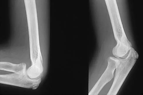
Figures 29a and 29b show the radiographs of a 13-year-old competitive gymnast who has had elbow pain for the past 2 weeks. The pain is worse with tumbling activities. Examination reveals a mild effusion and slight limitation of extension and forearm rotation with no locking. Initial management should consist of
Explanation
Question 2
A 12-year-old boy who has had a 1-month history of right thigh pain and a limp reports worsening of the pain after a fall, and he can no longer walk or bear weight on the involved extremity. Radiographs of the pelvis reveal a slipped capital femoral epiphysis with moderate to severe displacement. While positioning the patient on the fracture table for screw fixation, partial reduction of the slip is achieved. No further reduction maneuvers are attempted, and the epiphysis is stabilized with a single cannulated screw. What complication is most likely to develop following this procedure?
Explanation
Question 3
Figure 30 shows the AP radiograph of a 9-month-old girl who has been referred for evaluation of unequal leg lengths. Examination reveals symmetrical abduction of the hips. When the hips are flexed 90 degrees, the right knee height is greater than the left knee. The girth of the right thigh and calf is larger than the contralateral side. There are no cutaneous lesions, and examination of the spine is normal. The infant is moving all extremities equally and spontaneously. Management should consist of
Explanation
Question 4
What is the mechanism of action of an intramuscular injection of botulinum type A toxin in reducing spasticitiy?
Explanation
Question 5
A 5-year-old boy has had right hip pain and a limp for the past 3 months. Examination of the right hip reveals irritability and restricted abduction and internal rotation. AP and lateral radiographs of the hips are shown in Figures 31a and 31b. Initial management should consist of
Explanation
Question 6
Hamstring lengthening and posterior transfer of the rectus femoris will be most successful in a patient with cerebral palsy who has which of the following gait abnormalities?
Explanation
Question 7
Figures 32a and 32b show the radiographs of a 13-year-old boy who sustained a fracture while playing football 1 week ago. Management at the time of injury included application of a cast and the use of crutches. A follow-up office visit reveals a normal neurologic examination, and the patient reports no discomfort with the cast and crutches. Management should now include
Explanation
Question 8
A 14-year-old patient with an L3 myelomeningocele underwent anterior and posterior spinal fusion for a curve of 50 degrees. Follow-up examination 1 week after the procedure now reveals persistent drainage from the posterior wound. Results of laboratory cultures show Streptococcus viridans, Staphylococcus aureus, and Enterococcus. In addition to IV antibiotics, surgical irrigation, and debridement, management should include
Explanation
Question 9
What is the primary mechanism of injury for the fracture shown in Figures 33a and 33b?
Explanation
Question 10
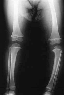
Figure 34 shows the standing AP radiograph of a 2-year-old girl who has a left bowleg deformity. Her mother states that she first noticed the problem when the child began walking at age 10 months, and the deformity has worsened over the past 6 months. Examination reveals a definite lateral thrust of the knee during the stance phase of gait. Management should consist of
Explanation
Question 11
Figures 35a and 35b show the radiographs of a 7-year-old patient who has progressive deformity of the right thigh accompanied by a dull persistent pain radiating to the knee. Examination reveals an obvious bulge in the right thigh, with flexion of the hip beyond 50 degrees only if the hip is allowed to externally rotate. Management should consist of
Explanation
Question 12
Figures 36a and 36b show the MRI scans of a 15-year-old girl who has had pain and recurrent hemarthrosis in the knee for the past year. Plain radiographs are normal. What is the most likely diagnosis?
Explanation
Question 13
A 2-year-old child has marked hypotonia and depressed reflexes. History reveals that the child was normal at birth and developed normally for the first year. The child also began to ambulate, but lost this ability during the next 6 months. Laboratory studies show a creatine phosphokinase level that is within the normal range. DNA testing confirms a deletion in the survival motor neuron (SMN) gene. What is the most likely diagnosis?
Explanation
Question 14
A 13-year-old boy sustains a valgus stress injury to the knee while playing football, and he is unable to bear weight after the injury. Examination reveals tenderness medially superior to the joint line. The knee is held in flexion, and he has a large effusion and localized medial swelling. Plain radiographs show no obvious fracture. What is the next diagnostic step?
Explanation
Question 15
Figure 37 shows the clinical photograph of a 1-day-old infant who weighed 10.25 lb at birth. Examination reveals an absent right Moro reflex and limited active motion of the right shoulder, elbow, and wrist, but flexion of the fingers. Passive range of motion of the shoulder and elbow is normal. What is the most likely diagnosis?
Explanation
Question 16
Figure 38 shows the radiograph of a 5-year-old child who sustained a type III supracondylar fracture. Examination reveals the absence of a radial pulse, but an otherwise well-perfused hand. Following closed reduction and percutaneous pinning, the radial pulse remains absent; however, the hand is pink and well perfused. Management should now include
Explanation
Question 17
Figures 39a and 39b show the radiographs of an otherwise healthy 10-year-old boy who has had thigh pain and a limp for the past 9 months. Examination reveals that the left lower extremity is 1 cm shorter, with reduced flexion, abduction, and internal rotation on the left side. The patient is at the 50th percentile for height and the 90th percentile for weight. Serum studies will most likely show
Explanation
Question 18
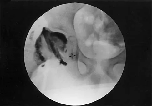
A 7-year-old patient has had a painless limp for several months. Examination reveals pain and spasm with internal rotation, and abduction is limited to 10 degrees on the involved side. Management consists of 1 week of bed rest and traction, followed by an arthrogram. A maximum abduction/internal rotation view is shown in Figure 40a, and abduction and adduction views are shown in Figures 40b and 40c. The studies are most consistent with
Explanation
Question 19
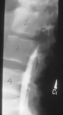
A 14-year-old football player has had thigh pain and weakness following a full-contact scrimmage 24 hours ago. He recalls that he felt a sharp pain in his back after colliding with a much heavier player. Examination reveals that the spine is minimally tender to palpation in the upper lumbar region. Motor testing reveals quadriceps weakness bilaterally, and a reverse straight leg raising test is positive. Plain radiographs of the thoracolumbar spine are normal. A myelogram, a CT scan with contrast, and an MRI scan are shown in Figures 41a through 41c. What is the most likely diagnosis?
Explanation
Question 20
Figure 42 shows the radiograph of a 12-year-old boy who has a limp and pain in the left hip with athletic activity. Examination reveals decreased abduction and internal rotation of the left hip, with pain at the extremes of motion and a 1-cm limb-length discrepancy. Management should consist of
Explanation
Question 21
The mother of a 5-year-old child reports that he has had a fever of 103 degrees F (39.4 degrees C), leg swelling, and has been unwilling to bear weight on his right lower leg for the past 7 days. Examination reveals point tenderness at the distal femur. Aspiration at the metaphysis yields 10 mL of purulent fluid, and a Gram stain reveals gram-positive cocci. In addition to hospital admission, management should include
Explanation
Question 22
Figure 43 shows the lateral radiograph of a 12-year-old boy with mild osteogenesis imperfecta who injured his left elbow after pushing his brother. Treatment should consist of
Explanation
Question 23
Figure 44 shows the radiograph of an 11-year-old girl who has hip pain. Further diagnostic workup should include
Explanation
Question 24
Figure 45 shows the radiograph of a 2-year-old patient who has progressive lumbar scoliosis as the result of hemivertebra. Examination reveals no associated cutaneous lesions, and an MRI scan shows no associated intraspinal anomalies. Treatment should consist of
Explanation
Question 25
A 10-year-old girl with a history of an obstetrical brachial plexus palsy has been referred for evaluation. Examination reveals a severe adduction internal rotation contracture of the shoulder and a mild flexion contracture of the elbow. Hand function is normal. Radiographs show mild glenohumeral joint incongruity. To achieve the best functional outcome, management should consist of
Explanation
Question 26
A 6-week-old female infant is currently being treated with a Pavlik harness for Developmental Dysplasia of the Hip (DDH). At her 2-week follow-up, the ultrasound reveals that the left hip remains dislocated. During the physical examination, you note that she has an absent patellar reflex on the left side and decreased spontaneous extension of the left knee. What is the most appropriate next step in management?
Explanation
Question 27
A 6-year-old boy sustains a completely displaced Gartland type III extension-type supracondylar humerus fracture. On initial presentation, his hand is pink but the radial pulse is not palpable. Following urgent closed reduction and percutaneous pinning, the fracture is anatomically aligned, but the hand remains pink and pulseless. Capillary refill is less than 2 seconds. What is the most appropriate management?
Explanation
Question 28
An 8-month-old boy is evaluated for infantile idiopathic scoliosis. Radiographs demonstrate a 25-degree left thoracic curve. According to Mehta's criteria, a Rib-Vertebra Angle Difference (RVAD) greater than which of the following values is most predictive of curve progression?
Explanation
Question 29
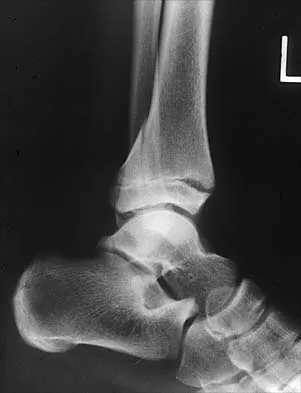
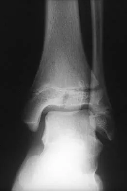
A 14-year-old boy presents with an ankle injury after an external rotation force during football. Radiographs and a subsequent CT scan reveal a Salter-Harris type III fracture of the anterolateral aspect of the distal tibial epiphysis, with 3.5 mm of displacement. What anatomic structure is responsible for the avulsion of this specific fracture fragment?
Explanation
Question 30
During an open reduction of a late-presenting Developmental Dysplasia of the Hip (DDH) via a medial (Ludloff) approach, several anatomical structures blocking reduction can be accessed. Which of the following pathological obstacles to reduction CANNOT be adequately addressed through this approach?
Explanation
Question 31
A 12-year-old boy with Duchenne muscular dystrophy presents with a progressive, sweeping thoracolumbar neuromuscular scoliosis measuring 45 degrees. He became wheelchair-bound 6 months ago. His forced vital capacity (FVC) is 45% of predicted. What is the most appropriate definitive management for his spinal deformity?
Explanation
Question 32
A 9-year-old boy undergoes closed reduction and casting for a midshaft both-bone forearm fracture. During healing, he develops an angular malunion. Loss of which of the following anatomic features is most likely to significantly restrict his functional supination and pronation?
Explanation
Question 33
A 35-year-old male presents with numbness in his small and ring fingers, along with intrinsic muscle weakness in his dominant hand. He reports a history of an elbow fracture treated non-operatively with a cast when he was 5 years old. Examination reveals significant valgus carrying angle at the elbow. Which of the following pediatric fracture patterns is most likely responsible for this late presentation?
Explanation
Question 34
In the evaluation of Adolescent Idiopathic Scoliosis using the Lenke classification, specific radiographic criteria are used to determine if a minor curve is structural. Which of the following findings correctly defines a proximal thoracic minor curve as structural?
Explanation
Question 35
A 4-week-old female infant with a breech presentation history is referred for a developmental dysplasia of the hip (DDH) ultrasound. When obtaining a standard Graf coronal view, the sonographer must ensure a standard plane is captured. Which bony landmark must be clearly visualized to confirm a true standard coronal view?
Explanation
Question 36
An 8-year-old, 32 kg boy sustains an isolated, length-stable midshaft transverse femur fracture and undergoes treatment with titanium elastic nails. Which of the following is the most frequently encountered complication specific to this fixation method in this age group?
Explanation
Question 37
A 2-year-old child is being evaluated for congenital scoliosis. Radiographs reveal multiple vertebral anomalies. Which of the following patterns of vertebral malformation carries the highest risk for rapid, unrelenting curve progression?
Explanation
Question 38
A 3-month-old female infant is being treated with a Pavlik harness for developmental dysplasia of the hip (DDH). During her 2-week follow-up, the parents report she has stopped kicking her left leg. On examination, there is an absence of active knee extension on the left, but she flexes the hip and moves the toes spontaneously. What is the most appropriate next step in management?
Explanation
Question 39
A 14-month-old boy presents with a left thoracic curve.
The physician is evaluating the curve to determine the risk of progression. Which of the following is the most reliable radiographic prognostic factor for progression in infantile idiopathic scoliosis?
Explanation
Question 40
A 6-year-old boy sustains a completely displaced, extension-type supracondylar humerus fracture.
He presents with an absent radial pulse but a well-perfused, pink hand. After closed reduction and percutaneous pinning, the hand remains pink, but the radial pulse remains non-palpable. Doppler signals are audible at the wrist. What is the most appropriate management?
Explanation
Question 41
A 12-year-old boy presents with right elbow deformity and a history of a fracture at age 4 that was treated non-operatively. Examination reveals severe cubitus valgus and numbness extending to the palmar aspect of the small finger and ulnar half of the ring finger. This condition most likely resulted from nonunion of which of the following fractures?
Explanation
Question 42
A 2-year-old child is diagnosed with congenital scoliosis. Which of the following vertebral anomalies carries the highest probability of rapid, unrelenting curve progression?
Explanation
Question 43
A 24-month-old girl presents with a painless limp. Examination demonstrates a positive Trendelenburg sign on the left.
Radiographs confirm a dislocated left hip with a false acetabulum and severe acetabular dysplasia. What is the most appropriate initial treatment?
Explanation
Question 44
A 13-year-old girl twists her ankle. Radiographs reveal a Salter-Harris III fracture of the anterolateral aspect of the distal tibia. This specific fracture pattern (Juvenile Tillaux fracture) is primarily due to which of the following anatomical factors?
Explanation
Question 45
A 12-year-old premenarchal girl has a right thoracic curve of 32 degrees on standing posteroanterior radiographs. Her Risser stage is 0. What is the most appropriate management?
Explanation
Question 46
A 14-year-old non-ambulatory male with spastic quadriplegic cerebral palsy presents with a 75-degree sweeping neuromuscular scoliosis and marked pelvic obliquity, causing difficulty with wheelchair seating. What is the recommended surgical intervention?
Explanation
Question 47
In an ultrasound evaluation of a 4-week-old infant's hip for developmental dysplasia,
what specific anatomic feature is quantified by the alpha angle?
Explanation
Question 48
A 3-year-old boy is brought to the emergency department after a fall while running, resulting in a closed spiral fracture of the femoral shaft. Non-accidental trauma has been definitively ruled out. What is the most appropriate definitive treatment?
Explanation
Question 49
A 10-year-old boy presents with a stable slipped capital femoral epiphysis (SCFE) of the right hip. Under which of the following conditions is prophylactic pinning of the contralateral, asymptomatic hip most strongly indicated?
Explanation
Question 50
A 14-year-old boy sustains a triplane fracture of the distal tibia following a fall. By definition, this fracture complex involves fracture lines propagating through three distinct planes. Which of the following accurately describes these planes?
Explanation
Question 51
A 9-year-old girl with Neurofibromatosis Type 1 (NF-1) has a sharp 45-degree thoracic kyphoscoliosis.
Radiographs demonstrate vertebral scalloping, spindling of the transverse processes, and penciling of the ribs. What is the most appropriate surgical treatment?
Explanation
Question 52
A 6-year-old boy sustains a completely displaced, overriding fracture of the distal third of the radius and ulna. Closed reduction under conscious sedation achieves 15 degrees of dorsal angulation and 1 cm of bayonet apposition in a well-molded long-arm cast. What is the most appropriate next step?
Explanation
Question 53
When treating developmental dysplasia of the hip (DDH) with closed reduction and spica casting, maintaining the hip in forced, extreme abduction ('frog-leg' position) significantly increases the risk of which of the following complications?
Explanation
Question 54
A 6-year-old boy sustains a completely displaced extension-type supracondylar humerus fracture. On presentation, the hand is pink but the radial pulse is absent. After closed reduction and percutaneous pinning, the fracture is anatomically reduced, the hand remains pink, but the radial pulse is still absent. What is the most appropriate next step in management?
Explanation
Question 55
A 4-month-old female with developmental dysplasia of the hip (DDH) is being treated with a Pavlik harness. During a follow-up visit, the parents report she has stopped kicking her right leg. Examination reveals decreased active extension of the right knee, but she withdraws to pain. What is the most likely cause, and what is the appropriate management?
Explanation
Question 56
Which of the following factors represents the highest risk for curve progression in a 12-year-old girl with Adolescent Idiopathic Scoliosis (AIS)?
Explanation
Question 57
A 5-year-old boy sustained a pediatric lateral condyle fracture of the distal humerus 2 years ago, which was treated non-operatively. He now presents with progressive cubitus valgus. Which of the following tardy nerve palsies is most likely to develop?
Explanation
Question 58
A newborn girl with arthrogryposis multiplex congenita is found to have bilateral rigid, irreducible teratologic hip dislocations. What is the most appropriate management?
Explanation
Question 59
A 9-month-old boy is diagnosed with infantile idiopathic scoliosis. Radiographs show a 25-degree left thoracic curve. Which of the following radiographic parameters best predicts whether this curve will progress or spontaneously resolve?
Explanation
Question 60
A 13-year-old obese boy undergoes in-situ pinning for a unilateral stable slipped capital femoral epiphysis (SCFE). Which of the following is the strongest indication for prophylactic pinning of the contralateral hip?
Explanation
Question 61
A 2-year-old boy sustains a closed, isolated midshaft femur fracture. He is treated with early spica casting. Which of the following acceptable radiographic parameters is correct for this age group?
Explanation
Question 62
In the Lenke classification for Adolescent Idiopathic Scoliosis, a curve is considered "structural" if it fails to reduce to less than what Cobb angle on side-bending radiographs?
Explanation
Question 63
An 11-year-old boy presents with progressive ulnar deviation of the wrist 18 months after sustaining a distal radius Salter-Harris II fracture. Radiographs show a complete premature closure of the distal radius physis, with continued growth of the distal ulna (ulnar plus variance). What is the most appropriate surgical treatment?
Explanation
Question 64
On an AP pelvis radiograph of a 6-month-old infant evaluated for DDH, the femoral head ossific nucleus is absent. Which of the following describes the normal expected position of the proximal femoral metaphysis?
Explanation
Question 65
A 9-year-old girl sustains a Delbet Type II (transcervical) femoral neck fracture after falling from a tree. She is treated with urgent open reduction and internal fixation with cannulated screws. Which of the following complications occurs at the highest rate in this specific injury pattern?
Explanation
Question 66
A 14-year-old boy sustains a twisting injury to his ankle. Radiographs show a Salter-Harris III fracture of the anterolateral aspect of the distal tibial epiphysis. What is the anatomical rationale for this specific fracture pattern?
Explanation
Question 67
A 12-year-old boy with spastic quadriplegic cerebral palsy has a progressive 80-degree neuromuscular scoliosis with marked pelvic obliquity. He is non-ambulatory and has difficulty sitting in his wheelchair. Surgical planning should most likely involve:
Explanation
Question 68
A 6-year-old girl sustains a closed fracture of the distal third of the radius and ulna. Following closed reduction and casting, the radiographs show 15 degrees of apex volar angulation. What is the most appropriate management?
Explanation
Question 69
A 15-year-old boy presents with back pain and increased thoracic kyphosis. Standing lateral radiographs reveal a thoracic kyphosis of 65 degrees and anterior wedging of 3 consecutive vertebrae of 6 degrees each. What is the most appropriate initial management?
Explanation
Question 70
A 6-week-old female is treated with a Pavlik harness for developmental dysplasia of the hip (DDH). At her 2-week follow-up, the mother notes that the child is no longer kicking her leg on the affected side. On examination, the infant has decreased active knee extension but normal ankle movements. What is the most appropriate next step in management?
Explanation
Question 71
A 5-year-old boy falls on an outstretched hand and sustains a lateral condyle fracture of the distal humerus. Initial radiographs show 1 mm of displacement, and he is placed in a long arm cast. At 1-week follow-up, radiographs show 3 mm of displacement. What is the most appropriate management?
Explanation
Question 72
A 12-year-old premenarchal girl presents with adolescent idiopathic scoliosis. Standing radiographs demonstrate a right thoracic curve of 32 degrees. Her Risser stage is 0. What is the most appropriate management?
Explanation
Question 73
A 6-year-old boy sustains a completely displaced posteromedial supracondylar humerus fracture. On presentation, the hand is pink and warm, but the radial pulse is absent. Capillary refill is 2 seconds. What is the most appropriate initial management?
Explanation
Question 74
An 18-month-old girl is brought to the clinic for a waddling gait. Examination shows restricted hip abduction on the left side and a positive Galeazzi sign. Radiographs confirm a dislocated left hip with a dysplastic acetabulum (acetabular index 38 degrees). What is the most appropriate initial surgical management?
Explanation
Question 75
A 3-year-old boy is diagnosed with congenital scoliosis secondary to a fully segmented hemivertebra at T8. Which of the following screening tests must be routinely obtained to evaluate for associated conditions?
Explanation
Question 76
A 10-year-old boy sustains a Salter-Harris II fracture of the distal femur. It is treated with closed reduction and percutaneous pinning. Which of the following is the most common complication associated with this specific injury?
Explanation
Question 77
On an anteroposterior pelvis radiograph of a 6-month-old infant being evaluated for DDH, the proximal femoral ossific nucleus is located in the upper outer quadrant formed by Hilgenreiner's and Perkin's lines. What does this radiographic finding indicate?
Explanation
Question 78
A 14-year-old boy with spastic quadriplegic cerebral palsy presents with a 75-degree thoracolumbar scoliosis and severe pelvic obliquity. He is non-ambulatory and has lost the ability to sit comfortably in his custom wheelchair. Which of the following surgical strategies is most appropriate?
Explanation
Question 79
A 13-year-old boy complains of ankle pain after a twisting injury. Radiographs show a fracture of the distal tibia that appears as a Salter-Harris III on the AP view and a Salter-Harris II on the lateral view (Triplane fracture). What is the normal closure pattern of the distal tibial physis that predisposes adolescents to this injury?
Explanation
Question 80
During closed reduction and spica casting for a 9-month-old girl with DDH, the hip reduces at 40 degrees of abduction and re-dislocates at 20 degrees of abduction. The safe zone of Ramsey is 20 to 60 degrees. What is the primary risk of immobilizing the hip in excessive abduction (>60 degrees)?
Explanation
Question 81
A 10-month-old infant presents with infantile idiopathic scoliosis measuring 25 degrees. The rib-vertebral angle difference (RVAD) of Mehta is 28 degrees, and phase 2 rib-vertebral overlap is present on the convex side. What is the most likely natural history of this condition without intervention?
Explanation
Question 82
A 7-year-old girl sustains a both-bone forearm fracture. Closed reduction is performed in the emergency department. Which of the following post-reduction radiographic parameters is considered unacceptable and requires surgical fixation or remanipulation?
Explanation
Question 83
A 4-week-old boy is fitted with a Pavlik harness for an Ortolani-positive right hip. To ensure proper positioning and minimize neurovascular or ischemic complications, how should the harness straps be adjusted?
Explanation
Question 84
A 13-year-old female with adolescent idiopathic scoliosis presents for routine evaluation. Radiographs reveal a 22-degree right thoracic curve. Evaluation of the iliac apophysis demonstrates ossification covering the anterior 75% of the iliac crest, but not yet reaching the posterior superior iliac spine (PSIS). What Risser stage does this represent?
Explanation
Question 85
A 4-month-old girl has been treated with a Pavlik harness for 4 weeks for an irreducible developmental dysplasia of the hip (DDH). Repeat ultrasound demonstrates a persistently dislocated left hip without significant improvement in the alpha angle. What is the most appropriate next step in management?
Explanation
Question 86
A 6-year-old boy presents with a left-sided thoracic curve of 25 degrees. His neurological examination is unremarkable. What is the most appropriate next diagnostic step before considering orthotic management?
Explanation
Question 87
A 6-year-old boy sustains a completely displaced, extension-type supracondylar humerus fracture. On presentation, the hand is pink but the radial pulse is absent. After successful closed reduction and percutaneous pinning, the hand remains pink and capillary refill is brisk, but the radial pulse remains unpalpable. What is the most appropriate next step?
Explanation
Question 88
An 18-month-old girl is newly diagnosed with developmental dysplasia of the right hip (DDH). The hip is completely dislocated but reducible on examination. Which of the following is the most appropriate initial treatment?
Explanation
Question 89
A 12-year-old girl presents with adolescent idiopathic scoliosis. She is premenarchal and has a Risser stage of 0. Standing radiographs demonstrate a right thoracic curve of 32 degrees. What is the most appropriate management?
Explanation
Question 90
A 3-year-old boy sustains a low-energy, isolated spiral fracture of the midshaft femur. Child abuse has been thoroughly ruled out. What is the gold standard of treatment for this injury?
Explanation
Question 91
Which of the following congenital spinal anomalies has the highest risk of rapid curve progression and requires the earliest surgical intervention?
Explanation
Question 92
A 13-year-old boy sustains an intra-articular fracture of the distal tibia. Radiographs show a fracture line traversing the lateral epiphysis and extending superiorly through the lateral physis. What is the classic mechanism of this specific injury pattern?
Explanation
Question 93
An ultrasound of the hip is performed on a 6-week-old infant suspected of having DDH. Which of the following sonographic measurements is most diagnostic of a dysplastic hip?
Explanation
Question 94
A 14-year-old girl with adolescent idiopathic scoliosis presents for follow-up. Standing full-spine radiographs show a right thoracic curve of 55 degrees. She is post-menarchal and Risser 4. What is the most appropriate treatment recommendation?
Explanation
Question 95
An 8-year-old boy falls and sustains a closed diaphyseal both-bone forearm fracture. What is the maximum acceptable angulation in the middle third of the radius and ulna to allow for a satisfactory functional outcome with non-operative treatment?
Explanation
Question 96
An infant is born with bilateral dislocated hips and severe contractures of multiple joints consistent with arthrogryposis multiplex congenita. What is the expected outcome regarding the management of these teratologic hip dislocations?
Explanation
Question 97
A 14-year-old non-ambulatory boy with Duchenne muscular dystrophy develops a progressive neuromuscular scoliosis of 45 degrees. Which of the following is an established principle regarding surgical treatment for this patient?
Explanation
Question 98
A 12-year-old boy presents with an acute ankle injury. Radiographs reveal a distal tibia fracture that appears as a Salter-Harris III injury on the AP view and a Salter-Harris II injury on the lateral view. What is the most appropriate anatomical classification of this fracture?
Explanation
Question 99
During the application of a spica cast for DDH, the hip is forcefully placed in a position of extreme abduction (the "frog-leg" position). What is the most devastating complication associated with this specific positioning?
Explanation
Question 100
A 5-year-old child sustains a widely displaced (3 mm) lateral condyle fracture of the humerus. If left untreated and a nonunion develops, which of the following complications is most likely to present years later?
Explanation
None
