العربية
Part of the Master Guide

AAOS Orthopedic MCQs (Set 1): Spine Disorders, Sports Medicine & Arthroplasty | 2026 Board Review

Orthopedic Spine 2026 MCQs: Board Review Questions & Answers (Part 4)

27 Apr 2026 88 min read 82 Views
Figure for Spine 2006 MCQs - Part 4 - Question 76

Key Takeaway

Discover the latest medical recommendations for Orthopedic Spine 2026 MCQs: Board Review Questions & Answers (Part 4). Top-rated Orthopedic Spine 2026 MCQs bank. Practice with clinical case questions, orthopedic surgery board review, and evidence-based answers updated for 2026.

Orthopedic Spine 2026 MCQs: Board Review Questions & Answers (Part 4)

Comprehensive 100-Question Exam


00:00

Start Quiz

Question 1

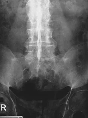
Figures 25a and 25b show the radiograph and MRI scan of a 48-year-old man who reports increasing unsteadiness in his gait and hand clumsiness. Examination reveals a positive Hoffmann's reflex bilaterally, positive clonus, and a spastic gait. Management should consist of


Explanation

25b The patient has a congenitally small spinal canal with secondary multilevel degenerative changes causing stenosis and cord compression across multiple segments, including directly posterior to the vertebral bodies. A multilevel diskectomy may address the cord compression at the disk level, but not posterior to the bodies, and most likely would be inadequate. The patient has significant stenosis distal to C5, necessitating a more extensive surgical approach than simply C3-C5. Because the patient's cervical lordosis is preserved, a posterior procedure such as laminoplasty or laminectomy would allow the cord to fall away from the anterior pathology and afford decompression. Cervical myelopathy does not tend to resolve, and there is a significant risk for progression; therefore, surgical management usually is recommended. Edwards CC II, Riew KD, Anderson PA, et al: Cervical myelopathy: Current diagnostic and treatment strategies. Spine J 2003;3:68-81.

Question 2

Lumbar disk replacement has been shown to offer which of the following results?


Explanation

There is no clear evidence that disk replacement results in pain relief that is superior to fusion. Pain relief appears to be equivalent with these two procedures. No study has clearly demonstrated that normal segmental motion has been consistently restored. Preexisting facet arthropathy is considered to be a contraindication to disk replacement. Comparative long-term data demonstrating a reduced incidence of adjacent segment disease compared to fusion are not yet available. Geisler FH, Blumenthal SL, Guyer RD, et al: Neurological complications of lumbar artificial disc replacement and comparison of clinical results with those related to lumbar arthrodesis in the literature. J Neurosurg Spine 2004;1:143-154.

Question 3

When performing the exposure for an anterior approach to the cervical spine, the surgical dissection should not enter the plane between the trachea and the esophagus and excessive retraction should be avoided to prevent injury to the


Explanation

The recurrent laryngeal nerve lies between the trachea and the esophagus. The vagus nerve lies in the carotid sheath. The sympathetic trunk lies anterior to the longus colli muscles. The hypoglossal and superior laryngeal nerves are both at risk during the exposure but are not located between the trachea and the esophagus. Flynn TB: Neurologic complications of anterior cervical interbody fusion. Spine 1982;7:536-539.

Question 4

A 39-year-old man reports low back pain, lower extremity numbness, and urinary retention after being injured in a motor vehicle accident 1 day ago. He is able to walk but is in pain. A straight leg raise results in increased back pain, and examination reveals that perianal sensation is decreased. Placement of a urinary catheter results in 500 mL of urine. What is the next most appropriate step in management?


Explanation

Acute cauda equina syndrome, including saddle hypesthesia and bowel/bladder incontinence, is a red flag that demands emergent evaluation with MRI and urgent surgery if compression is confirmed. Results appear to be improved if surgery is performed within 48 hours. The other treatment approaches listed are not indicated if a cauda equina syndrome is present. Ahn UM, Ahn NU, Buchowski JM, et al: Cauda equina syndrome secondary to lumbar disc herniation: A meta-analysis of surgical outcomes. Spine 2000;25:1515-1522. Shapiro S: Medical realities of cauda equina syndrome secondary to lumbar disc herniation. Spine 2000;25:348-351.

Question 5

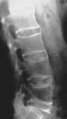
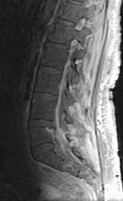
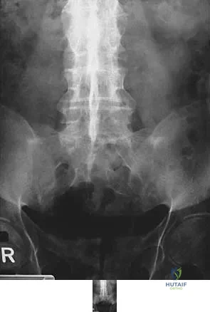
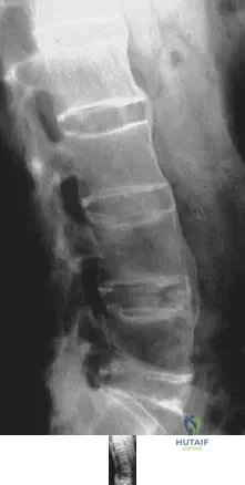
Figures 26a and 26b show the radiograph and MRI scan of an 18-year-old man who fell from a trampoline. Examination reveals exquisite local tenderness at the thoracolumbar junction, but he is neurologically intact. Management should consist of


Explanation

26b Based on the radiographic findings of marked disruption of the posterior ligamentous complex with a relatively small anterior bony fracture, the patient has a classic Chance-type ligamentous flexion-distraction injury. The pathology is mostly in soft tissues with limited healing potential. The treatment of choice is posterior reconstruction of the tension band with a short segment fusion with instrumentation. Casting or bracing may result in a painful kyphosis with ligamentous insufficiency. The anterior bony column is mostly intact, so anterior reconstruction is not necessary. Carl AL: Adult spine trauma, in DeWald RL (ed): Spinal Deformities: A Comprehensive Text. New York, NY, Thieme, 2003, pp 406-423.

Question 6

A 17-year-old high school football player is seen for follow-up after sustaining an injury 3 days ago. He reports that he tackled a player, felt numbness throughout his body, and could not move for approximately 15 seconds. A spinal cord injury protocol was initiated on the field. Evaluation in the emergency department revealed a normal neurologic examination and full painless neck motion. He states that he has no history of a similar injury. An MRI scan of the cervical spine is normal. During counseling, the patient and his family should be informed that he has sustained


Explanation

The long-term effect of transient quadriplegia is unknown. Based on a history of one brief episode of transient quadriplegia and normal examination and MRI findings, the risk of permanent spinal cord injury with a return to play is low. There is a risk of recurrent episodes of transient quadriplegia after the initial episode. Morganti C, Sweeney CA, Albanese SA, et al: Return to play after cervical spine injury. Spine 2001;26:1131-1136. Odor JM, Watkins RG, Dillin WH, et al: Incidence of cervical spinal stenosis in professional and rookie football players. Am J Sports Med 1990;18:507-509. Torg JS, Naranja RJ Jr, Palov H, et al: The relationship of developmental narrowing of the cervical spinal canal to reversible and irreversible injury of the cervical spinal cord in football players. J Bone Joint Surg Am 1996;78:1308-1314.

Question 7

Which of the following is considered a contraindication to cement injection techniques, such as kyphoplasty or vertebroplasty, in the treatment of osteoporotic compression fractures?


Explanation

When retropulsion of the posterior vertebral wall is present, nothing prohibits the cement from following the path of least resistance into the canal or from pushing a bone fragment further into the canal; most clinicians consider it a contraindication to these techniques. Patient age itself is not a contraindication as long as there are no medical contraindications to surgery. An acute fracture in a patient who remains immobile and hospitalized because of pain may be a good indication for such a technique. Prior compression fracture and older compression fractures are not contraindications, but pain relief may be less predictable. Phillips FM, Pfeifer BA, Leiberman IH, et al: Minimally invasive treatment of osteoporotic vertebral compression fractures: Vertebroplasty and kyphoplasty. Instr Course Lect 2003;52:559-567. Truumees E, Hilibrand A, Vaccaro AR: Percutaneous vertebral augmentation. Spine J 2004;4:218-229.

Question 8

Chronic anterior donor site pain following the harvest of autologous iliac crest bone graft for use during anterior cervical diskectomy and fusion is reported by approximately what percent of patients?


Explanation

Four years after surgery, more than 90% of patients are satisfied with the cosmetic appearance of the iliac donor site scar. Approximately 25% still have pain and/or functional difficulty, including 12.7% who still report difficulty with ambulation, 11.9% difficulty with recreational activities, 7.5% with sexual intercourse, and 11.2% require pain medication for iliac donor site symptoms. Silber JS, Anderson DG, Daffner SD, et al: Donor site morbidity after anterior iliac crest bone harvest for single-level anterior cervical discectomy and fusion. Spine 2003;28:134-139.

Question 9

When treating osteoporosis with alendronate, what is the most common side effect?


Explanation

Alendronate is a second-generation bisphosphonate, and it can cause epigastric distress in up to 30% of patients. This side effect can be minimized by gradually building up to therapeutic doses over a period of 4 to 8 weeks. Marshall JK, Rainsford KD, James C, et al: A randomized controlled trial to assess alendoronate-associated injury of the upper gastrointestinal tract. Aliment Pharmacol Ther 2000;14:1451-1457.

Question 10

Figures 27a and 27b show the radiographs of a 32-year-old woman who was involved in a high-speed motor vehicle accident. She is neurologically intact. After stabilization and assessment, treatment should consist of


Explanation

27b The radiographs show a fracture-dislocation with translation in both the coronal and sagittal planes, evidence of significant instability requiring surgical stabilization. Anterior instrumentation is not as effective as posterior instrumentation in restoring stability, and because there is little bony destruction, the anterior column can be successfully reconstructed with simple realignment. The treatment of choice is multisegment posterior fusion with instrumentation. Lewandrowski KU, McLain RF: Thoracolumbar fractures: Evaluation, classification, and treatment, in Frymoyer JW, Wiesel SW (eds): The Adult and Pediatric Spine. Philadelphia, PA, Lippincott Williams and Wilkins, 2004, pp 817-843.

Question 11

Figures 28a through 28c show the MRI scans of a 30-year-old woman who weighs 290 lb and has low back and left leg pain. She also reports frequent urinary dribbling, which her gynecologist has advised her may be related to obesity. Examination will most likely reveal


Explanation

28b 28c The patient will most likely exhibit ipsilateral weakness of the tibialis anterior. Gaenslen's test is designed to detect sacroiliac inflammation as a source of low back pain. Beevor's sign tests the innervation of the rectus abdominus and paraspinal musculature (L1 innervation). The extensor hallucis longus is predominantly innervated by L5. The peroneals are predominantly innervated by S1. Hoppenfeld S: Physical Examination of the Spine and Extremities. Appleton, WI, Century-Crofts, 1976.

Question 12

Which of the following statements regarding conus medullaris syndrome is most accurate?


Explanation

Conus medullaris syndrome most frequently occurs as a result of trauma or with a disk herniation at L1, resulting in a lower motor neuron syndrome but with a poor prognosis for recovery of bowel and bladder dysfunction. The conus region, as the termination of the spinal cord, contains the motor cell bodies of the sacral roots. The syndrome is usually a sacral level neural injury; therefore, lower extremity weakness is uncommon. Haher TR, Felmly WT, O'Brien M: Thoracic and lumbar fractures: Diagnosis and management, in Bridwell KH, Dewald RL, Hammerberg KW, et al (eds): The Textbook of Spinal Surgery, ed 2. New York, NY, Lippincott Williams & Wilkins, 1977, pp 1773-1778.

Question 13

Which of the following factors has the greatest effect on the pull-out strength of a lumbar pedicle screw?


Explanation

All of the factors listed contribute to some extent to the pull-out strength of lumbar pedicle screws, but bone mineral density correlates most precisely. Wittenberg RH, Shea M, Swartz DE, et al: Importance of bone mineral density in instrumented spine fusions. Spine 1991;16:647-652.

Question 14

An inverted radial reflex is associated with


Explanation

An inverted radial reflex is a hypoactive brachioradialis reflex in combination with involuntary finger flexion. It is a spinal cord "release" sign and is associated with upper motor neuron pathology as seen in cervical stenosis with myelopathy. Radiculopathy is characterized by a diminished reflex but no finger flexion. Peripheral neuropathy is not associated with any reflex change. Parsonage-Turner syndrome is an idiopathic brachial neuritis. Clark CR (ed): The Cervical Spine, ed 3. Philadelphia, PA, Lippincott Williams & Wilkins, 1998, p 762.

Question 15

Figures 29a and 29b show the radiograph and CT scan of a 48-year-old man who has diffuse spinal pain. What is the most likely diagnosis?


Explanation

29b The studies show marginal syndesmophyte formation characteristic of ankylosing spondylitis. These patients typically have diffuse ossification of the disk space without large osteophyte formation. DISH typically presents with large osteophytes, referred to as nonmarginal syndesmophytes. In this patient, the zygoapophyseal joints are fused rather than degenerative as would be seen in rheumatoid arthritis, and the costovertebral joints are also fused. Osteopetrosis does not normally ankylose the disk space. McCullough JA, Transfeldt EE: Macnab's Backache, ed 3. Baltimore, MD, Williams and Wilkins, 1997, pp 190-194.

Question 16

The cervical disk herniation shown in the MRI scans in Figures 30a and 30b will most likely create which of the following constellations of symptoms?


Explanation

30b The MRI scans reveal a right-sided C5-6 herniated nucleus pulposus. A disk herniation in this region encroaches on the C6 root and is accompanied by a sensory change along the thumb and index finger, alterations in the brachioradialis reflex, and possible wrist extension weakness. Although the nerve root associated with the vertebral body passes above the pedicles such that the C6 root passes above the C6 pedicle, it is still the C6 root that is encroached on because the herniation affects the exiting root rather than the traversing root as seen in the lumbar spine. Klein JD, Garfin SR: Clinical evaluation of patients with suspected spine problems, in Frymoyer JW (ed): Adult Spine: Principles and Practice, ed 2. Philadephia, PA, Lippincott-Raven, 1997, pp 319-330.

Question 17

A 21-year-old man has had posterior neck discomfort for the past 6 months. A whole-body bone scan and a cervical single-photon emission CT reveal increased activity at the C7 spinous process. MRI reveals multifocal involvement of the spinous process lamina and facet of C7. A CT-directed needle biopsy reveals osteoblastoma. What is the best course of action?


Explanation

En bloc excision is the recommended treatment of osteoblastoma. Treatment should consist of en bloc removal of the lamina, facet, and spinous process. Facet removal would necessitate fusion. Radiation therapy is not recommended. Intralesional curettage has a high rate of recurrence. Bridwell KH, Ogilvie JW: Primary tumors of the spine, in Bridwell KH, DeWald RL (eds): The Textbook of Spinal Surgery. Philadelphia, PA, JB Lippincott, 1991, vol 2, pp 1143-1174.

Question 18

What is the most likely consequence of a vertebral compression fracture associated with osteoporosis?


Explanation

After an osteoporotic vertebral compression fracture, the risk of subsequent fractures at adjacent levels increases. This is felt to be the result of a shifting of the sagittal alignment more anteriorly, putting more stress on the osteopenic vertebral bodies and their anterior cortices. Pain generally resolves with rest, but this may take weeks or months. It has been demonstrated experimentally that osteoporotic vertebral bodies are actually less stiff and weaker after a compression fracture; therefore, deformity predisposes to further deformity. The extensor musculature often fatigues over time and usually does not hypertrophy. Frontal plane deformity is a rare development. Heaney RP: The natural history of vertebral osteoporosis: Is low bone mass an epiphenomenon? Bone 1992;13:S23-S26.

Question 19

What is the most appropriate treatment for a chordoma involving the sacrum?


Explanation

Chordomas are very radio- and chemotherapy resistant; therefore, en bloc resection with a negative margin is the preferred treatment. Lesions at or below S3 can be resected without compromising pelvis stability, and continence usually is maintained. The mean survival rate for patients with sacral chordomas is approximately 7 years. Patients with chordoma of the mobile (cervical, thoracic, or lumbar) spine have a mean survival rate of approximately 5 years. This difference is most likely the result of an earlier diagnosis. Fardin DF, Garfin SR, Abitbol J, et al (eds): Orthopaedic Knowledge Update: Spine 2. Rosemont, IL, American Academy of Orthopaedic Surgeons, 2002, pp 123-133. Stener B, Gunterberg B: High amputation of the sacrum for extirpation of tumors: Principles and technique. Spine 1978;3:351-366.

Question 20

A 62-year-old woman has back pain and right L2 radicular pain. MRI scans suggest a neoplastic lesion at L2, and a bone scan is negative except at L2. History reveals that she was treated for breast cancer without known metastatic disease 12 years ago and is thought to be free of disease. What is the next most appropriate step in management?


Explanation

Because of the long disease-free interval, it cannot be assumed that this is breast cancer. The lesion could represent metastasis from a new primary tumor or could itself be a primary tumor. CT-guided biopsy will most effectively identify the lesion and guide treatment options. Depending on the specific diagnosis, any of the other options may be appropriate.

Question 21

A 60-year-old woman with rheumatoid arthritis has atlanto-axial instability and basilar invagination. What MRI findings would suggest the need for cervical fusion?


Explanation

The cervical medullary junction should be 135 degrees or greater. An angle of 125 degrees suggests compression of the cervical medullary junction. Other findings supporting surgical intervention include a cord diameter in flexion of less than 6 mm or less than 13 mm of space available for the cord. Clark CR (ed): The Cervical Spine, ed 3. Philadelphia, PA, Lippincott Williams & Wilkins, 1998, pp 700-701. Monsey RB: Rheumatoid arthritis of the cervical spine. J Am Acad Orthop Surg 1997;5:240-248.

Question 22

Which of the following statements is most accurate regarding undetected intraoperative surgical glove perforation?


Explanation

The incidence of undetected intraoperative surgical glove perforation has been demonstrated at approximately 8.5%, occurring most frequently on the index finger or left hand of the assistant surgeon. The frequency of glove perforation is higher in surgeries lasting longer than 3 hours. Al-Habdan I, Sadat-Ali M: Glove perforation in pediatric orthopaedic practice. J Pediatr Orthop 2003;23:791-793.

Question 23

Which of the following is NOT considered a risk factor for nonunion of a type II odontoid fracture?


Explanation

Although obesity can make brace or halo wear difficult, it has not been associated with an increased risk for nonunion. Carson GD, Heller JG, Abitbol JJ, et al: Odontoid fractures, in Levine AM, Eismont FJ, Garfin SR, et al (eds): Spine Trauma. Philadelphia, PA, WB Saunders, 1998, pp 235-238.

Question 24

A 27-year-old woman has a bilateral C5-C6 facet dislocation and quadriparesis after being involved in a motor vehicle accident. Initial management consisted of reduction with traction, but she remains a Frankel A quadriplegic. To facilitate rehabilitation, surgical stabilization and fusion is planned. From a biomechanical point of view, which of the following techniques is the LEAST stable method of fixation?


Explanation

In two different biomechanical studies performed in both bovine and human cadaveric spines, all posterior techniques of stabilization were found to be superior to anterior plating in flexion-distraction injuries of the cervical spine. These injuries usually have an intact anterior longitudinal ligament that allows posterior fixation to function as a tension band. Anterior plating with grafting destroys this last remaining stabilizing structure and does not allow for a tension band effect because all of the posterior stabilizing structures have been destroyed with the injury. In clinical practice, however, anterior plating can be effective in the treatment of this injury with appropriate postoperative orthotic management. Sutterlin CE III, McAfee PC, Warden KE, et al: A biomechanical evaluation of cervical spine stabilization methods in a bovine model: Static and cyclical loading. Spine 1988;13:795-802.

Question 25

Which of the following findings is considered a poor prognostic factor for postoperative neurologic recovery in patients with rheumatoid arthritis?


Explanation

When markedly diminished space available for the cord (demonstrated by a posterior atlantoaxial interval of less than 10 mm) is seen, there is a poor prognosis for recovery (25% of Ranawat class IIIb patients) following surgery. A posterior atlantoaxial interval of 14 mm or less is a predictor of increased risk of paralysis, but patients with an interval between 10 mm and 14 mm have a greater chance of recovery. Space available for the cord that is at least 14 mm is not associated with an increased risk of neurologic deficit. Boden SD, Dodge LD, Bohlman HH, et al: Rheumatoid arthritis of the cervical spine: A long-term analysis with predictors of paralysis and recovery. J Bone Joint Surg Am 1993;75:1282-1297.

Question 26

A 65-year-old man presents with progressive gait difficulties and loss of fine motor skills over the past 18 months. Examination reveals positive Hoffmann and Babinski signs. Figure 1 shows a sagittal T2-weighted MRI of his cervical spine.

What is the most critical clinical prognostic factor regarding his expected functional recovery following surgical decompression?


Explanation

In cervical spondylotic myelopathy (CSM), the duration of symptoms prior to surgery is the most consistently reported clinical predictor of postoperative functional recovery. A shorter duration of symptoms (typically less than 12 months) correlates strongly with better neurological outcomes. While T2 hyperintensity is common, T1 hypointensity (indicating myelomalacia) is a stronger imaging prognosticator. Surgical approach and number of levels do not dictate functional recovery as strongly as preoperative symptom duration.

Question 27

An 82-year-old woman falls from a standing height and presents with localized neck pain. She is neurologically intact. A CT scan of the cervical spine reveals a Type II odontoid fracture with 2 mm of posterior displacement. She has a history of severe COPD, osteoporosis, and congestive heart failure. What is the most appropriate definitive management?


Explanation

In elderly patients (typically >80 years) with multiple comorbidities, halo vest immobilization is associated with high morbidity and mortality (up to 26%). Anterior odontoid screw fixation is contraindicated due to osteoporosis. While posterior C1-C2 fusion provides highest union rates, surgical risks must be weighed against nonoperative management. For a minimally displaced Type II odontoid fracture in an elderly, highly comorbid patient, a rigid cervical collar provides the best balance of safety and acceptable outcomes. Even if a fibrous non-union occurs, it is usually clinically stable and asymptomatic.

Question 28

When planning corrective surgery for adult spinal deformity, achieving optimal sagittal balance has been shown to strongly correlate with improved health-related quality of life (HRQOL) scores. According to the SRS-Schwab classification, which of the following is a primary radiographic target for sagittal realignment?


Explanation

The SRS-Schwab classification of adult spinal deformity emphasizes three key sagittal modifiers that correlate closely with pain and disability: 1) Sagittal vertical axis (SVA) < 50 mm, 2) Pelvic Tilt (PT) < 20 degrees, and 3) Mismatch between Pelvic Incidence and Lumbar Lordosis (PI - LL) ≤ 10 degrees. Achieving a PI-LL mismatch of less than 10 degrees is a critical surgical target to restore proper spinopelvic harmony.

Question 29

A 72-year-old male complains of bilateral leg and buttock pain that progressively worsens with walking and is promptly relieved by sitting or leaning forward over a shopping cart. Pedal pulses are 2+ bilaterally. An axial MRI of his lumbar spine is shown in Figure 2.

Which of the following anatomic structures is primarily responsible for the neural compression observed in the lateral recess?


Explanation

The patient's clinical presentation is classic for neurogenic claudication secondary to lumbar spinal stenosis. Lateral recess stenosis is most commonly caused by hypertrophy of the superior articular facet and infolding or hypertrophy of the ligamentum flavum. The lateral recess is bounded laterally by the pedicle, posteriorly by the superior articular facet and ligamentum flavum, and anteriorly by the vertebral body and intervertebral disc.

Question 30

A 14-year-old female gymnast presents with persistent lower back pain for 8 months. She has no radiating leg pain and a normal neurologic examination. Radiographs reveal a Grade I isthmic spondylolisthesis at L5-S1. She has exhausted 6 months of nonoperative management, including bracing and physical therapy. What is the most appropriate surgical intervention?


Explanation

For an adolescent with symptomatic Grade I isthmic spondylolisthesis at L5-S1 that has failed extensive nonoperative management, an in situ L5-S1 posterior instrumented fusion with autogenous bone grafting is the gold standard of treatment. A direct pars repair (e.g., Buck's or Scott's wiring) is typically reserved for symptomatic L1-L4 spondylolysis without a significant slip. Laminectomy alone in a pediatric patient is contraindicated as it exacerbates instability.

Question 31

A 35-year-old male is evaluated after falling from a ladder. Examination demonstrates completely intact motor and sensory function in his bilateral lower extremities, with normal rectal tone. CT scan reveals a T12 burst fracture with 30% canal compromise and 10 degrees of focal kyphosis. MRI confirms an intact posterior ligamentous complex (PLC). According to the Thoracolumbar Injury Classification and Severity (TLICS) system, what is the patient's score and the recommended treatment?


Explanation

The TLICS system assigns points based on three categories: 1) Morphology: Burst fracture = 2 points. 2) Neurologic status: Intact = 0 points. 3) Posterior Ligamentous Complex (PLC): Intact = 0 points. The total score is 2. A TLICS score of 3 or less is typically managed non-operatively (e.g., TLSO brace). A score of 4 is indeterminate (surgeon's choice), and a score of 5 or more dictates operative intervention.

Question 32

A 55-year-old man with a 20-year history of severe ankylosing spondylitis presents to the emergency department after a low-speed motor vehicle collision. He complains of severe lower cervical pain but exhibits no neurologic deficits. Plain radiographs of the cervical spine appear unchanged from previous films, showing diffuse syndesmophytes and a 'bamboo spine' appearance. What is the most appropriate next step in management?


Explanation

Patients with ankylosing spondylitis have highly rigid, osteopenic spines that act like long bones. They are exceptionally susceptible to highly unstable fractures, even from low-energy trauma. Plain radiographs are frequently inadequate and notoriously insensitive for detecting fractures in this population due to the altered, obscured anatomy. A CT scan of the spine is mandatory to rule out occult fractures. Flexion-extension views are strictly contraindicated due to the high risk of catastrophic neurologic injury.

Question 33

A 60-year-old diabetic male presents with 5 days of severe midthoracic back pain, low-grade fevers, and new-onset bilateral lower extremity weakness (motor grade 3/5). MRI with gadolinium confirms a posterior epidural abscess from T6 to T9 causing severe spinal cord compression and cord signal change. What is the most appropriate and definitive management?


Explanation

This patient presents with a spinal epidural abscess accompanied by an acute, progressive neurologic deficit (spinal cord compression). This constitutes a surgical emergency. Emergent posterior laminectomy with decompression and debridement is required to prevent irreversible paralysis. Nonoperative management (IV antibiotics alone or needle aspiration) is only considered for patients who are neurologically intact with no impending cord compression, or those who are medically unfit for surgery.

Question 34

A 45-year-old man presents with severe, burning anterior thigh pain, accompanied by weakness in knee extension. Examination reveals a diminished patellar tendon reflex and a positive femoral nerve stretch test. MRI of the lumbar spine demonstrates a far lateral (extraforaminal) disc herniation at the L3-L4 level. Which specific nerve root is most likely compressed by this pathology?


Explanation

In the lumbar spine, a paracentral or posterolateral disc herniation typical affects the traversing nerve root (e.g., an L3-L4 paracentral herniation affects the L4 root). However, a far lateral (extraforaminal) disc herniation impinges upon the exiting nerve root at the same level. Therefore, an L3-L4 far lateral disc herniation will compress the exiting L3 nerve root, manifesting as L3 radiculopathy (anterior thigh pain, weak quadriceps, decreased patellar reflex).

Question 35

A 12-year-old girl with adolescent idiopathic scoliosis (AIS) undergoes a posterior spinal fusion from T4 to L1. On postoperative day 4, she develops acute abdominal pain, bilious vomiting, and significant abdominal distension. Upright abdominal radiographs reveal marked dilation of the stomach and proximal duodenum, with an abrupt cutoff in the third portion of the duodenum. What is the primary pathophysiologic mechanism of this complication?


Explanation

The clinical presentation is classic for Superior Mesenteric Artery (SMA) syndrome, also known as Cast syndrome. It is a well-documented complication following corrective spinal surgery for scoliosis. The acute lengthening of the spine during deformity correction decreases the aortomesenteric angle, which mechanically compresses the third portion of the duodenum between the aorta and the superior mesenteric artery, leading to proximal obstruction.

Question 36

A 72-year-old male sustains a trauma to the neck after a fall from a standing height. Radiographs and CT imaging demonstrate a Type II odontoid fracture with 6 mm of posterior displacement.

If surgical intervention is considered, which of the following findings is an absolute contraindication to anterior odontoid screw fixation?


Explanation

Anterior odontoid screw fixation relies on an intact transverse atlantal ligament (TAL) to maintain C1-C2 stability after the dens fracture is reduced and fixed. If the TAL is ruptured, the C1 ring can still translate anteriorly relative to C2, rendering isolated anterior screw fixation mechanically insufficient. In cases of TAL rupture, a posterior C1-C2 fusion is indicated. Advanced age and posterior displacement are risk factors for nonunion but not absolute contraindications for anterior screw fixation, though bone density must be considered.

Question 37

A 65-year-old woman presents with worsening back pain and a progressive forward-stooping posture. Standing full-length spine radiographs reveal a 'flatback' deformity.

Her Pelvic Incidence (PI) is measured at 62 degrees. To achieve a harmonious sagittal profile and minimize the risk of adjacent segment disease and mechanical failure after a long-segment fusion, her post-operative Lumbar Lordosis (LL) should ideally be targeted within what range?


Explanation

In adult spinal deformity surgery, achieving proper sagittal balance is critical for good clinical outcomes and preventing implant failure or adjacent segment disease. The Schwab criteria dictate that the pelvic incidence (PI) and lumbar lordosis (LL) should be matched within 10 degrees (PI - LL < 10°). For a patient with a PI of 62 degrees, the ideal LL should be approximately 52 to 72 degrees. Attempting to under-correct or over-correct outside of this matching range leads to compensatory mechanisms like increased pelvic tilt or knee flexion, which result in poor outcomes.

Question 38

A 25-year-old male sustains a cervical spine injury following a diving accident. On examination in the trauma bay, he has 0/5 motor strength in his lower extremities and 2/5 strength in the C5 and C6 muscle groups bilaterally. He has absent pinprick and light touch sensation below the T4 dermatome. A digital rectal examination reveals no voluntary anal contraction, but deep anal pressure (sensory) is intact. According to the American Spinal Injury Association (ASIA) Impairment Scale, what is his correct grade?


Explanation

The ASIA Impairment Scale classifies spinal cord injuries based on motor and sensory findings. ASIA A is a complete injury with no sensory or motor function preserved in the sacral segments (S4-S5). ASIA B is an incomplete injury where sensory function is preserved below the neurologic level (including S4-S5, such as deep anal pressure) but no motor function is preserved below the neurologic level, and there is no voluntary anal contraction. Since this patient has deep anal pressure but no voluntary anal contraction and no motor function below the level of injury, he is classified as ASIA B. If he had voluntary anal contraction or any motor function >3 levels below the motor level, he would be motor incomplete (ASIA C or D).

Question 39

A 48-year-old man presents with sharp, shooting neck pain radiating down his right arm that has persisted for 6 weeks despite conservative management. On physical examination, he demonstrates weakness in elbow extension and wrist flexion. His triceps reflex is 1+ (diminished compared to the contralateral side), and he has decreased sensation to light touch over the middle finger of his right hand. Which of the following cervical nerve roots is most likely compressed?


Explanation

This clinical scenario describes a classic C7 radiculopathy. The C7 nerve root primarily supplies motor innervation to the triceps (elbow extension), flexor carpi radialis (wrist flexion), and extensor digitorum communis (finger extension). It is also responsible for the triceps reflex. Sensory innervation for C7 covers the middle finger. In contrast, C6 radiculopathy typically affects wrist extension and elbow flexion (biceps/brachioradialis), with sensory loss over the thumb and index finger. C8 radiculopathy affects finger flexion and interossei, with sensory loss over the ulnar side of the hand.

Question 40

A 14-year-old female gymnast presents with progressive low back pain and tight hamstrings. Standing lateral lumbar radiographs reveal an isthmic spondylolisthesis at L5-S1 with a 60% slip (Meyerding Grade III). Which of the following radiographic parameters is the most significant predictor of further slip progression in this patient?


Explanation

In pediatric and adolescent isthmic or dysplastic spondylolisthesis, a high slip angle (also known as lumbosacral kyphosis) is the most significant radiographic predictor for the risk of further progression of the slip. A high slip angle indicates severe local kyphotic deformity at the lumbosacral junction, which alters the biomechanical shear forces, making progressive anterior translation highly likely. Other risk factors for progression include high pelvic incidence, age (immature skeleton), and female gender, but slip angle remains the strongest radiographic predictor.

Question 41

A 62-year-old male with a long-standing history of ankylosing spondylitis presents to the emergency department complaining of new-onset lower neck pain after a low-speed rear-end motor vehicle collision. He has no neurological deficits. Initial plain radiographs of the cervical spine (AP, lateral, and odontoid views) are interpreted by the radiologist as 'no acute fracture or dislocation.' What is the most appropriate next step in the management of this patient?


Explanation

Patients with ankylosing spondylitis have highly brittle, osteoporotic, and fused spines that behave mechanically like long bones. Even trivial or low-energy trauma can cause highly unstable, through-and-through fractures (often extension-distraction injuries). These fractures are notoriously difficult to visualize on plain radiographs due to altered anatomy, osteopenia, and baseline deformity. The standard of care for any patient with ankylosing spondylitis presenting with neck or back pain after trauma, regardless of normal-appearing plain films, is a CT scan of the entire involved spine to rule out an occult fracture.

Question 42

A 35-year-old roofer falls 15 feet, sustaining an L1 burst fracture. On physical examination in the emergency department, his neurological examination is completely intact (ASIA E). A CT scan and MRI demonstrate 30% loss of anterior vertebral body height, 15 degrees of focal kyphosis, retropulsion of bone into the spinal canal narrowing it by 20%, and an intact posterior ligamentous complex (PLC). According to the Thoracolumbar Injury Classification and Severity (TLICS) score, what is his total score and the recommended management?


Explanation

The Thoracolumbar Injury Classification and Severity (TLICS) score guides treatment based on three categories: injury morphology, neurological status, and posterior ligamentous complex (PLC) integrity. In this scenario: Morphology is a burst fracture = 2 points. Neurological status is intact = 0 points. PLC is intact = 0 points. The total score is 2. According to TLICS, a score of 3 or less is an indication for non-operative management (e.g., TLSO brace). A score of 4 is indeterminate (either operative or non-operative), and a score of 5 or greater is an indication for operative management.

Question 43

Twelve hours following an elective C4-C5 anterior cervical discectomy and fusion (ACDF), a 55-year-old male patient suddenly develops progressive difficulty swallowing, stridor, and significant anterior neck swelling. His oxygen saturation drops to 86% on room air, and he exhibits suprasternal retractions. What is the most critical and appropriate immediate next step in management?


Explanation

The patient is presenting with a life-threatening, rapidly expanding post-operative retropharyngeal hematoma causing acute airway compromise. In a post-ACDF patient presenting with stridor, hypoxia, and acute respiratory distress, the immediate life-saving maneuver is to open the surgical incision down to the fascial layer at the bedside to evacuate the hematoma and relieve the extrinsic compression on the airway. Waiting for a CT scan, transferring to the OR, or attempting complex intubations without decompressing the neck can result in anoxic brain injury or death. Once the hematoma is evacuated and the airway is secured, the patient can be safely transported to the OR for formal exploration and hemostasis.

Question 44

A 55-year-old male with a history of intravenous drug use presents with 2 weeks of severe mid-thoracic back pain, fevers, and new-onset inability to void. Examination reveals 3/5 motor strength in both lower extremities, diminished sensation below the umbilicus, and hyperreflexia at the knees and ankles. MRI of the spine reveals a large, peripherally enhancing fluid collection in the dorsal epidural space spanning T6 to T9, causing severe spinal cord compression. What is the most appropriate definitive management?


Explanation

This patient has a spinal epidural abscess (SEA) presenting with progressive neurological deficits and cauda equina/conus/cord symptoms (inability to void, paraparesis). The presence of a neurological deficit is an absolute indication for emergent surgical decompression and evacuation of the abscess. Because the abscess is located in the dorsal epidural space, a posterior approach (laminectomy) is the standard and most direct method for decompression and evacuation. If the abscess were purely ventral, an anterior approach (like a corpectomy) might be necessary to avoid destabilizing the spine or to adequately reach the pathology without manipulating the spinal cord.

Question 45

A 42-year-old woman presents to the emergency department with the sudden onset of bilateral perineal numbness, loss of voluntary bowel control, and symmetric distal lower extremity weakness (bilateral foot drop). On examination, her patellar reflexes are 2+ (normal) bilaterally, but her Achilles reflexes are absent. She also reports acute sexual dysfunction. Given her symmetric presentation and mixed upper/lower motor neuron-like signs, the pathology is most likely compressing which anatomical level of the neural axis?


Explanation

The clinical picture describes Conus Medullaris Syndrome, which typically localizes to the T12-L1 vertebral level where the spinal cord terminates. Hallmarks of conus medullaris syndrome include sudden onset, bilateral and symmetric symptoms, early and prominent sphincter (bowel/bladder) dysfunction, saddle anesthesia, and a mix of upper and lower motor neuron signs (e.g., preserved knee jerks from intact L4 roots above the lesion, but absent ankle jerks from affected sacral segments). In contrast, Cauda Equina Syndrome (typically L2-sacrum) presents with gradual onset, asymmetric/unilateral radicular pain, and late sphincter dysfunction with purely lower motor neuron signs.

Question 46

A 65-year-old man undergoes a C3-C7 posterior cervical laminectomy and fusion for severe cervical spondylotic myelopathy. On postoperative day 1, he complains of new-onset right shoulder weakness. Examination reveals 2/5 strength in the right deltoid and biceps, while his grip strength and lower extremity motor function remain at baseline. What is the most widely accepted etiology of this specific postoperative complication?


Explanation

Postoperative C5 palsy is a well-documented complication of cervical decompression, particularly following posterior laminectomy and fusion. The most widely accepted mechanism is the 'tethering effect' on the C5 nerve root. Because the C5 root is relatively short and runs a more horizontal course, posterior drift of the spinal cord following decompression places tension on the root, leading to palsy. An epidural hematoma or cord contusion would typically present with more global or long-tract signs rather than an isolated single-root motor deficit.

Question 47

In planning corrective surgery for an adult patient with significant sagittal spinal deformity, restoring appropriate spinopelvic parameters is critical to surgical success. If a 68-year-old woman presents with severe back pain and a forward-leaning posture and is found to have a pelvic incidence (PI) of 62 degrees, her postoperative lumbar lordosis (LL) should ideally be reconstructed to fall within what range to minimize the risk of mechanical failure?


Explanation

In the surgical management of adult spinal deformity, restoring sagittal balance is highly correlated with improved patient-reported outcomes. A key radiographic target is achieving a Pelvic Incidence to Lumbar Lordosis (PI-LL) mismatch of less than 10 degrees. Therefore, for a patient with a pelvic incidence of 62 degrees, the ideal reconstructed lumbar lordosis should be between 52 and 72 degrees.

Question 48

A 78-year-old man with a history of severe COPD, ischemic heart disease, and osteoporosis sustains a Type II odontoid fracture after a ground-level fall. Radiographs demonstrate 2 mm of posterior displacement. He is neurologically intact. What is the most appropriate initial management?


Explanation

In elderly patients with Type II odontoid fractures, rigid cervical collar immobilization is increasingly favored as initial management over halo vest immobilization due to the high rates of morbidity and mortality associated with halo application in this demographic (e.g., respiratory decline, pin-site infections). Although surgical fixation (like posterior C1-C2 fusion) offers higher radiographic union rates, the perioperative risks are substantial. Current evidence supports collar immobilization as a safe strategy, as many elderly patients achieve a stable, asymptomatic fibrous nonunion.

Question 49

According to the results of the Spine Patient Outcomes Research Trial (SPORT) evaluating the treatment of degenerative spondylolisthesis with symptomatic spinal stenosis, which of the following statements most accurately reflects the study's long-term findings?


Explanation

The SPORT trial for degenerative spondylolisthesis demonstrated an as-treated analysis showing that patients treated surgically maintained significantly greater improvements in pain and function at 4 years (and beyond) compared to those managed non-operatively. The trial specifically looked at decompression and fusion vs. non-operative care.

Question 50

A 60-year-old man with known cervical spondylosis presents after a hyperextension injury in a motor vehicle collision. Examination reveals 3/5 strength in the upper extremities, predominantly affecting hand intrinsics, and 4/5 strength in the lower extremities.

MRI shows multi-level cervical stenosis and cord edema without fracture. What is the most appropriate initial management parameter for his neurological injury?


Explanation

This patient presents with Central Cord Syndrome, characterized by a disproportionately greater motor impairment in the upper extremities compared to the lower extremities, typically following a hyperextension injury in a stenotic cervical spine. Current guidelines strongly recommend strict blood pressure augmentation, maintaining the mean arterial pressure (MAP) between 85-90 mmHg for 5-7 days to optimize spinal cord perfusion and reduce secondary ischemic injury. Steroids are no longer standard of care due to complication profiles, and emergent surgery is not universally mandated unless there is progressive deficit or gross instability.

Question 51

A 55-year-old male with poorly controlled diabetes mellitus presents with 2 weeks of worsening mid-back pain. He now reports new-onset inability to walk and urinary retention. Examination demonstrates 2/5 strength in the bilateral iliopsoas and quadriceps. MRI of the thoracic spine reveals a T8-T9 discitis with an anterior epidural abscess causing severe spinal cord compression. What is the most appropriate next step in management?


Explanation

Spinal epidural abscess presenting with acute, profound, or progressive neurological deficits (such as myelopathy, severe weakness, and bowel/bladder dysfunction) constitutes an absolute surgical emergency. Emergent surgical decompression and debridement are required to relieve cord compression and maximize the chance of neurological recovery. While antibiotics are essential, relying on them alone in the face of profound acute neurological deficit is inappropriate.

Question 52

A 30-year-old construction worker falls from scaffolding. A CT of the lumbar spine reveals an L1 burst fracture with 40% loss of anterior vertebral body height and 30% canal compromise. The posterior elements are intact. He is neurologically intact (ASIA E), and a subsequent MRI confirms an intact posterior ligamentous complex (PLC). Using the Thoracolumbar Injury Classification and Severity (TLICS) system, what is his total score and the generally recommended treatment?


Explanation

The TLICS score is calculated based on morphology, neurological status, and PLC integrity. For this patient: Morphology = Burst fracture (2 points); Neurological status = Intact (0 points); PLC = Intact (0 points). Total score = 2. A TLICS score of 3 or less suggests non-operative management. A score of 4 is indeterminate, and a score of 5 or more suggests operative intervention.

Question 53

A 14-year-old elite female gymnast presents with progressive, activity-limiting lower back pain and tight hamstrings. Radiographs reveal a Grade II isthmic spondylolisthesis at L5-S1. Despite 6 months of comprehensive conservative management including rest, bracing, and targeted physical therapy, her pain remains debilitating. What is the most appropriate surgical intervention?


Explanation

In adolescents with symptomatic Grade I or II isthmic spondylolisthesis that is recalcitrant to conservative measures, the gold standard surgical treatment is an L5-S1 posterolateral fusion (with or without instrumentation). Pars repair is generally reserved for patients with a pars defect (spondylolysis) without significant listhesis, typically at L4 or above. Laminectomy alone is contraindicated in pediatric isthmic spondylolisthesis as it significantly increases the risk of progressive slip.

Question 54

A 45-year-old woman presents with severe right-sided neck and arm pain. Physical examination demonstrates a positive Spurling's test reproducing pain radiating down the posterior aspect of her right arm into her middle finger. Motor testing reveals weakness with elbow extension, and her triceps reflex is absent. Which cervical nerve root is most likely compressed, and at which intervertebral disc level does this typically occur?


Explanation

The clinical signs of pain radiating to the middle finger, weakness in elbow extension (triceps), and an absent or diminished triceps reflex are classic pathognomonic findings of a C7 radiculopathy. In the cervical spine, the exiting nerve roots are named for the pedicle below them. Therefore, the C7 nerve root exits through the C6-C7 intervertebral foramen, making a C6-C7 disc herniation the most common cause of C7 radiculopathy.

Question 55

A 72-year-old man with a known history of Diffuse Idiopathic Skeletal Hyperostosis (DISH) presents to the emergency department after a minor mechanical fall. He complains of severe neck pain but exhibits no neurological deficits. Initial plain radiographs of the cervical spine show flowing anterior bridging osteophytes but no obvious fracture.

What is the most critical next step in his management?


Explanation

Patients with ankylosing spinal disorders such as DISH or Ankylosing Spondylitis possess highly rigid spines that act as long lever arms, making them extremely susceptible to highly unstable fractures even from low-energy mechanisms. These fractures are notoriously difficult to visualize on plain radiographs due to the altered bony anatomy. Therefore, a CT scan of the spine is mandatory in these patients following any trauma, even minor, to rule out an occult, highly unstable fracture. Dynamic radiographs are contraindicated in the acute trauma setting for this population due to the risk of iatrogenic neurological injury.

Question 56

A 42-year-old male presents with acute onset of bilateral lower extremity weakness, saddle anesthesia, and urinary retention that began 12 hours ago. MRI confirms a massive L4-L5 central disc herniation. What is the most critical prognostic factor for full recovery of bowel and bladder function following surgical decompression?


Explanation

The duration and severity of the preoperative neurological deficit, specifically urinary dysfunction (e.g., retention vs. incontinence), are the most significant predictors of recovery in cauda equina syndrome. Urgent decompression is generally recommended, as prolonged compression worsens the prognosis for sphincter recovery.

Question 57

A 55-year-old female undergoes a C4-C7 Anterior Cervical Discectomy and Fusion (ACDF). Postoperatively, she develops severe and progressive dysphagia, requiring reintubation. Which of the following factors is most strongly associated with an increased risk of severe, life-threatening postoperative dysphagia and airway edema in this setting?


Explanation

The use of rhBMP-2 in the anterior cervical spine is associated with a significantly increased risk of severe prevertebral soft tissue swelling, which can lead to life-threatening airway compromise and severe dysphagia. The FDA issued a public health warning in 2008 regarding this specific off-label use due to these catastrophic complications.

Question 58

A 16-year-old male presents with worsening mid-back pain and a noticeable hyperkyphosis.

To meet the classic Sorensen radiographic criteria for Scheuermann's kyphosis, his standing lateral spine radiograph must demonstrate which of the following?


Explanation

The classic Sorensen criteria for diagnosing Scheuermann's disease include a thoracic kyphosis greater than 40 degrees accompanied by anterior wedging of at least 5 degrees in three or more consecutive vertebrae. Schmorl's nodes and endplate irregularities are common but are not the defining geometric criteria.

Question 59

A 35-year-old male is involved in a high-speed motor vehicle collision.

A CT scan of the cervical spine demonstrates a bilateral pars interarticularis fracture of C2 with severe angulation and > 3mm of translation of C2 on C3, accompanied by bilateral C2-C3 facet dislocation. According to the Levine and Edwards classification, what is the most appropriate management?


Explanation

This describes a Type III Hangman's fracture (bilateral pars fractures with bilateral C2-C3 facet dislocation). These are highly unstable injuries. Cervical traction is strictly contraindicated as it may exacerbate the displacement and stretch the spinal cord. The recommended treatment is surgical open reduction and internal fixation (typically a posterior C2-C3 instrumented fusion).

Question 60

In the surgical evaluation of adult spinal deformity, achieving appropriate sagittal balance is strongly correlated with favorable health-related quality of life (HRQOL) outcomes. According to the Schwab classification, what is the target goal for the relationship between Pelvic Incidence (PI) and Lumbar Lordosis (LL)?


Explanation

The Schwab classification for adult spinal deformity established that a PI-LL mismatch of less than 10 degrees (ideally PI = LL ± 9 degrees) is a primary goal to optimize sagittal alignment, improve patient-reported HRQOL scores, and minimize the risk of adjacent segment disease and hardware failure.

Question 61

A 14-year-old gymnast presents with chronic lower back pain that worsens significantly with extension. Radiographs show a Meyerding Grade II L5-S1 isthmic spondylolisthesis. If she subsequently develops radicular symptoms, which nerve root is most likely to be compressed, and what is the typical anatomical site of compression?


Explanation

In isthmic spondylolisthesis at L5-S1, the primary defect is in the pars interarticularis. The exiting L5 nerve root is most commonly compressed within the L5-S1 neural foramen by fibrocartilaginous hypertrophic tissue at the pars defect (Gill nodule) or by the descent of the L5 pedicle as the vertebra slips forward. S1 radiculopathy is more characteristic of a paramedian disc herniation.

Question 62

A 65-year-old male with a history of widely metastatic renal cell carcinoma presents with progressive bilateral leg weakness and hyperreflexia.

MRI demonstrates an epidural metastatic lesion at T8 causing severe, high-grade spinal cord compression without mechanical instability. The tumor is known to be radioresistant. What is the most appropriate next step in management based on the NOMS framework?


Explanation

The NOMS framework assesses Neurologic, Oncologic, Mechanical, and Systemic factors. This patient has high-grade cord compression from a radioresistant tumor (RCC). SRS is effective for radioresistant tumors but cannot be safely delivered to targets directly abutting the spinal cord. 'Separation surgery' creates a safe margin between the tumor and the spinal cord, allowing subsequent high-dose SRS to be delivered safely.

Question 63

A 78-year-old female sustains an acute T12 osteoporotic vertebral compression fracture without neurological deficits. She reports debilitating mechanical pain. According to the American Academy of Orthopaedic Surgeons (AAOS) Clinical Practice Guidelines, what is the recommendation regarding the use of vertebroplasty for this condition?


Explanation

The AAOS Clinical Practice Guidelines strongly recommend against the use of vertebroplasty for patients presenting with osteoporotic spinal compression fractures. High-quality, randomized, double-blind, sham-controlled trials have consistently demonstrated no significant benefit of vertebroplasty over a sham procedure in terms of pain relief or functional improvement.

Question 64

A 52-year-old active intravenous drug user presents with severe back pain. MRI reveals L3-L4 pyogenic spondylodiscitis with a 1 cm epidural abscess, but there is no evidence of spinal cord or cauda equina compression. The neurological examination is entirely normal. Blood cultures grow methicillin-sensitive Staphylococcus aureus (MSSA), and targeted intravenous antibiotics are initiated. What is the strongest indication to abandon medical management and proceed with surgical debridement and stabilization?


Explanation

Most cases of pyogenic spondylodiscitis, including those with small epidural abscesses that do not compress neural elements, can be successfully managed with IV antibiotics. Absolute indications for surgery include the development of progressive neurological deficits, significant epidural compression of the spinal cord or cauda equina, progressive spinal instability/deformity, or definitive failure of medical management (intractable pain and unremitting infection despite prolonged, appropriate antibiotic therapy).

Question 65

A 24-year-old male is brought to the trauma bay after a diving accident. He is awake, alert, cooperative, and complains of severe neck pain. His neurological examination is completely normal.

A lateral cervical radiograph demonstrates a unilateral C5-C6 facet dislocation. What is the most appropriate initial step in management?


Explanation

In an awake, alert, and cooperative patient with a cervical facet dislocation, rapid closed reduction using cranial traction with serial neurological examinations is the standard of care. An MRI is not mandated prior to closed reduction because the patient can reliably communicate any neurological changes during traction. If the patient is obtunded or uncooperative, an MRI is required prior to reduction to evaluate for a herniated disc that could cause catastrophic spinal cord compression during reduction.

Question 66

A 55-year-old Asian male presents with progressive hand clumsiness and gait disturbance. Examination reveals a positive Hoffmann's sign and bilateral upgoing plantars. Imaging demonstrates continuous ossification along the posterior aspect of the vertebral bodies from C3 to C6.

During preoperative planning for an anterior decompression, which of the following radiographic findings is the most significant predictor of an intraoperative dural tear?


Explanation

The 'double-layer' sign on a CT scan in patients with Ossification of the Posterior Longitudinal Ligament (OPLL) is highly specific for dural ossification. It appears as an anterior and posterior hyperdense rim separated by a central hypodense area of non-ossified ligament. Its presence alerts the surgeon to a significantly increased risk of dural tear and cerebrospinal fluid (CSF) leak during anterior cervical corpectomy and decompression. While K-line status is important for choosing between an anterior versus posterior approach, the double-layer sign is the specific predictor for dural ossification and tearing.

Question 67

A 78-year-old man sustains a Type II odontoid fracture after a ground-level fall. He complains of neck pain but is neurologically intact. Computed tomography (CT) shows 2 mm of posterior displacement.

If nonoperative management is selected, what is the most significant disadvantage of utilizing a halo vest orthosis compared to a rigid cervical collar in this specific patient demographic?


Explanation

In the elderly population (typically >65-70 years old), halo vest immobilization is associated with significantly increased morbidity and mortality compared to a rigid cervical collar. Complications such as pneumonia, pin-site infections, and respiratory distress are prominent. Multiple studies have shown that while rigid cervical collars have a higher rate of fibrous nonunion for Type II odontoid fractures, the overall survival and complication profile in the elderly strongly favor collar immobilization over halo placement.

Question 68

A 52-year-old male with end-stage renal disease on hemodialysis presents with isolated severe midthoracic back pain. He has a low-grade fever. Neurological examination is completely normal with full strength, intact sensation, and normal reflexes. MRI reveals a large ventral epidural abscess spanning T5-T10. His ESR is 110 mm/hr. What is the most appropriate initial management?


Explanation

In a patient with a spinal epidural abscess who is completely neurologically intact, the standard of care is medical management with close neurological monitoring. Immediate surgical decompression is typically reserved for patients presenting with or progressing to neurological deficits, spinal instability, or failure of medical management. Therefore, obtaining cultures (blood or CT-guided aspirate) and starting prompt empiric IV antibiotics is the most appropriate initial step. A posterior laminectomy alone for a purely ventral abscess can also lead to destabilization and inadequate decompression.

Question 69

A 25-year-old male presents after falling 15 feet from a roof. He complains of back pain but has no motor or sensory deficits. CT imaging reveals an L1 burst fracture.

According to the Thoracolumbar Injury Classification and Severity (TLICS) score, which of the following isolated findings is sufficient to strongly recommend operative rather than nonoperative management?


Explanation

The TLICS system scores based on injury morphology, posterior ligamentous complex (PLC) integrity, and neurological status. A score of ≤3 suggests nonoperative management, 4 is a 'surgeon's choice' tie, and ≥5 suggests operative management. A burst fracture (morphology) scores 2 points. If the patient is neurologically intact, that is 0 points. Definitive PLC disruption scores 3 points. Therefore, a burst fracture (2) + PLC disruption (3) = 5 points, which pushes the recommendation unequivocally to operative intervention. Canal compromise and loss of height are not independent drivers of surgery in the TLICS system if neurology is intact and PLC is intact.

Question 70

A 65-year-old female presents with severe neurogenic claudication that limits her walking to 1 block. Radiographs show a grade I degenerative spondylolisthesis at L4-L5.

Based on the findings of the Spine Patient Outcomes Research Trial (SPORT) regarding degenerative spondylolisthesis, what outcome should be expected when comparing surgical (decompression and fusion) versus nonoperative treatment at 4-year follow-up?


Explanation

The SPORT trial for degenerative spondylolisthesis demonstrated a clear, statistically significant advantage of surgical treatment (decompression with fusion) over nonoperative treatment in terms of pain relief, physical function, and patient satisfaction at 4-year follow-up (and extending to 8 years). Cauda equina progression in nonoperative treatment is exceptionally rare. The trial also had a very high crossover rate from the nonoperative arm to the surgical arm.

Question 71

A 40-year-old man with a 15-year history of ankylosing spondylitis presents to the emergency department after a low-energy fall from a standing height. He complains of severe neck pain. Neurological examination is normal. Routine plain lateral radiographs of the cervical spine are obscured at the cervicothoracic junction but the visible segments are interpreted as normal.

What is the most appropriate next step in management?


Explanation

Patients with ankylosing spondylitis have a rigid, osteopenic spine that acts as a long lever arm, making them extremely susceptible to highly unstable fractures (often extension-type) even from low-energy trauma. Plain radiographs are notoriously inadequate for diagnosing these fractures due to altered anatomy, osteopenia, and obscured junctions. A CT scan of the entire cervical spine (and extending into the thoracic spine) is mandatory for any patient with ankylosing spondylitis presenting with neck or back pain after trauma, regardless of normal-appearing plain films.

Question 72

A 14-year-old female presents with Adolescent Idiopathic Scoliosis. Radiographs demonstrate a main thoracic curve of 55 degrees and a lumbar curve of 35 degrees. On dynamic side-bending films, the lumbar curve corrects to 15 degrees. The proximal thoracic curve measures 20 degrees and corrects to 10 degrees on side bending.

According to the Lenke classification system, what is the curve type?


Explanation

In the Lenke classification, a curve is considered 'structural' if it fails to correct to <25 degrees on side-bending films. Here, the main thoracic curve is 55 degrees (major curve). The proximal thoracic corrects to 10 degrees (<25, so non-structural) and the lumbar curve corrects to 15 degrees (<25, so non-structural). Because only the Main Thoracic curve is structural, this is a Lenke Type 1 curve.

Question 73

A 60-year-old male sustains a hyperextension injury to his neck in a motor vehicle collision. On physical examination, he demonstrates profound weakness in his upper extremities (deltoids, biceps, hand intrinsics) but is able to move his lower extremities against gravity. He has variable sensory loss below the lesion. Which of the following accurately describes the anatomy and expected recovery of this specific spinal cord injury syndrome?


Explanation

The patient is presenting with Central Cord Syndrome, classic for an older patient with cervical spondylosis who sustains a hyperextension injury. The injury disproportionately affects the central gray matter and the most medial fibers of the lateral corticospinal tracts, which topographically correspond to the upper extremities (especially the hands). Therefore, upper extremity motor deficit is worse than lower extremity deficit. Recovery generally follows a pattern where the lower extremities recover first, followed by bowel/bladder function, proximal upper extremities, and finally the distal upper extremities (hands), which often have residual deficits.

Question 74

A 68-year-old man presents with severe lower back pain and a forward-stooped posture. Standing full-length spine radiographs are obtained to evaluate his adult spinal deformity. His measured pelvic incidence (PI) is 60 degrees.

To achieve optimal sagittal balance postoperatively and minimize the risk of adjacent segment disease, his lumbar lordosis (LL) should be surgically corrected to approximately which of the following values?


Explanation

In adult spinal deformity surgery, achieving proper sagittal balance is paramount for good clinical outcomes and minimizing hardware failure/adjacent segment disease. The classic rule formulated by Schwab et al. is that the patient's Lumbar Lordosis (LL) should be matched to their fixed Pelvic Incidence (PI) within 9 degrees (Target LL = PI ± 9°). Since the patient's PI is 60 degrees, the ideal target for surgical correction of his LL is approximately 60 degrees.

Question 75

A 45-year-old woman undergoes a C5-C6 anterior cervical discectomy and fusion (ACDF) via a right-sided approach. Postoperatively, she complains of a newly hoarse voice. Direct laryngoscopy reveals a unilateral right vocal cord paralyzed in the paramedian position. Injury to the recurrent laryngeal nerve (RLN) is suspected. Which of the following statements regarding the relevant surgical anatomy is most accurate?


Explanation

The right recurrent laryngeal nerve loops under the right subclavian artery and ascends in a more variable and oblique path toward the tracheoesophageal (TE) groove. In contrast, the left RLN loops under the aortic arch and ascends vertically, protected within the TE groove. Because of its oblique, less protected path, the right RLN is more vulnerable to direct injury or stretch during a right-sided lower cervical approach. Additionally, a non-recurrent laryngeal nerve (arising directly from the vagus) occurs in roughly 1% of patients and is almost exclusively found on the right side. Lower cervical approaches (e.g., C6-C7) carry a higher risk of RLN injury than upper cervical approaches.

Question 76

A 65-year-old man presents with progressive hand clumsiness and difficulty buttoning his shirt. He has a wide-based gait and a positive Hoffman's sign. MRI of the cervical spine is obtained as shown in Figures 1 and 2.


Which of the following MRI findings is most strongly associated with failure to improve neurologically following surgical decompression?


Explanation

T1 hypointensity in the spinal cord represents myelomalacia, cystic cavitation, or permanent cord damage, and is a strong predictor of poor neurologic recovery after surgical decompression for cervical spondylotic myelopathy. A T2 hyperintensity alone indicates edema or gliosis and has a more variable prognostic value. Therefore, combined T1 hypointensity and T2 hyperintensity indicates a poorer prognosis than T2 hyperintensity alone.

Question 77

An 82-year-old woman with a history of osteoporosis falls from a standing height and sustains an isolated Type II odontoid fracture with 2 mm of posterior displacement. She is neurologically intact. In this specific patient population (older than 80 years), what is the primary advantage of utilizing a rigid cervical collar rather than halo vest immobilization?


Explanation

Halo vest immobilization in the elderly population (older than 80 years) is associated with a significantly increased mortality rate (up to 40% in some series) primarily due to respiratory complications (e.g., pneumonia) and falls. A rigid cervical collar is associated with lower mortality, and although it carries a higher rate of fibrous nonunion compared to surgery or rigid fixation, most elderly patients remain asymptomatic with a fibrous union.

Question 78

A 55-year-old man with a history of intravenous drug use presents with severe back pain, fevers, and acute onset of bilateral lower extremity weakness (motor strength 3/5 in L4-S1 distributions) along with urinary retention over the last 12 hours. MRI demonstrates a large dorsal epidural abscess from L2 to L5. Which of the following is the most appropriate next step in management?


Explanation

A spinal epidural abscess presenting with acute, progressive neurologic deficits (weakness, cauda equina syndrome) is an absolute indication for emergent surgical decompression and debridement. Non-operative management with antibiotics or CT-guided drainage is strictly reserved for patients without neurologic deficits who are clinically stable or those who are medically unfit for surgery.

Question 79

A 45-year-old man with advanced ankylosing spondylitis presents to the emergency department after a low-speed motor vehicle collision. He complains of severe lower neck pain. Neurologic examination is unremarkable. Lateral radiograph and CT scan


show a highly unstable extension-distraction injury at C7-T1. What is the most appropriate definitive management?


Explanation

Fractures of the ankylosed spine act like long-bone fractures due to the fused segments acting as long lever arms. They are highly unstable and frequently involve all three columns (often extension-distraction injuries). The treatment of choice is typically posterior long-segment instrumented fusion (at least 3 points of fixation above and below the injury). Anterior-only constructs or short-segment fusions often fail due to poor bone quality and immense biomechanical forces. Conservative management has high complication rates and fails to control the fracture.

Question 80

A 15-year-old boy presents with progressive mid-back pain and a noticeable cosmetic deformity. Standing lateral radiographs demonstrate a thoracic kyphosis of 80 degrees. Radiographic criteria (Sorensen's criteria) for typical Scheuermann's kyphosis includes anterior wedging of at least:


Explanation

Sorensen's criteria for the diagnosis of Scheuermann's disease include an abnormally increased thoracic kyphosis (>40 degrees) and anterior wedging of 5 degrees or more in at least three consecutive vertebrae. Other radiographic findings often include Schmorl's nodes, endplate irregularities, and disc space narrowing.

Question 81

A 62-year-old woman with a history of breast cancer presents with severe axial back pain that worsens with movement. Imaging reveals a lytic metastasis in the L3 vertebral body involving 60% of the vertebral body and the left pedicle.

There is a kyphotic deformity of 15 degrees but no epidural spinal cord compression. According to the Spinal Instability Neoplastic Score (SINS), what does a score of 13-18 indicate?


Explanation

The Spinal Instability Neoplastic Score (SINS) is used to assess spinal stability in patients with neoplastic disease. A score of 0-6 represents a stable spine; 7-12 is potentially unstable; and 13-18 denotes definite instability requiring surgical consultation for stabilization. The SINS score evaluates location, pain with movement, bone lesion quality, radiographic alignment, vertebral body collapse, and posterolateral involvement. It does not evaluate life expectancy or tumor response to radiation (which are assessed by frameworks like NOMS).

Question 82

A 42-year-old man presents with acute back pain, bilateral sciatica, and saddle anesthesia. He reports urinary incontinence that started 12 hours ago. A post-void residual is 400 mL. MRI demonstrates a massive L4-L5 disc herniation compressing the cauda equina. Which of the following factors is most predictive of the recovery of bladder function following emergent surgical decompression?


Explanation

In cauda equina syndrome, the most critical predictor of postoperative neurologic recovery, including bladder and bowel function, is the duration and severity of the autonomic dysfunction before surgical decompression. Decompression within 24 to 48 hours of symptom onset is generally recommended to maximize the chances of recovery.

Question 83

A 55-year-old man undergoes an L4-L5 transforaminal lumbar interbody fusion (TLIF) for degenerative spondylolisthesis.

Five years later, he presents with new-onset neurogenic claudication. Imaging reveals symptomatic spinal stenosis at L3-L4. Which of the following is the most significant surgeon-controlled risk factor for the development of adjacent segment disease (ASD) in this patient?


Explanation

Adjacent segment disease (ASD) refers to new degenerative changes causing symptoms at a spinal level adjacent to a previous fusion. While patient factors like age and pre-existing degeneration play a role, the most significant surgeon-controlled risk factor for ASD is postoperative sagittal imbalance (especially failure to restore lumbar lordosis and pelvic incidence-lumbar lordosis mismatch). Violation of the adjacent facet capsule during screw placement is another major modifiable risk factor.

Question 84

A 38-year-old male construction worker presents with persistent lower back pain and right-sided L5 radiculopathy that has failed 6 months of conservative treatment. Radiographs

demonstrate a Grade 2 L5-S1 isthmic spondylolisthesis. What is the most common anatomic source of the L5 nerve root compression in this specific condition?


Explanation

In adult isthmic spondylolisthesis at L5-S1, the L5 nerve root (exiting root) is most commonly compressed in the neural foramen by a fibrocartilaginous mass known as a Gill body, which forms at the site of the pars interarticularis nonunion. Treatment typically involves decompression of the root by removing the Gill body along with a stabilization procedure.

Question 85

During a revision anterior cervical discectomy and fusion (ACDF) at C6-C7 on the right side, the surgeon notes postoperative hoarseness in the patient. Indirect laryngoscopy confirms a vocal cord paralysis. Which of the following best describes the anatomical basis for the variable risk to the recurrent laryngeal nerve (RLN) during a right-sided versus left-sided anterior cervical approach?


Explanation

The right recurrent laryngeal nerve (RLN) loops under the right subclavian artery and ascends into the neck with a more variable, oblique course before entering the tracheoesophageal groove. This makes it more susceptible to surgical injury or retractor stretch during a right-sided anterior cervical approach, particularly at the lower cervical levels (C6-T1). The left RLN loops under the aortic arch and ascends vertically within the protective tracheoesophageal groove, making its location more predictable.

Question 86

A 48-year-old man presents with acute bilateral radicular leg pain, severe lower back pain, saddle anesthesia, and overflow urinary incontinence following a heavy lifting injury. An MRI confirms a massive central disc herniation at L4-L5 compressing the cauda equina. Emergent surgical decompression is planned. Which of the following is the most consistent and significant predictor of the extent of his postoperative bladder function recovery?


Explanation

While early decompression (ideally within 24-48 hours) is critical in Cauda Equina Syndrome (CES), multiple studies have shown that the preoperative degree of bladder dysfunction—specifically whether the patient has progressed to overflow incontinence (CES-Retention complete) versus purely retention with preserved sphincter tone (CES-Incomplete)—is the single most important prognostic factor for eventual bladder recovery. Patients with complete loss of voluntary control and overflow incontinence have a significantly poorer prognosis for full sphincter recovery regardless of exact surgical timing.

Question 87

A 62-year-old Japanese male presents with progressive hand clumsiness and broad-based gait. Imaging confirms cervical myelopathy secondary to multi-level Ossification of the Posterior Longitudinal Ligament (OPLL). The surgeon is considering a posterior cervical laminoplasty. Which of the following preoperative radiographic findings is the strongest predictor of a poor neurologic outcome if a posterior-only motion-preserving operation (laminoplasty) is performed?


Explanation

The K-line is a straight line connecting the midpoints of the spinal canal at C2 and C7 on a neutral lateral radiograph. It evaluates cervical alignment and the size of the OPLL mass. If the OPLL mass crosses the K-line (a 'negative' K-line), a posterior procedure like laminoplasty will not allow sufficient posterior shift of the spinal cord to decompress it adequately over the anterior mass. These patients often require an anterior approach or a posterior decompression with fusion to halt kyphosis. A positive K-line indicates the OPLL does not cross the line, making laminoplasty a viable option.

Question 88

Based on the 4-year and 8-year follow-up data from the Spine Patient Outcomes Research Trial (SPORT) for degenerative spondylolisthesis, which of the following statements most accurately describes the outcomes comparing surgical intervention to non-operative treatment?


Explanation

The SPORT trial for degenerative spondylolisthesis demonstrated that patients who underwent surgical decompression and fusion had significantly greater improvements in pain and function (SF-36, ODI) compared to those treated non-operatively, and this advantage was maintained at 4-year and 8-year follow-ups. While intent-to-treat analyses were often confounded by high crossover rates, the 'as-treated' analysis robustly favored surgical intervention.

Question 89

A 42-year-old male is involved in a motor vehicle collision and sustains a fracture of the L1 vertebra. A CT scan demonstrates a burst morphology with retropulsion into the spinal canal. Neurological examination reveals 3/5 strength in the bilateral tibialis anterior and extensor hallucis longus, with intact sensation. An MRI confirms the posterior ligamentous complex (PLC) is entirely intact. According to the Thoracolumbar Injury Classification and Severity (TLICS) score, what is this patient's total score and the recommended management category?


Explanation

The TLICS system scores three categories: Morphology, Neurologic Status, and PLC integrity. For this patient: Morphology is Burst = 2 points. Neurologic status is Incomplete spinal cord / cauda equina injury = 3 points. PLC is Intact = 0 points. Total Score = 5 points. A score of less than 4 recommends non-operative treatment, exactly 4 is indeterminate (surgeon's choice), and 5 or more recommends operative management. Therefore, the score is 5, making it an operative indication.

Question 90

A 68-year-old woman presents with progressive severe mechanical low back pain, early satiety, and an inability to stand upright for more than 10 minutes without supporting herself on a walker (flatback syndrome). Standing full-length radiographs are obtained. Her pelvic incidence (PI) is 60 degrees, and her pelvic tilt (PT) is 35 degrees. To achieve optimal sagittal spinopelvic balance during a planned multi-level adult spinal deformity reconstruction, the postoperative goal for her lumbar lordosis (LL) should be approximately:


Explanation

In adult spinal deformity surgery, achieving appropriate sagittal balance is critical to prevent adjacent segment failure and improve patient outcomes. The key relationship is that Lumbar Lordosis (LL) should be matched to the patient's Pelvic Incidence (PI) to within approximately 9 to 10 degrees (PI - LL < 10°). Since this patient's PI is 60 degrees, the target LL should be approximately 50-60 degrees. Therefore, 60 degrees is the most appropriate target among the choices provided.

Question 91

A 16-year-old elite male gymnast presents with a 9-month history of mechanical low back pain that has not improved despite rigorous non-operative management, including bracing, physical therapy, and rest. Imaging reveals an L4 bilateral isthmic spondylolysis with no measurable spondylolisthesis. An MRI shows healthy, well-hydrated discs at L3-L4, L4-L5, and L5-S1. What is the most appropriate surgical treatment?


Explanation

Direct pars repair is indicated in young patients (typically adolescents or young adults) with symptomatic isthmic spondylolysis who have failed conservative treatment, provided there is minimal or no spondylolisthesis (Grade 1 or less) and no significant disc degeneration on MRI. It is particularly successful for defects at L1 to L4. At L5, the biomechanical forces make direct repair more prone to failure, though it is sometimes still attempted; however, at L4 with a healthy adjacent disc, a direct pars repair preserves motion segments and avoids the need for a fusion.

Question 92

A 28-year-old male is brought to the trauma bay obtunded and intubated after falling from a 20-foot scaffold. Cervical spine radiographs and a non-contrast CT demonstrate a right-sided unilateral C5-C6 facet dislocation. His Glasgow Coma Scale is 3T, and he cannot participate in a neurological examination. What is the most appropriate next step in the management of his cervical spine injury?


Explanation

In a patient with a cervical facet dislocation who is unexaminable (e.g., obtunded, intubated, or intoxicated), it is critical to obtain an MRI prior to attempting either closed or open reduction. This is to rule out a herniated disc behind the vertebral body. Reducing a dislocated facet in the presence of a large extruded disc can result in the disc being dragged into the canal, causing an iatrogenic spinal cord injury. If an awake, alert, and examinable patient were presented, a closed reduction could be attempted prior to MRI with serial neurological exams after every weight addition.

Question 93

A 60-year-old man with poorly controlled diabetes mellitus and chronic kidney disease presents with a 2-week history of unrelenting mid-back pain, low-grade fevers, and night sweats. Neurologic examination of his bilateral lower extremities is entirely normal. Blood tests reveal an erythrocyte sedimentation rate (ESR) of 90 mm/hr and a C-reactive protein (CRP) of 15 mg/L. MRI of the thoracic spine demonstrates a well-circumscribed posterior epidural abscess from T7 to T9, with no significant spinal cord compression. What is the most appropriate initial management step?


Explanation

This patient has a spinal epidural abscess (SEA) without neurologic deficits and without significant cord compression. Current guidelines (e.g., IDSA) support medical management for SEA in neurologically intact and clinically stable patients. However, obtaining a precise microbiological diagnosis is critical before initiating prolonged antibiotics, as blood cultures are negative in up to 40% of cases. Therefore, a CT-guided aspiration of the fluid collection is recommended prior to initiating antibiotics, provided the patient does not deteriorate neurologically. Emergent surgical decompression is reserved for cases with developing neurologic deficits, spinal instability, or failure of medical management.

Question 94

During an anterior cervical discectomy and fusion (ACDF) at the C6-C7 level, the surgeon must decide whether to approach the spine from the left or the right side. While right-handed surgeons often prefer a right-sided approach for ergonomics, many surgeons specifically advocate for a left-sided approach in the lower cervical spine to minimize the risk of injury to the recurrent laryngeal nerve (RLN). Which anatomical feature best explains this preference?


Explanation

The left recurrent laryngeal nerve (RLN) loops under the arch of the aorta and ascends into the neck with a very consistent, protected, and vertical course within the tracheoesophageal groove. In contrast, the right RLN loops under the right subclavian artery and ascends towards the larynx in a more oblique and variable course, crossing from lateral to medial. This oblique orientation on the right makes it more vulnerable to traction injury during retractor placement, especially at lower cervical levels (C6-T1). Furthermore, the rare 'non-recurrent' laryngeal nerve (approx. 1% of the population) almost exclusively occurs on the right side.

Question 95


A 45-year-old man presents with acute onset of severe left-sided radiating leg pain following a twisting injury. Physical examination demonstrates 4/5 weakness in left knee extension, a diminished left patellar reflex, and decreased pinprick sensation over the medial aspect of the left lower leg. An MRI of the lumbar spine confirms a single-level extraforaminal (far lateral) disc herniation. Given the clinical presentation, what is the most likely location of the herniation?


Explanation

The patient's physical examination indicates an L4 radiculopathy (weakness in knee extension/quadriceps, diminished patellar reflex, medial lower leg sensory loss). In the lumbar spine, a paracentral disc herniation typically impinges on the traversing nerve root (e.g., a paracentral L3-L4 herniation compresses the L4 root). Conversely, a far lateral (extraforaminal) disc herniation impinges on the exiting nerve root at the same level. Therefore, a far lateral disc herniation at L4-L5 would compress the exiting L4 nerve root, causing the described L4 radiculopathy.

Question 96

In a patient with cervical myelopathy secondary to ossification of the posterior longitudinal ligament (OPLL), the 'K-line' is frequently utilized on sagittal imaging to guide surgical decision-making. Which of the following statements is true regarding a 'K-line negative' cervical spine?


Explanation

The K-line is a straight line connecting the midpoints of the spinal canal at C2 and C7 on a neutral lateral radiograph or sagittal CT/MRI. In a 'K-line negative' spine, the OPLL mass exceeds the K-line (crosses it posteriorly), which is typically seen in kyphotic alignment or cases with a very large OPLL mass. Under these conditions, the spinal cord will not shift posteriorly enough following a posterior decompression (like laminoplasty), resulting in inadequate cord decompression and poor neurological recovery. Therefore, an anterior decompression or a combined/posterior approach with deformity correction is favored.

Question 97

An 82-year-old man falls from a standing height and sustains a Type II odontoid fracture with 3 mm of posterior displacement. He is neurologically intact. He has a past medical history of severe chronic obstructive pulmonary disease (COPD) and ischemic heart disease. What is the most appropriate initial management?


Explanation

In the elderly population (especially those >80 years old) with significant medical comorbidities, the morbidity and mortality associated with operative intervention or halo vest immobilization are exceedingly high. Halo vests, in particular, severely restrict respiration and are poorly tolerated by patients with COPD, often leading to pneumonia or respiratory failure. For Type II odontoid fractures in frail, elderly patients, nonoperative management with a rigid cervical collar is recommended. Although the nonunion rate is higher with a collar than with surgery, the nonunions are typically stable fibrous unions that are well-tolerated, thus safely avoiding perioperative complications.

Question 98

A 45-year-old man underwent an L4-L5 microdiscectomy 6 weeks ago with complete resolution of his preoperative radicular symptoms. He now presents with recurrent right leg pain in the L5 distribution following a violent sneezing episode. MRI with contrast demonstrates a recurrent focal disc extrusion at L4-L5 on the right. Physical examination reveals 4/5 weakness in the extensor hallucis longus and severe pain refractory to oral medications. Flexion-extension radiographs show no evidence of spondylolisthesis or instability. What is the most appropriate surgical management?


Explanation

For a patient experiencing a recurrent symptomatic lumbar disc herniation in the early postoperative period accompanied by progressive motor weakness or intractable pain, a revision microdiscectomy is the procedure of choice. Lumbar fusion is generally reserved for patients presenting with predominant mechanical back pain, documented spinal instability, or multiple herniation recurrences with significant disc height collapse and foraminal stenosis. A single early recurrence presenting with pure radiculopathy and an extruded disc fragment can be safely and effectively treated with a repeat discectomy.

Question 99

A 65-year-old man with pre-existing cervical spondylosis sustains a hyperextension injury to his neck in a low-speed motor vehicle collision. He presents with severe bilateral upper extremity weakness (motor score 1/5 in hands, 3/5 in shoulders) and mild lower extremity weakness (motor score 4/5). He has patchy sensory loss below the shoulders but maintains intact perianal sensation. Which of the following best describes the expected prognosis and rationale for early (<24 hours) versus delayed surgery?


Explanation

This patient's presentation is classic for acute traumatic central cord syndrome (CCS), typically occurring after a hyperextension injury in a stenotic cervical spine. Recent literature, including the STASCIS trial and subsequent subgroup analyses, suggests that early surgical decompression (<24 hours) is safe, may provide improved neurological recovery, and often reduces hospital length of stay compared to delayed surgery. However, unlike acute complete cord injuries with ongoing compression, the absolute timing of surgery for CCS remains debated and lacks a universally mandated timeline. The prognosis for regaining ambulation in CCS is generally favorable, although fine motor function in the hands often remains permanently impaired.

Question 100

A 16-year-old elite gymnast presents with a 3-month history of insidious-onset, activity-related lower back pain that worsens with lumbar extension. Neurological examination is unremarkable. Plain standing anterior-posterior, lateral, and oblique radiographs demonstrate no obvious pars interarticularis defect or spondylolisthesis. What is the most appropriate next imaging modality to evaluate for an acute or active spondylolysis?


Explanation

In pediatric and adolescent patients with suspected acute pars interarticularis stress reactions or fractures (spondylolysis) who have normal plain radiographs, MRI of the lumbar spine is currently the advanced imaging modality of choice. STIR or T2 fat-suppressed MRI sequences provide high sensitivity for detecting bone marrow edema in the pars interarticularis, indicating an active stress reaction. MRI avoids the significant ionizing radiation exposure associated with CT and SPECT scans, which is a critical consideration in the pediatric population.

None

Clinic OS
Medically Verified Content by
Prof. Clinic OS
Consultant Orthopedic & Spine Surgeon
Chapter Index