العربية
Part of the Master Guide

AAOS Orthopedic MCQs (Set 1): Spine Disorders, Sports Medicine & Arthroplasty | 2026 Board Review

Orthopedic Spine 2026 MCQs (Part 4): Deformity, Trauma & Degenerative Conditions | AAOS & ABOS Board Review

27 Apr 2026 63 min read 130 Views
Figure for Spine 2009 MCQs - Part 4 - Question 77

Key Takeaway

This high-yield Orthopedic Spine MCQ set (Part 4) prepares you for AAOS/ABOS exams. It covers critical areas like spinal deformity management, traumatic spine injuries, degenerative conditions, and spinal tumor pathology, emphasizing diagnosis, surgical indications, and treatment algorithms.

Orthopedic Spine 2026 MCQs (Part 4): Deformity, Trauma & Degenerative Conditions | AAOS & ABOS Board Review

Comprehensive 100-Question Exam


00:00

Start Quiz

Question 1

During the application of halo skeletal fixation, the most appropriate position for the placement of the anterior halo pins is approximately 1 cm above the superior orbital rim and


Explanation

Halo fixation is the most rigid form of cervical orthosis, but complications can arise from improper placement of the initial halo ring. A relatively safe zone for anterior pin placement is located 1 cm above the orbital rim and superior to the lateral two thirds of the orbit. This position avoids the supraorbital and supratrochlear nerves and arteries over the medial one third of the orbit. The more lateral positions in the temporal fossa have very thin bone and can interfere with the muscles of mastication. Posterior pin site locations are less critical; positioning on the posterolateral aspect of the skull, diagonal to the contralateral anterior pins, is generally desirable. Botte MJ, Byrne TP, Abrams RA, et al: Halo skeletal fixation: Techniques of application and prevention of complications. J Am Acad Orthop Surg 1996;4:44-53.

Question 2

Figures 28a and 28b show the sagittal and axial lumbar MRI scans of a 72-year-old man who reports dull aching back pain that spreads to his legs, calves, and buttocks. He has had the pain for several years and it is precipitated by standing and walking and relieved by sitting. His symptoms have been worsening over the past year and he notes that he is leaning forward while walking to help relieve his symptoms. He has had no treatment to date. What is his prognosis if he chooses to pursue nonsurgical management for this condition?


Explanation

28b The patient has lumbar spinal stenosis and the MRI scans reveal the pathology at L4-5, which is secondary to posterior disk bulging and hypertrophy and infolding of the ligamentum flavum, as well as degenerative facet arthrosis. The degree of spinal stenosis is moderate and his symptoms are positional in nature. Tadokoro and associates reported on a prospective study of 89 patients older than 70 years of age who underwent nonsurgical management for lumbar spinal stenosis. They found the prognosis to be relatively good with patients scoring at "excellent" or "good" for activities of daily living at final follow-up. However, they did note that patients with a complete block on myelography did not respond favorably to nonsurgical management. Amundsen and associates reported on a 10-year prospective study comparing surgical care to nonsurgical management. They concluded that, while the long-term results largely favored surgical treatment, more than half of the nonsurgically managed patients had a satisfactory outcome. They also concluded that a delay of surgery for some months did not worsen the prognosis. Therefore, their recommendation was for an initial primarily nonsurgical approach. Amundsen T, Weber H, Nordal HJ, et al: Lumbar spinal stenosis: Conservative or surgical management? A prospective 10-year study. Spine 2000;25:1424-1435. Hilibrand AS, Rand N: Degenerative lumbar stenosis: Diagnosis and management. J Am Acad Orthop Surg 1999;7:239-249.

Question 3

Which of the following vertebrae has the smallest pedicle isthmic width in a nondeformity patient?


Explanation

The smallest pedicle isthmic width is at L1, whereas T12 has the largest pedicle width in the upper lumbar and lower thoracic spine. Although smaller in diameter than T12, both T10 and T11 have larger pedicle widths than L1.

Question 4

Which of the following represents a contraindication for interspinous process decompression for the treatment of lumbar spinal stenosis?


Explanation

Kondrashov and associates noted stable good outcomes at 4 years in 14 of 18 patients treated with X-STOP interspinous process decompression as defined as an improvement over preoperative Oswestry scores of 15 points or more. Similar results were seen after 1 year in a European study by Siddiqui and associates. Exclusion and inclusion criteria for these studies varied somewhat, but cauda equina syndrome was the only exclusion criteria listed in both studies. All of the other choices did not represent exclusion criteria in either study. Kondrashov DG, Hannibal M, Hsu KY, et al: Interspinous process decompression with the X-STOP device for lumbar spinal stenosis: A 4-year follow-up study. J Spinal Disord Tech 2006;19:323-327.

Question 5

Which of the following statements about hoarseness due to vocal cord paralysis after anterior cervical diskectomy and fusion is most accurate?


Explanation

It has been traditionally taught that a left-sided approach to the anterior cervical spine is associated with a lower incidence of injury compared to the right-sided approach. This is due in part to the anatomic differences in the path the recurrent laryngeal nerve (RLN) takes on the right as compared to the left. Both nerves ascend in the tracheoesophageal groove after branching off the vagus nerve in the upper thorax. The left-sided RLN loops around the aortic arch and stays relatively medial as compared to the right-sided RLN which loops around the right subclavian artery and is somewhat more lateral at this point, and therefore is theoretically more vulnerable as it ascends toward the larynx before becoming protected in the tracheoesophageal groove. Furthermore, the variant of a nonrecurrent inferior laryngeal nerve branching directly off the vagus nerve at the level of the midcervical spine is much more common on the right than the left. Despite this reasoning, there has been no clinical evidence to suggest that laterality of approach for anterior cervical surgery makes any difference in the incidence of vocal cord paralysis. Furthermore, two recent studies have shown that the incidence of RLN injury and vocal cord paralysis is equal with either side of approach. Beutler WJ, Sweeney CA, Connolly PJ: Recurrent laryngeal nerve injury with anterior cervical spine surgery risk with laterality of surgical approach. Spine 2001;26:1337-1342.

Question 6

A 23-year-old man is involved in a motor vehicle accident. An AP radiograph is shown in Figure 29a, and axial and sagittal CT scans are shown in Figures 29b and 29c. Neurologic examination shows 1/5 strength of his quadriceps and iliopsoas on the right, with 1/5 quadriceps function on the left. Definitive treatment of his injury should consist of


Explanation

29b 29c The imaging studies show a fracture-dislocation. Surgical treatment of this injury consists of a decompression reduction, stabilization, and fusion. A posterolateral decompression can also be performed as necessary. An isolated anterior procedure in this type of injury is contraindicated. The anterior longitudinal ligament is most likely intact; therefore, an anterior procedure further destabilizes the spine. Reduction by an anterior approach would also be difficult. Nonsurgical management of the neurologic injury in this patient is not indicated. Theiss SM: Thoracolumbar and lumbar spine trauma, in Stannard JP, Schmidt AH, Kregor PJ (eds): Surgical Treatment of Orthopaedic Trauma. New York, NY, Thieme, 2007, pp 179-207.

Question 7

Surgical treatment for symptomatic disk herniations is associated with which of the following?


Explanation

The recently published SPORT trial verifies that surgical treatment of symptomatic disk herniations is associated with early and sustained pain relief. The trial also verifies that nonsurgical management is associated with improved symptoms as well. Nerve root injury, recurrent herniation, and diskitis are known complications of surgery, but all are less common than described above.

Question 8

A 25-year-old man is unresponsive at the scene of a high-speed motor vehicle accident and remains obtunded. Initial evaluation in the emergency department reveals a left-sided femoral shaft fracture and a right-sided humeral shaft fracture. The cervical spine remains immobilized in a semi-rigid cervical collar, and the initial AP and lateral radiographs obtained in the emergency department are unremarkable. What is the most appropriate management at this time?


Explanation

Clearance of the cervical spine can be difficult in the obtunded or unresponsive patient. Various trauma series have been reported to detect up to 95% of cervical fractures but only when ideal imaging views have been obtained, which is not often possible in the unresponsive or uncooperative patient. Passively performed cervical flexion-extension under live fluoroscopy has been suggested but is not without inherent risk in the potentially unstable cervical spine. CT of the cervical spine has gained acceptance for the evaluation of these patients given the excellent evaluation of the osseous anatomy and for the common availability in most emergency departments. Sanchez and associates, using a protocol to evaluate for cervical spine injuries after blunt trauma, were able to detect 99% of cervical fractures with 100% specificity. Chiu WC, Haan JM, Cushing BM, et al: Ligamentous injuries of the cervical spine in unreliable blunt trauma patients: Incidence, evaluation, and outcome. J Trauma 2001;50:457-463. Sanchez B, Waxman K, Jones T, et al: Cervical spine clearance in blunt trauma: Evaluation of a computed tomography-based protocol. J Trauma 2005;59:179-183.

Question 9

A 55-year-old woman undergoes an anterior cervical diskectomy and fusion at C5-C6 through a left-sided approach. One year later, she requires an anterior cervical diskectomy and fusion on another level. Which of the following is considered a contraindication to performing a right-sided approach for the revision procedure?


Explanation

When attempting a revision anterior cervical approach from the side opposite the original approach, it is important to evaluate the function of the vocal cords. If this evaluation reveals dysfunction of the vocal cord on the side of the original approach, then an approach on the contralateral side should not be attempted. Injury to the stellate ganglion, which causes a Horner's syndrome, should not preclude an approach on the contralateral side. While the side of the symptomatology can influence the surgeon's choice as to the side of an anterior approach, it does not preclude a certain approach. When approaching the lower cervical spine from the right side, the recurrent laryngeal nerve can cross the surgical field and should be preserved. Excessive intraoperative pressure on the esophagus can increase the incidence of dysphagia, but its incidence is no different with either approach. Spivak JM, Connolly PJ (eds): Orthopaedic Knowledge Update: Spine 3. Rosemont, IL, American Academy of Orthopaedic Surgeons, 2006, pp 387-394.

Question 10

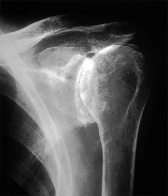
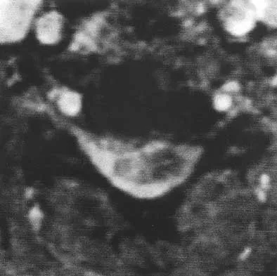
A 56-year-old woman sustained the fracture shown in Figures 30a and 30b in a motor vehicle accident. What mechanism is most likely responsible for the injury?


Explanation

30b The CT scans show a burst fracture that results from an axial load injury. The radiographic hallmark of a burst fracture is compression of the posterior cortex of the vertebral body with retropulsion of bone into the spinal canal. AP radiographs often show widening of the interpedicular distance with a fracture of the lamina. Theiss SM: Thoracolumbar and lumbar spine trauma, in Stannard JP, Schmidt AH, Kregor PJ (eds): Surgical Treatment of Orthopaedic Trauma. New York, NY, Thieme, 2007, pp 179-207.

Question 11

In providing culturally competent care to a Muslim woman with a cervical spine injury, which of the following most accurately describes the steps a male orthopaedist should take to respect her religious beliefs during his examination?


Explanation

In examining a traditional Muslim woman, a male physician should have another woman present, and the patient's husband, if possible. Only the affected limb or area needing examination should be exposed.

Question 12

Figure 31 shows the radiograph of a 64-year-old woman who is seen in the emergency department following a motor vehicle accident. She has no voluntary motor function in her distal upper extremities or lower extremities. She does not have a bulbocavernosus reflex. She has a blood pressure of 80/50 mm Hg with a pulse of 50/min. Her hypotension does not improve with initial fluid resuscitation. Further treatment of her hypotension should consist of


Explanation

The hallmark of neurogenic shock is hypotension without tachycardia. It is associated most commonly with high cervical spinal cord injuries and results from loss of function of the sympathetic nervous system. Because the peripheral vasculature is dilated due to loss of its sympathetic tone, continued rapid administration of fluid corrects the hypotension and can quickly lead to fluid overload and congestive heart failure. Therefore, neurogenic shock is best treated by the use of pressors. Cardioversion or administration of antibiotics or systemic steroids is not appropriate treatment for this patient's hypotension. Spivak JM, Connolly PJ (eds): Orthopaedic Knowledge Update: Spine 3. Rosemont, IL, American Academy of Orthopaedic Surgeons, 2006, pp 179-187.

Question 13

What is the typical axial plane transverse angulation of the thoracic pedicles?


Explanation

Thoracic pedicles typically are angled 25 degrees medially at T1 so the starting point is more lateral. T2 angles about 15 degrees, and then the pedicles average about 5 to 7 degrees down to T10. At T11 and 12, the angulation is minimal. Weinstein L: Pediatric Spine Principles and Practice. New York, NY, Raven Press, 1994, pp 1659-1681.

Question 14

What muscle is most often encountered during surgical approaches to C5-6?


Explanation

The omohyoid muscle crosses the surgical field from inferior lateral to anterior superior traveling from the scapula to the hyoid bone and may need to be transected. The posterior digastric crosses the field as well but higher near C3-4. The other muscles run longitudinally. Chang U, Lee MC, Kim DH: Anterior approach to the midcervical spine, in Kim DH, Henn JS, Vaccaro AR, et al (eds): Surgical Anatomy and Techniques to the Spine. Philadelphia, PA, Saunders Elsevier, 2006, pp 45-56.

Question 15

Which of the following lumbar disk components has the highest tensile modulus to resist torsional, axial, and tensile loads?


Explanation

The annulus fibrosis has a multilayer lamellar architecture mode of type I collagen fibers. Each successive layer is oriented at 30 degrees to the horizontal in the opposite direction, leading to a "criss-cross" type pattern. This composition allows the annulus, which has the highest tensile modulus, to resist torsional, axial, and tensile loads.

Question 16

When comparing the overall outcomes of surgical versus nonsurgical treatment of stable thoracolumbar burst fractures in patients without neurologic injury, 5 years following injury, the principle differences lie in


Explanation

When patients are compared at 5 years follow-up, there are no statistically significant differences between the two groups with respect to kyphosis, the degree of retropulsed bone resorption, pain and function levels, or the ability to return to work. Nonsurgical management of stable neurologically intact burst fractures has a very low incidence of complications. Wood K, Butterman G, Mehbod A, et al: Operative compared with nonoperative treatment of a thoracolumbar burst fracture without neurological deficit: A prospective, randomized study. J Bone Joint Surg Am 2003;85:773-781.

Question 17

A 42-year-old woman who has had an 18-month history of severe low back pain is referred to your office for surgical evaluation. She reports that the pain initially began with right lower extremity pain and management consisted of oral analgesics, nonsteroidal anti-inflammatory drugs, and muscle relaxants. She has seen a chiropractor as well as a pain management specialist and she is status-post epidural steroid injections. She has also completed exhaustive physical therapy, as she is a certified athletic trainer and runs a health fitness program at a community hospital. Currently, she denies lower extremity pain and her pain is isolated to her low back and is subjectively graded as 8/10, with 10 being the worst pain she has ever experienced. The pain is interfering with her activities of daily living and she is seeking definitive treatment. Figures 32a through 32c show current MRI scans. Based on the current available medical literature, what is the most appropriate treatment?


Explanation

32b 32c The MRI scans reveal advanced degenerative disk disease at L5-S1. Nonsurgical management has failed to provide relief and the patient is quite debilitated as a result of her back pain. Fritzell and associates demonstrated that in a well-informed and selected group of patients with severe low back pain, lumbar fusion can diminish pain and decrease disability more efficiently than commonly used nonsurgical treatments. In a recent updated Cochrane Review of surgery for degenerative lumbar spondylosis, it was noted that while Fritzell and associates appeared to provide strong evidence in favor of fusion, a more recent trial by Brox and associates demonstrated no difference between those patients undergoing lumbar fusion compared to those receiving cognitive intervention and exercise. The Cochrane Review suggests that this may reflect a difference between the control groups. Fritzell and associates compared lumbar fusion to standard 1990s "usual care," whereas Brox and associates compared lumbar fusion to a "modern rehabilitation program." Bear in mind that this patient is a certified athletic trainer and runs a hospital health fitness department; therefore, at least for purposes of this question, it can be assumed that she has participated in a "modern rehabilitation program." The Cochrane Review goes on to state that preliminary results of three small trials of intradiskal electrotherapy suggest that it is ineffective and that preliminary data from three trials of disk arthroplasty do not permit firm conclusions. Gibson JN, Waddell G: Surgery for degenerative lumbar spondylosis: Updated Cochrane Review. Spine 2005;30:2312-2320. Fritzell P, Hagg O, Wessberg P, et al: 2001 Volvo Award Winner in Clinical Studies: Lumbar fusion versus nonsurgical treatment for chronic low back pain: A multicenter randomized controlled trial from the Swedish Lumbar Spine Study Group. Spine 2001;26:2521-2532.

Question 18

Figure 33 shows the MRI scan of a 55-year-old woman who has had a 6-week history of back and leg pain. Which of the following clinical scenarios is most consistent with the MRI scan findings at L4-L5?


Explanation

The MRI scan reveals a L4-L5 foraminal disk herniation originating from the L4-5 disk space that has migrated up into the foramen, compressing the left L4 nerve root. There is normal distribution of the roots in the cerebrospinal fluid, excluding arachnoiditis as a diagnosis, and disk herniation in this location would not result in cauda equina syndrome or myelopathy.

Question 19

Intradiskal electrothermal therapy (IDET) uses an intradiskal catheter to deliver controlled thermal energy to the inner periphery of the annulus fibrosis of a chronically painful intervertebral disk. Lumbar diskography is used diagnostically to identify the presumed pain generator to be targeted with IDET. Based on the medical literature, what can be said about the current status of IDET?


Explanation

Intradiskal electrothermal therapy (IDET) initial clinical results were reported in 2000. The early case series were quite encouraging with reported therapeutic success rates of 60% to 80%. Early enthusiasm was high as IDET provided a nonsurgical treatment option for an otherwise complex and difficult clinical entity, chronic diskogenic low back pain. The actual mechanism of action was not well understood, and while the theoretic explanation made good sense, it did not hold up under laboratory testing. Soon clinical results from the field did not meet the high expectations set by the developers of the technique. Since those early case studies, a few level I evidence studies have been conducted, one by Freeman and associates and one by Pauza and associates. These randomized, placebo-controlled trials demonstrated no significant benefit of IDET over the placebo. Freeman BJ, Fraser RD, Cain CM, et al: A randomized, double-blind, controlled trial: Intradiscal electrothermal therapy versus placebo for the treatment of chronic discogenic low back pain. Spine 2005;30:2369-2377. Pauza KJ, Howell S, Dreyfuss P, et al: A randomized, placebo-controlled trial of intradiscal electrothermal therapy for the treatment of discogenic low back pain. Spine J 2004;4:27-35.

Question 20

A 56-year-old mechanic has had pain in the hypothenar region of his dominant right hand for the past 6 months. He reports weakness in his grip and pain is worse with activity. Which of the following examination findings is most suggestive of a cervical etiology?


Explanation

Hypothenar atrophy is a nonspecific sign that can be seen in ulnar neuropathy, C8 radiculopathy, or even cervical myelopathy; however, the atrophy usually is not unilateral and includes other muscle groups. The Spurling test is an excellent method of eliciting cervical radicular pain but involves hyperextension and ipsilateral rotation of the cervical spine, resulting in nerve root compression by reducing the cross-sectional area of the ipsilateral neuroforamen. Tinel's sign at the levator scapulae, if present, is indicative of an upper cervical (C3 or C4) radiculopathy. A subluxable ulnar nerve at the cubital tunnel, while often asymptomatic, points toward cubital tunnel syndrome as an etiology for this patient's pain. The shoulder abduction relief (SAR) sign (relief of upper extremity pain with shoulder abduction) is virtually pathognomic of cervical radiculopathy because this maneuver results in relaxation of a compressed and/or inflamed cervical nerve root. The SAR sign is the converse analog of the straight leg raising sign in the lumbar examination for lumbar radiculopathy, as it relieves tension in the nerve root, thereby relieving symptoms. Ducker TB, Zeidman SM: Neurologic and functional evaluation, in Clark CR (ed): The Cervical Spine, ed 3. Philadelphia, PA, Lippincott Raven, 1998, pp 143-161. An HS: Clinical presentation of discogenic neck pain, radiculopathy, and myelopathy, in Clark CR (ed): The Cervical Spine, ed 3. Philadelphia, PA, Lippincott Raven, 1998, pp 755-764.

Question 21

A 35-year-old woman reports an 8-week history of neck pain radiating to her right upper extremity. She denies any history of trauma or provocative event. Examination reveals decreased pinprick sensation in her right middle finger, otherwise sensation is intact bilaterally. Finger flexors and interossei demonstrate 5/5 motor strength bilaterally. Finger extensors are 4/5 on the right and 5/5 on the left. The triceps reflex is 1+ on the right and 2+ on the left. The most likely diagnosis is a herniated nucleus pulposus at what level?


Explanation

The patient's neurologic examination is consistent with a C7 radiculopathy on the right side. In a patient with this symptom complex in the absence of trauma, a cervical disk herniation is the most common etiology for a C7 radiculopathy. There are eight cervical nerve roots and the C7 nerve exits at the C6-7 disk space and is most frequently impinged by a disk herniation at this level. Houten JK, Errico TJ: Cervical spondylotic myelopathy and radiculopathy: Natural history and clinical presentation, in Clark CR (ed): The Cervical Spine, ed 4. Philadelphia, PA, Lippincott Williams & Wilkins, 2005, pp 985-990.

Question 22

What is the most common nonanesthetic-related reversible cause of changes in intraoperative neurophysiologic monitoring data?


Explanation

Patient positioning that results in local nerve compression, plexus traction, or improper neck alignment is the most common nonanesthetic-related cause of changes in intraoperative neurophysiologic monitoring data during spinal surgery. Jones SC, Fernau R, Woeltjen BL: Use of somatosensory evoked potentials to detect peripheral ischemia and potential injury resulting from positioning of the surgical patient: Case reports and discussion. Spine J 2004;4:360-362.

Question 23

During a left-sided transforaminal lumbar interbody fusion at the L4-5 level, the surgeon notes a significant amount of bleeding that cannot be controlled while using a pituitary rongeur. What anatomic structure has been injured?


Explanation

The surgeon perforated the anterior longitudinal ligament and injured the common iliac artery. Bingol and associates described injuries to the vascular structures during lumbar disk surgery. The common iliac artery was most commonly affected and constituted 76.9% of injuries.

Question 24

Six weeks after onset, what is the most clearly accepted indication for surgical management for lumbar disk herniation?


Explanation

In the absence of a cauda equina syndrome or progressive weakness, the best indication for surgical management is refractory radicular pain. Surgical decision-making should not be based on the size of the herniation. Large extruded herniations tend to resolve more predictably than smaller herniations. Stable motor weakness and numbness resolve similarly in both surgical and nonsurgical management, although surgery hastens the process. When intractable radicular pain is the strict indication for surgery, surgical intervention provides substantial and more rapid pain relief than nonsurgical care. Rhee JM, Schaufele M, Abdu WA: Radiculopathy and the herniated lumbar disc: Controversies regarding pathophysiology and management. J Bone Joint Surg Am 2006;88:2070-2080.

Question 25

A 45-year-old woman has idiopathic scoliosis. Surgery is to include an anterior thoracic release through an open left thoracotomy. The thoracotomy will have what effect on the patient's pulmonary function postoperatively?


Explanation

A thoracotomy in an adult with idiopathic scoliosis causes a reduction in pulmonary function that often does not return to preoperative levels. What pulmonary function that does recover, recovers over many months. Long-term improvement in pulmonary function, compared to preoperative function, is rarely seen. This should be considered in planning surgical intervention in adults with scoliosis. Graham EJ, Lenke LG, Lowe TG, et al: Prospective pulmonary function evaluation following open thoracotomy for anterior spinal fusion in adolescent idiopathic scoliosis. Spine 2000;25:2319-2325.

Question 26

An 82-year-old man presents with a Type II odontoid fracture after a ground-level fall. He has severe COPD and coronary artery disease. He is neurologically intact. What is the most appropriate initial management considering his age and fracture type?


Explanation

In elderly patients with Type II odontoid fractures, rigid cervical collar immobilization is often preferred initially. This avoids the high morbidity and mortality associated with halo vests and surgical intervention in a frail population with multiple medical comorbidities.

Question 27

When evaluating a 65-year-old female for adult spinal deformity correction, which of the following radiographic parameters correlates most strongly with an improvement in health-related quality of life (HRQOL) scores postoperatively?


Explanation

Achieving a PI-LL mismatch of less than 10 degrees is a primary goal in adult spinal deformity surgery. Along with an SVA < 5 cm and Pelvic Tilt < 20 degrees, this strongly correlates with improved postoperative HRQOL.

Question 28

According to the Thoracolumbar Injury Classification and Severity (TLICS) score, which of the following injury patterns warrants a score of 3 for the morphology category?


Explanation

In the TLICS system, morphology is scored as follows: compression (1), burst (2), translation/rotation (3), and distraction (4). These points are added to neurologic status and posterior ligamentous complex integrity to guide operative vs nonoperative management.

Question 29

A 60-year-old man presents with deteriorating hand dexterity and a broad-based gait. On examination, rapid tapping of the distal phalanx of the middle finger elicits involuntary flexion of the thumb and index finger. This clinical sign indicates dysfunction in which of the following tracts?


Explanation

The described exam finding is a positive Hoffmann sign, which indicates an upper motor neuron lesion such as cervical spondylotic myelopathy. Upper motor neuron signs result from compression or dysfunction of the corticospinal tract.

Question 30

A 45-year-old man presents with acute, severe right-sided anterior thigh pain and weakness in knee extension. MRI demonstrates a far lateral (extraforaminal) disc herniation at the L3-L4 level. Which nerve root is most likely compressed?


Explanation

A far lateral (extraforaminal) disc herniation compresses the exiting nerve root at that specific level. Therefore, an L3-L4 far lateral disc compresses the L3 nerve root, causing anterior thigh pain and quadriceps weakness.

Question 31

A 68-year-old man with known cervical stenosis sustains a hyperextension injury. He presents with profound bilateral upper extremity weakness but retains antigravity strength in his lower extremities. What is the most likely prognosis for his neurologic recovery?


Explanation

This patient has central cord syndrome. Most patients (>50%) regain the ability to ambulate, typically recovering lower extremity strength first, followed by bowel/bladder function, proximal upper extremity strength, and lastly fine hand dexterity.

Question 32

A newborn is diagnosed with congenital scoliosis secondary to a fully segmented hemivertebra at T8. What screening test is most critical to perform during the initial evaluation of this patient?


Explanation

Congenital scoliosis is highly associated with VACTERL anomalies, including renal and genitourinary abnormalities in up to 30% of patients. A renal ultrasound and an echocardiogram are mandatory standard screening tests.

Question 33

A 24-year-old male involved in a high-speed motor vehicle collision wearing a lap belt presents with a flexion-distraction injury of the L2 vertebra. Which of the following associated injuries is most highly correlated with this spinal fracture pattern?


Explanation

Flexion-distraction (Chance) fractures are highly associated with intra-abdominal hollow viscus injuries, particularly of the small bowel. This is due to the severe anterior shearing forces caused by lap belts.

Question 34

A patient sustains a displaced vertical fracture through the sacrum that extends medial to the sacral foramina, involving the central spinal canal. According to the Denis classification, what zone is this injury, and what is the associated risk?


Explanation

Denis Zone 3 sacral fractures involve the central sacral canal (medial to the foramina). They carry the highest risk (>50%) of neurologic deficit, specifically affecting bowel, bladder, and sexual function due to sacral root involvement.

Question 35

A 35-year-old male presents with bilateral jumped facets at C5-C6 after a diving accident. He is awake, alert, and cooperative but has 0/5 strength in his bilateral lower extremities. What is the most appropriate immediate step in management?


Explanation

In an awake, alert, and cooperative patient with a cervical facet dislocation and a neurologic deficit, emergent closed reduction with cranial traction is indicated. Waiting for an MRI delays critical decompression of the spinal cord.

Question 36

Which of the following radiographic criteria is strictly required to confirm the diagnosis of classic Scheuermann's disease?


Explanation

Sorensen's criteria for diagnosing Scheuermann's kyphosis require the presence of anterior wedging of 5 degrees or more in at least three consecutive apical vertebrae.

Question 37

A 70-year-old man with a long history of Ankylosing Spondylitis suffers a minor fall and complains of new neck pain. Initial plain radiographs are negative for fracture. What is the most appropriate next step in management?


Explanation

Patients with Ankylosing Spondylitis are at an extremely high risk for highly unstable, occult spinal fractures even after minor trauma. If plain films are negative, advanced imaging such as a CT scan or MRI is mandatory to rule out a fracture.

Question 38

Traumatic spondylolisthesis of the axis (Hangman's fracture) typically occurs through which of the following anatomic structures of C2?


Explanation

A Hangman's fracture is a bilateral fracture through the pars interarticularis of C2 (the axis). It is classically caused by a mechanism of hyperextension and axial loading.

Question 39

A 66-year-old female undergoes surgical planning for adult spinal deformity. Her pelvic incidence (PI) is measured at 62 degrees. To achieve optimal sagittal balance and minimize the risk of adjacent segment disease, what should be her approximate target for postoperative lumbar lordosis (LL)?


Explanation

The formula for sagittal balance dictates that lumbar lordosis (LL) should be matched to within 9-10 degrees of the pelvic incidence (PI). Therefore, a PI of 62 degrees requires an LL target of approximately 52 to 62 degrees.

Question 40

Pelvic incidence (PI) is a fundamental morphologic parameter in adult spinal deformity evaluation. What is the correct mathematical relationship between PI, pelvic tilt (PT), and sacral slope (SS)?


Explanation

Pelvic incidence is a fixed anatomical parameter defined as the algebraic sum of pelvic tilt and sacral slope (PI = PT + SS). It dictates the required lumbar lordosis for a patient, typically targeted at PI ± 9 degrees.

Question 41

A 65-year-old man with a known history of ankylosing spondylitis presents with severe neck pain following a ground-level fall. Neurological examination is intact. CT scan reveals a through-and-through fracture of the C6-C7 disc space extending into the posterior elements. What is the most appropriate management?


Explanation

Patients with ankylosing spondylitis are at high risk for unstable, highly displaced extension-distraction fractures even from minor trauma. Long-segment posterior instrumented fusion is required due to the long lever arms and poor bone quality.

Question 42

Which of the following is the most frequently encountered neurological complication following posterior cervical laminectomy and instrumented fusion for cervical spondylotic myelopathy?


Explanation

C5 nerve root palsy is the most common neurological complication following posterior cervical decompression (incidence ~5-10%), often presenting as deltoid and biceps weakness. It is thought to result from posterior spinal cord drift and nerve root tethering.

Question 43

According to the Thoracolumbar Injury Classification and Severity (TLICS) score, which of the following injury patterns strictly warrants surgical stabilization (score > 4)?


Explanation

A burst fracture (2 points) with posterior ligamentous complex disruption (3 points) yields a TLICS score of 5, which dictates operative management. A score of 4 can be treated operatively or non-operatively, while < 4 is managed non-operatively.

Question 44

A 68-year-old woman undergoes T10-pelvis fusion for adult spinal deformity. Six months postoperatively, she develops proximal junctional kyphosis (PJK). Which of the following is considered a significant risk factor for the development of PJK?


Explanation

Stopping a long fusion construct at the apex of thoracic kyphosis is a major risk factor for PJK. Construct endpoints should ideally fall in neutral or lordotic segments to avoid high biomechanical stress at the upper instrumented vertebra.

Question 45

A 62-year-old female presents with neurogenic claudication and an L4-L5 degenerative spondylolisthesis. She elects to undergo surgical intervention after failing conservative management. Based on the Spine Patient Outcomes Research Trial (SPORT), what is the expected long-term outcome comparing operative to non-operative treatment?


Explanation

The SPORT trial demonstrated that patients with symptomatic degenerative spondylolisthesis treated operatively had significantly greater improvement in pain and function compared to non-operative cohorts, a benefit maintained at long-term follow-up.

Question 46

A 24-year-old man arrives intubated and sedated after a high-speed motor vehicle collision. Radiographs demonstrate a bilateral facet dislocation at C5-C6. What is the most appropriate next step in management?


Explanation

In an unexaminable (intubated/sedated) patient with a cervical facet dislocation, an MRI must be obtained prior to any reduction maneuvers to rule out a herniated disc. If a herniation is present, an anterior decompression must precede reduction.

Question 47

When evaluating a patient with adolescent idiopathic scoliosis, which of the following defines the "stable vertebra"?


Explanation

The stable vertebra is defined as the most proximal (lowest) vertebra whose body is completely bisected by the central sacral vertical line (CSVL). It is a key anatomical landmark used to determine the lowest instrumented vertebra (LIV).

Question 48

A 45-year-old man presents with severe radiating right arm pain, weakness in wrist extension, and a diminished brachioradialis reflex. Sensation is decreased over the dorsal web space of the right hand. Which cervical nerve root is most likely affected?


Explanation

A C6 radiculopathy typically presents with weakness in wrist extension and elbow flexion, a diminished brachioradialis reflex, and sensory deficits in the thumb and index finger (radial aspect of the forearm/hand).

Question 49

An 85-year-old man sustains a Type II odontoid fracture with 3 mm of posterior displacement. He has multiple medical comorbidities, including severe COPD and coronary artery disease. What is the most appropriate initial management?


Explanation

In an elderly patient with significant comorbidities, a rigid cervical orthosis is the preferred initial management for a Type II odontoid fracture due to the unacceptably high morbidity and mortality associated with surgery or halo vest application.

Question 50

Which of the following radiographic criteria is strictly required to formally diagnose classic Scheuermann's kyphosis?


Explanation

Sorensen's criteria for the diagnosis of Scheuermann's kyphosis require anterior wedging of greater than 5 degrees in at least three consecutive vertebrae, typically accompanied by endplate irregularities and Schmorl's nodes.

Question 51

A 55-year-old man presents with progressive clumsiness in his hands and a broad-based gait. Imaging reveals continuous ossification of the posterior longitudinal ligament (OPLL) from C3 to C6, occupying 65% of the canal. His cervical alignment is lordotic. What is the preferred surgical approach?


Explanation

In patients with OPLL and preserved cervical lordosis, a posterior approach (laminoplasty or laminectomy and fusion) is preferred, especially when the ossified mass occupies >50-60% of the canal. Anterior approaches carry a severely elevated risk of dural tears.

Question 52

An 78-year-old woman with known severe cervical spondylosis presents after a hyperextension injury to her neck. She exhibits bilateral motor weakness in her upper extremities (grade 2/5) but retains 4/5 strength in her lower extremities. What is the most likely diagnosis?


Explanation

Central cord syndrome classically occurs after a hyperextension injury in a patient with pre-existing cervical stenosis. It presents with disproportionately greater motor impairment in the upper extremities compared to the lower extremities.

Question 53

A 65-year-old woman presents with severe back pain and a progressive inability to stand up straight following a previous L4-S1 fusion. Radiographs reveal a pelvic incidence (PI) of 65 degrees and a lumbar lordosis (LL) of 20 degrees. If surgical correction is planned, what is the generally accepted target lumbar lordosis to optimize her sagittal alignment?


Explanation

For optimal sagittal balance, the lumbar lordosis (LL) should be matched to within 10 degrees of the pelvic incidence (PI). With a PI of 65 degrees, the target LL should be approximately 55 degrees to prevent flatback syndrome and adjacent segment disease.

Question 54

A 78-year-old man with severe COPD sustains a Type II odontoid fracture with 3 mm of posterior displacement. He is neurologically intact. Compared to surgical intervention, nonoperative management with a rigid cervical collar in this patient demographic is associated with a higher rate of which of the following?


Explanation

In elderly patients, nonoperative management of Type II odontoid fractures using a rigid cervical collar is associated with high rates of pseudarthrosis (up to 40-50%). However, it is often favored due to the high perioperative morbidity and mortality associated with surgical fixation in patients with severe comorbidities.

Question 55

A 55-year-old man presents with progressive cervical myelopathy. Imaging reveals ossification of the posterior longitudinal ligament (OPLL) from C3 to C6, occupying 60% of the spinal canal, with an overall cervical kyphosis of 15 degrees.

What is the most appropriate surgical management?


Explanation

Anterior cervical corpectomy and fusion (ACCF) is preferred for OPLL when the canal compromise is >50% and the spine is kyphotic (K-line negative). Posterior-only indirect decompression (laminoplasty) is inadequate here because the kyphotic alignment prevents the spinal cord from drifting backward away from the anterior compression.

Question 56

According to the Lenke classification for adolescent idiopathic scoliosis, which of the following radiographic criteria defines a minor curve as "structural" and necessitates its inclusion in the fusion construct?


Explanation

In the Lenke classification, a minor curve is considered structural if it fails to bend out to less than 25 degrees on side-bending radiographs, or if there is a regional kyphosis of ≥ 20 degrees. Structural curves must be included in the operative fusion.

Question 57

A 40-year-old man falls from a height of 10 feet and sustains an L1 burst fracture. He is neurologically intact. MRI confirms an intact posterior ligamentous complex (PLC).

According to the Thoracolumbar Injury Classification and Severity (TLICS) score, what is the score and recommended management?


Explanation

The TLICS score assigns 2 points for a burst fracture morphology, 0 points for being neurologically intact, and 0 points for an intact PLC. A total score of 2 indicates nonoperative management with a brace.

Question 58

Which of the following anatomical variations is most strongly associated with the development of a degenerative spondylolisthesis at L4-L5 rather than an isthmic spondylolisthesis?


Explanation

Degenerative spondylolisthesis most commonly occurs at L4-L5 and is strongly associated with sagittally oriented facet joints, which provide less resistance to anterior shear forces. Isthmic spondylolisthesis involves a pars interarticularis defect and is most common at L5-S1.

Question 59

A 22-year-old woman is involved in a high-speed motor vehicle collision while wearing a lap belt. Radiographs show a flexion-distraction injury (Chance fracture) at L2. Which of the following concomitant injuries must be ruled out most urgently?


Explanation

Chance fractures (flexion-distraction injuries) are frequently caused by lap seatbelts and have a high association (up to 40-50%) with intra-abdominal injuries, particularly hollow viscus (bowel) rupture. Urgent general surgery evaluation is required.

Question 60

A 45-year-old man with ankylosing spondylitis presents with a severe chin-on-chest deformity, impairing his horizontal gaze and causing difficulty swallowing. Where is the most biomechanically appropriate and anatomically safe level to perform a posterior extension osteotomy to correct his cervical deformity?


Explanation

The cervicothoracic junction (C7-T1) is the standard and safest location for an extension osteotomy in ankylosing spondylitis. It provides a large spinal canal to accommodate cord shifting, good bone for fixation, and typically avoids the vertebral artery which usually enters the foramen transversarium at C6.

Question 61

Following a multi-level lumbar spinal fusion, adjacent segment disease (ASD) is a recognized complication. Which of the following surgical factors has been shown to most significantly increase the risk of developing symptomatic ASD at the proximal adjacent level?


Explanation

Postoperative sagittal malalignment, specifically hypolordosis (flatback), significantly alters the biomechanics of the unfused adjacent segments. This abnormal stress is a primary driver for the accelerated development of adjacent segment disease.

Question 62

A 3-year-old child is diagnosed with congenital scoliosis secondary to a fully segmented hemivertebra at T8. Which of the following screening strategies is most critical to perform routinely in this patient due to the high rate of associated systemic anomalies?


Explanation

Congenital scoliosis is highly associated with the VACTERL spectrum. Specifically, genitourinary anomalies occur in 20-30% of patients (requiring a renal ultrasound) and intraspinal anomalies like tethered cord occur in 20-40% (requiring total spine MRI).

Question 63

A 68-year-old man with underlying cervical spondylosis sustains a hyperextension injury. He presents with severe motor weakness in his hands and arms (deltoids 3/5, hand grip 1/5), with relatively preserved strength in his legs (hip flexion 4/5). Bladder function is intact. What is the most likely pathophysiological mechanism for this deficit?


Explanation

This is a classic presentation of Central Cord Syndrome, characterized by upper extremity weakness greater than lower extremity weakness. It results from hyperextension in a stenotic canal, causing a pinch on the cord leading to edema, ischemia, and hemorrhage primarily affecting the centrally located cervical motor tracts.

Question 64

During a routine L4-L5 microdiscectomy, a 3 mm incidental durotomy occurs with minor cerebrospinal fluid egress. The tear is repaired primarily with a 4-0 nonabsorbable suture, and a Valsalva maneuver confirms a watertight seal. What is the most appropriate postoperative management regarding mobilization?


Explanation

Current evidence demonstrates that after achieving a watertight primary repair of an incidental durotomy, early mobilization is safe and does not increase the risk of CSF leak or pseudomeningocele. It also reduces the morbidity associated with prolonged bed rest.

Question 65

A 35-year-old man falls from a 20-foot height. Pelvic CT demonstrates a highly displaced transverse fracture through the S2 vertebral body connecting bilateral longitudinal sacral fractures. He has perianal numbness and absent sphincter tone. What is this fracture pattern classically termed, and what is the required surgical intervention?


Explanation

A U-shaped sacral fracture represents a spinopelvic dissociation, meaning the axial spine is disconnected from the pelvic ring. Due to the high mechanical instability and neurologic injury, it requires robust lumbopelvic/spinopelvic fixation.

Question 66

A 15-year-old boy presents with progressive mid-back pain and a noticeable rounding of his upper back. Standing lateral radiographs reveal a thoracic kyphosis of 65 degrees. According to the Sorensen criteria, what specific radiographic finding confirms the diagnosis of Scheuermann's kyphosis?


Explanation

The classic Sorensen criteria define Scheuermann's disease radiographically as anterior wedging of 5 degrees or more in at least three consecutive vertebrae. Rigid kyphosis, Schmorl's nodes, and endplate irregularities are also supportive findings.

Question 67

A 42-year-old woman presents to the emergency department with acute onset of severe bilateral radicular leg pain, saddle anesthesia, and urinary retention with overflow incontinence that began 12 hours ago. MRI confirms a massive extruded herniated disc at L4-L5. To maximize the likelihood of recovering normal bladder function, surgical decompression must ideally occur within what maximum timeframe from symptom onset?


Explanation

Cauda equina syndrome is a surgical emergency. Decompression should ideally be performed within 24 to 48 hours from the onset of symptoms (especially autonomic dysfunction like urinary retention) to maximize the probability of significant neurologic and urologic recovery.

Question 68

A patient undergoing halo skeletal fixation complains of new-onset diplopia and lateral gaze palsy on the second day after application. Which cranial nerve is most likely affected by the traction?


Explanation

The abducens nerve (CN VI) is the most commonly injured cranial nerve during halo traction. It has a long intracranial course, making it uniquely susceptible to stretch, resulting in a lateral gaze palsy and diplopia.

Question 69

A 65-year-old man with a long-standing history of advanced ankylosing spondylitis presents after a low-energy ground-level fall. CT of the cervical spine reveals a hyperextension injury passing entirely through the C6-C7 disc space. What is the most appropriate management?


Explanation

Fractures in patients with ankylosing spondylitis are highly unstable due to the long, rigid lever arms of the fused spine. A combined anterior and posterior fusion is generally recommended to provide maximum biomechanical stability and prevent construct failure.

Question 70

A 65-year-old female presents with severe neurogenic claudication and an L4-L5 degenerative spondylolisthesis as demonstrated on her MRI.

She has failed conservative management. Which of the following factors most strongly supports performing a decompression with fusion rather than an isolated decompression?


Explanation

Decompression with fusion is indicated over decompression alone in degenerative spondylolisthesis when there is clear evidence of dynamic instability (e.g., >3 mm of translation on flexion/extension films) to prevent postoperative progression of the slip.

Question 71

A 19-year-old male presents with slowly progressive, asymmetric weakness and atrophy of his right hand and forearm. He reports no sensory deficits or lower extremity symptoms. An MRI of the cervical spine in neutral is unremarkable, but an MRI taken with the neck in flexion demonstrates forward displacement of the posterior dura with spinal cord compression against the vertebral bodies. What is the most likely diagnosis?


Explanation

Hirayama disease is a juvenile muscular atrophy of the distal upper extremities caused by dynamic cervical myelopathy. It is characterized by forward displacement of the posterior cervical dura during neck flexion, causing repetitive microtrauma to the anterior horn cells.

Question 72

In the evaluation of adult spinal deformity, which of the following sagittal radiographic parameters correlates most closely with poor health-related quality of life (HRQOL) scores?


Explanation

Positive sagittal imbalance is the most critical driver of disability in adult spinal deformity. A Sagittal Vertical Axis (SVA) greater than 5 cm is the most reliable radiographic predictor of clinical symptoms and poor HRQOL outcomes.

Question 73

A 24-year-old male is involved in a motor vehicle collision. Imaging reveals a fracture through the pars interarticularis of C2 with 4 mm of anterior translation and 12 degrees of angulation. What is the Levine-Edwards classification of this traumatic spondylolisthesis of the axis?


Explanation

Levine-Edwards Type II fractures (Hangman's fractures) involve >3 mm of anterior translation and significant angulation. They are typically caused by a hyperextension injury followed by a severe flexion/compression rebound.

Question 74

A 15-year-old boy presents with progressive mid-back pain and a notably rounded posture. Standing radiographs reveal a rigid thoracic kyphosis of 65 degrees. According to the classic Sorensen criteria, what specific radiographic finding is required to definitively diagnose Scheuermann's kyphosis?


Explanation

The classic Sorensen criteria for diagnosing Scheuermann's disease require the presence of anterior wedging of >5 degrees in at least three consecutive vertebrae, typically accompanied by a rigid kyphosis >45 degrees and irregular vertebral endplates.

Question 75

A 42-year-old male presents to the emergency department with acute onset saddle anesthesia, bilateral leg weakness, and urinary retention. MRI reveals a massive L4-L5 central disc herniation. To maximize the probability of full return of bladder and bowel function, surgical decompression should ideally be performed within what maximum timeframe from the onset of symptoms?


Explanation

While cauda equina syndrome is a surgical emergency, robust literature demonstrates that surgical decompression performed within 48 hours of symptom onset significantly improves the odds of recovering normal urologic and neurologic function.

Question 76

A 72-year-old man with known cervical spondylosis falls forward and strikes his chin. He presents with severe motor weakness in his bilateral hands and arms (1/5 strength), but maintains functional strength in his legs (4/5 strength). Sensation in the perianal area is intact. What is the most likely diagnosis?


Explanation

Central cord syndrome classically occurs following a hyperextension injury in patients with a pre-existing stenotic cervical canal. It presents with disproportionately greater motor weakness in the upper extremities compared to the lower extremities.

Question 77

A 68-year-old patient presents with bilateral calf and buttock pain when walking. Which of the following historical findings is most indicative of neurogenic claudication (due to lumbar spinal stenosis) rather than vascular claudication?


Explanation

Neurogenic claudication is characteristically relieved by lumbar flexion (e.g., sitting or leaning forward over a shopping cart), which increases the cross-sectional area of the spinal canal. Vascular claudication is typically relieved simply by resting or standing still.

Question 78

A 78-year-old woman presents after a ground-level fall. CT imaging demonstrates a displaced Type II odontoid fracture with 6 mm of posterior displacement. She has severe medical comorbidities precluding surgery. If treated nonoperatively with a hard cervical collar, what is the most likely complication?


Explanation

Type II odontoid fractures have a high rate of nonunion when managed conservatively, especially in elderly patients with displacement >5 mm or posterior angulation. This is due to the watershed vascular supply at the base of the dens.

Question 79

A 45-year-old male presents with severe right leg pain, weakness in ankle dorsiflexion, and decreased sensation over the medial aspect of the foot. His right patellar reflex is absent. Which nerve root is most likely compressed, and by what classic anatomical disc herniation?


Explanation

The L4 nerve root mediates the patellar reflex, sensation to the medial calf/foot, and assists in ankle dorsiflexion. It is classically compressed by a paracentral disc herniation at the L3-L4 level (affecting the traversing root).

Question 80

In the surgical planning for adolescent idiopathic scoliosis, a Lenke Type 1 curve is defined by which of the following structural characteristics?


Explanation

A Lenke Type 1 curve (Main Thoracic) dictates that the main thoracic curve is structural, while both the proximal thoracic and thoracolumbar/lumbar curves are nonstructural (they bend out to less than 25 degrees on side-bending radiographs).

Question 81

A 50-year-old male presents with progressive clumsiness in his hands and an unsteady gait. Cervical imaging reveals profound ossification of the posterior longitudinal ligament (OPLL) from C3 to C6, causing severe cord compression. Additionally, his cervical spine has 15 degrees of fixed kyphosis. What is the most appropriate surgical approach?


Explanation

In OPLL complicated by cervical kyphosis, posterior decompression (laminectomy or laminoplasty) is contraindicated because the spinal cord will remain draped over the anterior ossified mass (failure of cord 'drift back'). An anterior approach (e.g., corpectomy) is required.

Question 82

A 68-year-old female presents with severe back pain and inability to stand upright. Radiographs reveal a pelvic incidence (PI) of 62 degrees, pelvic tilt (PT) of 35 degrees, and lumbar lordosis (LL) of 20 degrees. When planning a corrective osteotomy for this adult spinal deformity, achieving a PI-LL mismatch of less than which of the following values is most highly correlated with favorable health-related quality of life (HRQOL) scores?


Explanation

The primary goal of sagittal realignment in adult spinal deformity is to restore an optimal spino-pelvic relationship. Achieving a Pelvic Incidence minus Lumbar Lordosis (PI-LL) mismatch of less than 10 degrees is strongly associated with improved HRQOL outcomes.

Question 83

Review the imaging study provided in the figure.

A 65-year-old female presents with neurogenic claudication. She is found to have grade 1 degenerative spondylolisthesis at L4-L5. Which of the following radiographic parameters on preoperative MRI is most indicative of dynamic instability, warranting a fusion rather than a decompression alone?


Explanation

The presence of facet joint effusions (fluid greater than 1.5 mm) on a supine MRI is a strong positive predictor of dynamic microinstability in degenerative spondylolisthesis, which can be confirmed on flexion-extension standing radiographs.

Question 84

A 14-year-old female with adolescent idiopathic scoliosis (AIS) has a main thoracic curve of 55 degrees and a lumbar curve of 40 degrees. On side-bending films, the lumbar curve corrects to 20 degrees. The center sacral vertical line (CSVL) lies completely medial to the medial border of the apical lumbar vertebra. According to the Lenke classification, what is the correct lumbar modifier for this curve pattern?


Explanation

In the Lenke classification, a lumbar modifier C is assigned when the CSVL falls completely medial to the pedicles of the apical lumbar vertebra. Modifier A means the CSVL lies between the pedicles, and B means it touches the medial border.

Question 85

A 72-year-old male with pre-existing cervical spondylosis presents after a hyperextension injury. He has 2/5 motor strength in his bilateral upper extremities and 4/5 strength in his lower extremities, while retaining bowel and bladder function. Which of the following anatomic factors best explains his disproportionate upper extremity weakness?


Explanation

Central cord syndrome often occurs following hyperextension in a spondylotic spine. The disproportionate upper extremity weakness is due to selective injury of the medial fibers of the lateral corticospinal tract, which topographically represent the upper extremities.

Question 86

A 60-year-old male presents with deteriorating handwriting and difficulty buttoning his shirts. Physical examination reveals a positive inverted brachioradialis reflex. This clinical finding most accurately localizes the predominant spinal cord pathology to which of the following levels?


Explanation

An inverted brachioradialis reflex occurs when tapping the brachioradialis tendon elicits finger flexion rather than the normal response. This indicates a lower motor neuron lesion at C5-C6 and an upper motor neuron lesion below, classic for C5-C6 cervical myelopathy.

Question 87

A 24-year-old male is involved in a motor vehicle collision. Lateral cervical spine radiographs show anterior subluxation of C5 on C6 by approximately 25%. A "bow-tie" or "bat-wing" appearance of the articular pillars is noted at the C5 level. Which of the following is the most likely mechanism of this injury?


Explanation

A unilateral facet dislocation typically results from a combined hyperflexion and rotation injury vector. This leads to a 25% anterior translation and the classic "bow-tie" sign on lateral radiographs due to rotation of the lateral masses.

Question 88

A 55-year-old male with long-standing ankylosing spondylitis presents to the emergency department after a low-energy ground-level fall. He reports severe neck pain but has no focal neurologic deficits. Initial plain radiographs of the cervical spine are difficult to interpret due to extensive ossification. Which of the following is the most appropriate next step in his imaging workup?


Explanation

Patients with ankylosing spondylitis are at extremely high risk for occult, highly unstable fractures even after minor trauma. Due to poor visualization on plain radiographs, a high-resolution CT scan of the entire cervical spine is mandatory.

Question 89

An MRI of the lumbar spine is shown in the figure.

A 45-year-old male presents with severe radicular leg pain. Imaging demonstrates a far lateral disc herniation at the L4-L5 level. Which of the following physical examination findings is most likely expected in this patient?


Explanation

Unlike paracentral disc herniations which affect the traversing root, a far lateral (extraforaminal) disc herniation at L4-L5 impinges the exiting L4 nerve root. This leads to L4 radiculopathy, characterized by quadriceps weakness and a diminished patellar reflex.

Question 90

A 68-year-old male undergoes a 10-hour posterior spinal fusion from T2 to the pelvis for adult spinal deformity while positioned prone. Postoperatively, he reports painless, bilateral visual loss with sluggish pupillary reflexes. What is the most likely etiology of his visual loss?


Explanation

Ischemic optic neuropathy (ION) is the most common cause of postoperative vision loss after prone spine surgery. Risk factors include prolonged operative time, significant estimated blood loss, hypotensive anesthesia, and a Wilson frame positioning.

Question 91

A 30-year-old female sustains a pelvic ring injury following a fall. CT imaging reveals a vertical fracture through the sacrum that extends through the central sacral canal. According to the Denis classification, what is the zone of this fracture and its associated hallmark neurologic complication?


Explanation

Denis Zone III sacral fractures involve the central sacral canal. Because they disrupt the sacral neural elements centrally, they have the highest incidence (up to 57%) of neurologic deficits, typically presenting as sphincter, bowel, and sexual dysfunction.

Question 92

A 62-year-old female is 4 years status post L4-S1 posterior spinal instrumented fusion. She now presents with a new-onset L3 radiculopathy. Imaging demonstrates adjacent segment degeneration with severe foraminal stenosis at L3-L4. Which of the following intraoperative factors from her index surgery most significantly increased her risk for developing adjacent segment disease?


Explanation

Iatrogenic disruption of the superior adjacent facet joint capsule (in this case, L3-L4) during pedicle screw insertion at the uppermost instrumented vertebra (L4) is a major biomechanical risk factor that rapidly accelerates adjacent segment disease.

Question 93

A 40-year-old male sustains a T12 burst fracture from a fall. He is neurologically intact. MRI demonstrates definitive disruption of the posterior ligamentous complex (PLC). Using the Thoracolumbar Injury Classification and Severity (TLICS) score, what is his total score and the corresponding treatment recommendation?


Explanation

In the TLICS system, a burst fracture morphology scores 2 points, definitive PLC injury scores 3 points, and intact neurology scores 0 points, yielding a total of 5 points. A score of 5 or greater indicates operative management.

Question 94

A 3-year-old child is being evaluated for congenital scoliosis. Which of the following specific vertebral anomalies carries the highest risk for rapid, unremitting curve progression and typically mandates early prophylactic surgical intervention?


Explanation

The combination of a unilateral unsegmented bar (tethering growth on one side) and a contralateral fully segmented hemivertebra (active growth pushing on the opposite side) causes a massive growth imbalance. This results in the most rapid and severe curve progression in congenital scoliosis.

None

Clinic OS
Medically Verified Content by
Prof. Clinic OS
Consultant Orthopedic & Spine Surgeon
Chapter Index