العربية
Part of the Master Guide

AAOS Orthopedic MCQs (Set 1): Spine Disorders, Sports Medicine & Arthroplasty | 2026 Board Review

AAOS & ABOS Anatomy MCQs (Set 1): Upper Limb, Lower Limb & Spine | 2025 Board Prep

23 Apr 2026 51 min read 141 Views
Anatomy 2005 MCQs - Part 1

Key Takeaway

This high-yield Set 1 question bank for the AAOS/ABOS exams covers critical musculoskeletal anatomy. Questions focus on the intricate structures of the upper limb, lower limb, and spine, including bones, muscles, nerves, and vessels. Perfect for solidifying foundational knowledge for board certification and residency training.

AAOS & ABOS Anatomy MCQs (Set 1): Upper Limb, Lower Limb & Spine | 2025 Board Prep

Comprehensive 100-Question Exam


00:00

Start Quiz

Question 1

A 65-year-old man with ankylosing spondylitis sustains an extension injury to his cervical spine. Two days later, a progressive neurologic deficit develops at the C6 level. An MRI scan is shown in Figure 1. What is the most likely diagnosis?


Explanation

It is common for patients with ankylosing spondylitis to sustain extension-type fractures, typically near the cervicothoracic junction. These fractures can be minimally displaced, making them difficult to diagnose. In addition, the vertebral bodies are vascular and their canals are relatively enclosed, making them vulnerable to epidural bleeding. The MRI scan shows an epidural hematoma posteriorly compressing the cord. Bohlman HH: Acute fractures and dislocations of the cervical spine. J Bone Joint Surg Am 1979;61:1119-1142. Weinstein PR, Karpman RR, Gall EP, et al: Spinal cord injury, spine fracture and spinal stenosis in ankylosing spondylitis. J Neurosurg 1982;57:609-616.

Question 2

What are the most common portals for arthroscopic surgery of the ankle?


Explanation

The most commonly used portals are the anterolateral, anteromedial, and posterolateral portals. They have been shown to be the safest areas for portal placement, allowing no penetration of neurovascular structures. All the other portals involve placing another structure at risk. The anterocentral portal is close to the deep peroneal nerve and anterior tibular artery. The trans-Achilles portal is not recommended because of its limited utility and potential to injure the Achilles tendon. The posteromedial portal is too close to the posterotibial artery and nerve, the flexor hallucis longus and flexor digitorum longus tendons, and the branches of the calcaneal nerve. Stetson WB, Ferkel RD: Ankle arthroscopy: I. Technique and complications. J Am Acad Orthop Surg 1996;4:17-23.


Question 3

A patient who underwent primary total hip arthroplasty 7 years ago that resulted in excellent pain relief and a normal gait now reports pain and a limp. Postoperative and current AP radiographs are shown in Figures 2a and 2b. What is the most likely cause of the pathology seen?


Explanation

Osteolysis in the trochanteric bed can result in weakening of the bone and fracture. Nonsurgical management will provide reasonable clinical and radiographic results in patients with limited fracture displacement. Claus MC, Hopper RH, Engh CA: Fractures of the greater trochanter induced by osteolysis with the anatomic medullary locking prosthesis. J Arthroplasty 2002;17:706-712.


Question 4

The safest surgical approach to the insertion of the tibial posterior cruciate ligament uses the interval between which of the following muscles?


Explanation

Burks and Schaffer described an approach to the tibial insertion of the posterior cruciate ligament that uses the interval between the semimembranosus and the medial gastrocnemius. The medial gastrocnemius muscle is retracted laterally and protects the neurovascular bundle. This approach is used to repair an avulsion of the posterior cruciate ligament tibial attachment or for performing a posterior cruciate ligament tibial inlay reconstruction. Berg EE: Posterior cruciate ligament tibial inlay reconstruction. Arthroscopy 1995;8:95-99.


Question 5

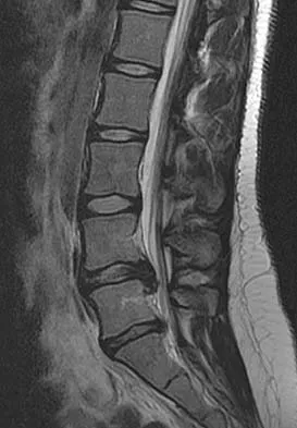
A 50-year-old man with no history of trauma reports new-onset back pain after doing some yard work the previous day. He reports pain radiating down his leg posteriorly and into the first dorsal web space of his foot. MRI scans are shown in Figures 3a through 3c. What nerve root is affected?


Explanation

The MRI scans clearly show an extruded L4-5 disk that is affecting the L5 nerve root on the left side. In addition, the L5 nerve root has a cutaneous distribution in the first dorsal web space. S1 affects the lateral foot. L4 affects the medial calf.


Question 6

A 19-year-old wrestler has numbness along the radial aspect of the forearm after undergoing an open Bankart repair through an anterior deltopectoral approach. Motor weakness would be expected along with what other finding?


Explanation

The musculocutaneous nerve may be injured by retracting the conjoined tendon medially. This nerve enters the coracobrachialis 5 cm distal to its origin. Its sensory distribution is the radial forearm, and its motor supply is to the biceps and brachialis. Bach BR, O'Brien SJ, Warren RF, et al: An unusual neurologic complication of the Bristow procedure. J Bone Joint Surg Am 1988;70:458-460.


Question 7

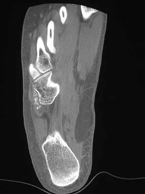
A 19-year-old man has had intermittent progressive knee pain with ambulation and pain at night following a rodeo accident 4 weeks ago. Figures 4a through 4e show the radiographs, a bone scan, CT scan, and T2-weighted MRI scan. What is the most likely diagnosis?


Explanation

The imaging studies reveal a predominantly blastic lesion in the distal femur with posterolateral periosteal changes. The bone scan shows increased uptake in the distal femur, beyond that expected with radiography. Cross-sectional imaging confirms the presence of a soft-tissue mass extending from the lateral aspect of the femur, with diffuse intramedullary signal changes. This aggressive presentation, particularly in this location and in a patient of this age, is most consistent with osteosarcoma. The mineralization in the soft tissue strongly suggests neoplasm, not the reactive bony changes seen in an infectious process. Menendez LR (ed): Orthopaedic Knowledge Update: Musculoskeletal Tumors. Rosemont, IL, American Academy of Orthopaedic Surgeons, 2002, pp 175-186.


Question 8

Figures 5a and 5b show the radiographs of an active 52-year-old man who has increasing knee pain and progressive varus deformity after undergoing total knee arthroplasty 7 years ago. Examination reveals a small effusion, but he has good motion and stability. What is the most likely diagnosis?


Explanation

The radiographs show narrowing of the medial joint space, which indicates polyethylene wear and progressive varus alignment. Wear particles incite osteolytic lesions like the one seen on the lateral radiograph. O'Rourke MR, Callaghan JJ, Goetz DG, et al: Osteolysis associated with a cemented modular posterior-cruciate-substituting total knee design. J Bone Joint Surg Am 2002;84:1362-1371.


Question 9

Which of the following best describes the course of the median nerve at the elbow?


Explanation

The median nerve courses superficial to the ulnar artery, deep to the fibrous arch of the superficialis muscle, and deep to the superficial head of the pronator teres muscle. The median nerve lies within the interval between the flexor digitorum superficialis muscle and the flexor digitorium muscle as it progresses toward the wrist. Hoppenfeld S, deBoer P: Surgical Exposures in Orthopaedics, ed 2. Philadelphia, PA, Lippincott-Raven, 1994, pp 118-131.


Question 10

A 62-year-old woman with soft-tissue calcifications and telangiectasia has severe pain in the left index, middle, ring, and little fingers. History reveals that she does not smoke. The clinical history and arteriogram shown in Figure 6 are consistent with which of the following conditions?


Explanation

The arteriogram shows generalized disease of all vascular structures. Even though the image was obtained following an infusion of nitroglycerin, little flow is present to the fingers. Based on the history of soft-tissue calcifications and telangiectasia, the most likely diagnosis is CREST (chondrocalcinosis, Raynaud's phenomenon, esophageal dysmotility, sclerodactyly and telangiectasias). The arteriogram reveals Raynaud's phenomenon or the "R" component of CREST. Buerger's disease, or thromboangiitis obliterans, is strongly associated with a history of smoking. Hypothenar hammer syndrome involves repetitive trauma to the ulnar artery at the wrist, resulting in well-defined filling defects in the superficial palmar arch of the hand. Although not well visualized in this patient, the superficial arch is narrowed, showing no evidence of aneurysmal dilation. Koman LA, Ruch DS, Patterson Smith B, et al: Vascular disorders, in Green DP, Hotchkiss RN, Pederson WC (eds): Green's Operative Hand Surgery, ed 4. New York, NY, Churchill Livingstone, 1999, vol 2, pp 2254-2302.


Question 11

During excision of a Baker cyst, the base or stalk is usually found between the


Explanation

Although there are several bursae in the posterior portion of the knee, the most prevalent one with a connection to the knee joint is the one in the interval between the semimembranosus and the medial head of the gastrocnemius muscle. The popliteus muscle and posterior cruciate ligament, the posterior cruciate ligament and lateral gastrocnemius muscle, and the medial gastrocnemius muscle and posterior cruciate ligament are all too lateral and uncommon. The semitendinosus and medial head of the gastrocnemius muscles do not come in contact in the posterior aspect of the knee. Resnick D: Diagnosis of Bone and Joint Disorders, ed 3. Philadelphia, PA, WB Saunders, 1995, p 379.


Question 12

A direct lateral (Hardinge) approach is used during total hip arthroplasty. The structure labeled A in Figure 7 is the


Explanation

The superior gluteal nerve is located approximately 7.82 cm above the tip of the greater trochanter as it courses through the gluteus medius. This anatomic consideration is relevant during a Hardinge approach to the hip, where excessive proximal dissection or retraction could result in nerve injury. A split of the gluteus medius of no more than 4 cm above the greater trochanter is considered safe. Hoppenfeld S, deBoer P: Surgical Exposures in Orthopaedics: The Anatomic Approach. Philadelphia, PA, JB Lippincott, 1984, pp 333-335.


Question 13

The dorsal digital cutaneous nerve of the great toe shown in Figure 8 is a branch of what nerve?


Explanation

The dorsal digital cutaneous nerve of the great toe is a branch of the medial branch of the superficial peroneal nerve. The deep peroneal nerve supplies the first web space. McMinn RMH, Hutchings RT, Logan BM: Color Atlas of Foot and Ankle Anatomy. Weert, Netherlands, Wolfe Medical Publications, 1982, p 50.


Question 14

In hip arthroplasty, the location of the medial femoral circumflex artery is best described as


Explanation

The obturator artery lies closest to the transverse acetabular ligament. The femoral artery is closest to the anterior rim of the acetabulum. No named vessel lies within the substance of the gluteus minimus or superior to the piriformis tendon. The medial femoral circumflex artery lies medial or deep to the quadratus femoris muscle. Hoppenfeld S, deBoer P: Surgical Exposures in Orthopaedics: The Anatomic Approach, ed 1. Philadelphia, PA, JB Lippincott, 1984, Figure 7-53, p 346.


Question 15

A 47-year-old man has acute right shoulder pain after falling off a ladder. The MRI scan shown in Figure 9 reveals


Explanation

The MRI scan reveals a full-thickness rotator cuff tear with retraction and increased signal in the subacromial space indicating joint fluid. Herzog RJ: Magnetic resonance imaging of the shoulder. Instr Course Lect 1998;47:3-20.


Question 16

The oblique radiograph of the foot and the CT scan shown in Figures 10a and 10b show a patient whose symptoms have failed to respond to rest and non-steroidal anti-inflammatory drugs. What is the best course of action?


Explanation

The radiograph and MRI scan show elongation and fragmentation of the os peroneum. Although casting, orthoses, and steroid injection may relieve symptoms, excision of the os peroneum and primary repair when necessary, with or without tenodesis of the peroneus longus to the peroneus brevis, have been shown to produce excellent results. Haddad SL: Disorders of tendons: Peroneal tendon dysfunction, in Coughlin MJ, Mann RA (eds): Surgery of the Foot and Ankle, ed 7. St Louis, MO, Mosby, 1999, pp 812-817.


Question 17

What is the typical MRI signal intensity of bone marrow affected by acute osteomyelitis?


Explanation

The classic MRI findings of osteomyelitis are a decrease in the normally high signal intensity of marrow on T1-weighted images and normal or increased signal intensity on T2-weighted images. This is the result of replacement of marrow fat by inflammatory cells and edema, which causes lower signal intensity than fat on T1-weighted images and higher signal intensity than fat on T2-weighted images. The addition of gadolinium to a T1-weighted sequence reveals increased signal intensity in the hyperemic marrow. Unger E, Moldofsky P, Gatenby R, et al: Diagnosis of osteomyelitis by MR imaging. Am J Roentgenol 1988;150:605-610. Dormans JP, Drummond DS: Pediatric hematogenous osteomyelitis: New trends in presentation, diagnosis and treatment. J Am Acad Orthop Surg 1994;2:333-341.


Question 18

Based on the appearance of the imaging studies shown in Figures 11a through 11c, what structure has most likely been injured?


Explanation

The radiographs reveal marked lateral subluxation of the patella in a patient who has recurrent patellar instability. The medial patellofemoral ligament is the main restraint to lateral subluxation of the patella. Boden BP, Pearsall AW: Patellofemoral instability: Evaluation and management. J Am Acad Orthop Surg 1997;5:47-57.


Question 19

In the anterior forearm approach to the distal radius (Henry approach), the radial artery is located between what two structures?


Explanation

The standard approach to the volar aspect of the distal radius is the Henry approach. Following incision of the skin and subcutaneous tissues, the forearm fascia is incised. The radial artery and venae comitantes lie in the interval between the tendons of the flexor carpi radialis muscle and the brachioradialis muscle. This interval is developed, and the radial artery and veins are retracted in a radial direction. Hoppenfeld S, deBoer P: Surgical Exposures in Orthopaedics, ed 2. Philadelphia, PA, Lippincott-Raven, 1994, pp 118-131.


Question 20

Following a radial nerve neurapraxia at or above the elbow, return of muscle function can be expected to start at the brachioradialis and return along which of the following progressions?


Explanation

Following a radial nerve neurapraxia above the elbow, muscle recovery can be expected in a predictable pattern. Although variations will occur, the return of function or reinnervation usually occurs in the following order: brachioradialis, extensor carpi radialis longus, supinator, extensor carpi radialis brevis, extensor carpi ulnaris, extensor digitorum comminus, extensor digiti minimi, extensor indicis proprious, extensor pollicis longus, abductor pollicis longus, and extensor pollicis brevis. Netter F: The Ciba Collection of Medical Illustrations: The Musculoskeletal System. Part 1, Anatomy, Physiology and Metabolic Disorders. West Caldwell, NJ, Ciba-Geigy, 1991, vol 8, p 53.


Question 21

To preserve blood supply to the fractured bone seen in Figures 12a and 12b, care should be taken when exposing which of the following areas?


Explanation

The blood supply to the adult capitellum and lateral trochlea comes from posterior vessels arising from the radial recurrent, radial collateral, and interosseous recurrent arteries. These arteries penetrate the distal humerus posterior and superior to the capitellum.


Question 22

An axial T1-weighted MRI scan of the pelvis is shown in Figure 13. The arrow is pointing to what muscle?


Explanation

The obturator internus muscle originates from the internal pelvic wall and passes laterally through the lesser sciatic foramen, banking around the ischium below the sacrospinous ligament before inserting on the medial aspect of the greater trochanter. Higuchi T: Normal anatomy and magnetic resonance appearance of the pelvis, in Takahashi HE, Morita T, Hotta T, Ogose A (eds): Operative Treatment of Pelvic Tumors. Tokyo, Japan, Springer-Verlag, 2003, pp 4-21.


Question 23

Which of the following radiographic views best depicts a Hill-Sachs defect?


Explanation

The Stryker notch view best shows this type of defect. An outlet view helps evaluate acromial shape, a true AP shows joint space narrowing, a serendipity view evaluates the sternoclavicular joint, and a Zanca view helps evaluate the acromioclavicular joint. An internal rotation AP may also depict a Hill-Sachs defect.


Question 24

What structure provides the major blood supply to the humeral head?


Explanation

The ascending branch of the anterior circumflex humeral artery provides the major blood supply to the humeral head. The posterior circumflex humeral artery supplies a much smaller portion of the proximal humerus. The nutrient humeral artery is the main blood supply for the humeral shaft. The thoracoacromial artery is primarily a muscular branch. The rotator cuff insertions contribute some blood supply to the tuberosities but not a major contribution. Beaty JH (ed): Orthopaedic Knowledge Update 6. Rosemont, IL, American Academy of Orthopaedic Surgeons, 1999, pp 271-286.


Question 25

Figure 14 shows an intra-articular gadolinium-enhanced MRI scan of a 52-year-old woman who has stopped playing tennis because of pain in her left shoulder while serving. What is the most likely diagnosis?


Explanation

The MRI scan shows increased signal intensity along the deep fibers of the supraspinatus near its insertion. This is typical of tendinosis and a probable partial-thickness rotator cuff tear. Herzog RJ: Magnetic resonance imaging of the shoulder. Instr Course Lect 1998;47:3-20.


Question 26

During a posterior approach to the hip (Moore or Southern), the short external rotators are tenotomized and reflected posteriorly. Which structure is most at risk if the piriformis is not carefully protected, specifically considering common anatomical variants?


Explanation

The sciatic nerve typically exits the pelvis inferior to the piriformis, but in up to 15% of the population, a portion (usually the common peroneal division) pierces or exits superior to the piriformis, placing it at high risk during tenotomy.

Question 27

When performing a direct lateral (deltoid-splitting) approach to the proximal humerus for open reduction and internal fixation, what is the maximum safe distance from the tip of the acromion to extend the split without risking denervation of the anterior deltoid?


Explanation

The axillary nerve courses from posterior to anterior on the deep surface of the deltoid approximately 5 to 7 cm distal to the lateral edge of the acromion. Splitting the deltoid beyond 5 cm puts the nerve at significant risk.

Question 28

During a lateral transpsoas approach (LLIF) to the lumbar spine at the L4-L5 level, the surgeon uses neuromonitoring to map the lumbar plexus. At this specific level, where is the lumbar plexus typically located relative to the psoas major muscle?


Explanation

The elements of the lumbar plexus migrate anteriorly as they travel caudally. At the L4-L5 disc space, the plexus is typically located within the posterior third of the psoas muscle, necessitating careful anterior retractor placement.

Question 29

When establishing a posteromedial portal for knee arthroscopy to address a posterior horn medial meniscus tear, which anatomical structure is at greatest risk of iatrogenic injury during portal creation?


Explanation

The saphenous nerve and great saphenous vein are located superficially in the posteromedial aspect of the knee. The posteromedial portal must be made carefully under direct visualization to avoid injuring these structures.

Question 30

During a volar (Henry) approach to the proximal radius, the surgeon supinates the forearm to protect the posterior interosseous nerve (PIN). Supination moves the PIN in which direction relative to the anterior surgical exposure?


Explanation

The PIN runs within the substance of the supinator muscle. Supinating the forearm winds the supinator laterally and posteriorly around the radius, moving the PIN safely away from the anterior dissection field.

Question 31

An extensile lateral approach to the calcaneus is planned for an intra-articular fracture. The surgeon must elevate a full-thickness flap to avoid wound necrosis. Which nerve is most at risk during the initial incision and flap elevation?


Explanation

The sural nerve travels posterior to the lateral malleolus and courses along the lateral aspect of the hindfoot. It must be included within the full-thickness flap to prevent iatrogenic transection or neuroma formation.

Question 32

In the normal anatomical course of the cervical spine, the vertebral artery typically enters the transverse foramen at which vertebral level?


Explanation

The vertebral artery classically arises from the subclavian artery and enters the transverse foramen at C6 in approximately 90% of individuals, bypassing the transverse foramen of C7.

Question 33

A 45-year-old cyclist presents with numbness in the little finger and weakness of the intrinsic hand muscles. Compression of the ulnar nerve is suspected in Guyon's canal. Which structure forms the floor of this canal?


Explanation

The floor of Guyon's canal is formed primarily by the transverse carpal ligament (flexor retinaculum) and the pisohamate ligament. The roof is formed by the volar carpal ligament and palmaris brevis.

Question 34

When performing an anterior approach to the hip (Smith-Petersen), the superficial internervous plane is between the sartorius and tensor fasciae latae (TFL). Which nerve innervates the muscle forming the medial border of this interval?


Explanation

The sartorius forms the medial border of the Smith-Petersen approach and is innervated by the femoral nerve. The lateral border is the TFL, which is innervated by the superior gluteal nerve.

Question 35

During an anatomic reconstruction of the posterolateral corner of the knee, the surgeon must accurately identify the fibular insertion of the lateral collateral ligament (LCL). Where does the LCL insert relative to the popliteofibular ligament (PFL)?


Explanation

On the fibular head, the LCL inserts accurately into a footprint that is situated anterior and distal to the insertion of the popliteofibular ligament (PFL) and the biceps femoris tendon.

Question 36

When placing a pedicle screw at the L5 level, the starting point is identified at the intersection of the pars interarticularis, superior articular process, and transverse process. What is the typical medial angulation required to safely traverse the L5 pedicle?


Explanation

The required medial angulation for pedicle screw placement increases caudally in the lumbar spine. L1 requires approximately 5-10 degrees, while L5 requires 25 to 30 degrees of medial trajectory.

Question 37

A patient presents with medial winging of the scapula after an axillary node dissection, indicating injury to the long thoracic nerve. From which cervical nerve roots does this nerve originate?


Explanation

The long thoracic nerve innervates the serratus anterior muscle and is formed by the ventral rami of the C5, C6, and C7 cervical nerve roots.

Question 38

Anterocentral portals for ankle arthroscopy are generally avoided due to the high risk of iatrogenic injury to which critical anatomical structure?


Explanation

The anterocentral portal is located centrally over the ankle joint and places the deep peroneal nerve and the dorsalis pedis artery at high risk of injury, hence it is rarely utilized.

Question 39

During an anterior approach to the elbow for a distal biceps tendon repair, the surgeon must carefully retract the lateral antebrachial cutaneous nerve. Between which two muscles does this nerve typically emerge in the distal arm?


Explanation

The lateral antebrachial cutaneous nerve is the terminal sensory branch of the musculocutaneous nerve. It emerges laterally in the distal arm between the biceps brachii and the brachialis muscles.

Question 40

During corrective spinal deformity surgery, the anterior spinal cord is vulnerable to ischemic injury. Which vascular territory is responsible for supplying the anterior spinal artery in the lower two-thirds of the spinal cord?


Explanation

The artery of Adamkiewicz, typically arising on the left side between T8 and L1, is the major blood supply to the lower two-thirds of the anterior spinal cord. Injury causes anterior spinal artery syndrome.

Question 41

A patient undergoes a two-incision fasciotomy for a tibial shaft fracture complicated by compartment syndrome. Which muscle group must be carefully identified and released to ensure adequate decompression of the deep posterior compartment?


Explanation

The deep posterior compartment of the leg contains the tibialis posterior, flexor digitorum longus, and flexor hallucis longus. This compartment is notoriously under-released if the soleus bridge is not adequately taken down.

Question 42

The recurrent motor branch of the median nerve innervates the thenar musculature. What is the most common anatomical relationship of this branch to the transverse carpal ligament?


Explanation

In approximately 50-70% of individuals, the recurrent motor branch is extraligamentous, branching off the median nerve distal to the transverse carpal ligament before curling back to innervate the thenar muscles.

Question 43

During a medial approach to the distal tibia for minimally invasive plating of a pilon fracture, the surgeon must protect the saphenous nerve. Which vascular structure closely travels with the saphenous nerve in this region?


Explanation

The great saphenous vein courses anterior to the medial malleolus alongside the saphenous nerve, making both structures highly susceptible to injury during medial approaches to the distal tibia and ankle.

Question 44

When placing half-pins for a humeral external fixator from a lateral approach, the radial nerve is at risk. At what location does the radial nerve predictably pierce the lateral intermuscular septum to enter the anterior compartment?


Explanation

The radial nerve transitions from the posterior to the anterior compartment by piercing the lateral intermuscular septum approximately 10 cm (range 10-12 cm) proximal to the radiocapitellar joint.

Question 45

A 35-year-old patient undergoes a lymph node biopsy in the posterior triangle of the neck. Postoperatively, the patient is unable to abduct the arm past 90 degrees and demonstrates scapular winging. Injury to which of the following nerves is the most likely cause?


Explanation

The spinal accessory nerve (CN XI) innervates the trapezius and is highly vulnerable during procedures in the posterior cervical triangle. Trapezius palsy leads to lateral scapular winging and inability to actively abduct the shoulder past 90 degrees.

Question 46

A 45-year-old sustains a displaced femoral neck fracture. Which of the following vessels provides the primary blood supply to the adult femoral head and is at greatest risk of disruption in this injury?


Explanation

The medial femoral circumflex artery, particularly its lateral epiphyseal branches, provides the predominant blood supply to the adult femoral head. Disruption of this vessel in displaced femoral neck fractures significantly increases the risk of avascular necrosis.

Question 47

During the placement of pedicle screws at the L4 level, the surgeon breaches the inferior wall of the L4 pedicle. Which nerve root is most directly at risk of injury due to this specific breach?


Explanation

The exiting L4 nerve root travels immediately inferior to the L4 pedicle. A breach of the inferior wall of the L4 pedicle puts the L4 exiting nerve root at high risk of iatrogenic injury.

Question 48

During a volar forearm fasciotomy for compartment syndrome, the distal skin incision is deliberately directed ulnar to the palmaris longus tendon at the wrist crease. This maneuver primarily avoids injury to which of the following structures?


Explanation

The palmar cutaneous branch of the median nerve arises proximal to the wrist and runs between the palmaris longus and flexor carpi radialis. Directing the fasciotomy incision ulnar to the palmaris longus protects this sensory branch.

Question 49

A surgeon is performing an extensile lateral approach to the calcaneus for an intra-articular fracture. Retraction of the full-thickness inferior flap places which of the following structures at highest risk?


Explanation

The sural nerve and lesser saphenous vein are located in the lateral subcutaneous tissues of the hindfoot. They must be carefully elevated and protected within the full-thickness inferior flap during an extensile lateral approach.

Question 50

During an anterior cervical discectomy and fusion (ACDF), the longus colli muscles are elevated to define the lateral margins. Overzealous lateral dissection and retraction past the longus colli places which structure at immediate risk?


Explanation

The cervical sympathetic trunk lies directly on the longus colli muscle, lateral to the medial border. Aggressive lateral dissection or lateral retractor blade placement can injure the trunk, resulting in Horner syndrome.

Question 51

During a trigger finger release, excessive division of the flexor sheath can lead to bowstringing of the flexor tendons. Which of the following pulleys is most critical to preserve to prevent this complication?


Explanation

The A2 and A4 pulleys are the major biomechanical restraints preventing bowstringing of the flexor tendons. While the A1 pulley is sectioned during a trigger finger release, the A2 pulley must be carefully protected.

Question 52

A patient presents with isolated varus instability at 30 degrees of knee flexion, but the knee is stable to varus stress at 0 degrees. Which of the following structures is the primary restraint to varus stress at 30 degrees of knee flexion?


Explanation

The fibular collateral ligament (LCL) is the primary restraint to varus stress at 30 degrees of knee flexion. Varus instability at both 0 and 30 degrees typically indicates combined LCL and cruciate ligament injury.

Question 53

During an arthroscopic rotator cuff repair, a portal is established posterior to the acromioclavicular joint. If the portal or dissection is extended more than 5 cm inferiorly from the lateral acromion edge, which nerve is at greatest risk of injury?


Explanation

The axillary nerve courses horizontally along the deep surface of the deltoid, approximately 5 cm distal to the lateral border of the acromion. Extending lateral portals or splits too far inferiorly places this nerve at significant risk.

Question 54

During an ilioinguinal approach to the acetabulum, severe hemorrhage is encountered while dissecting posterior to the superior pubic ramus. This is most likely due to an unligated vascular anastomosis between which two systems?


Explanation

The corona mortis is a prominent vascular anastomosis between the obturator vessels (internal iliac system) and the inferior epigastric vessels (external iliac system). It lies over the superior pubic ramus and can cause life-threatening bleeding if inadvertently torn.

Question 55

A patient undergoes a lateral transpsoas interbody fusion at L4-L5. Postoperatively, they display profound weakness in hip flexion and knee extension, along with anterior thigh numbness. Which nerve was most likely injured?


Explanation

The femoral nerve courses through the posterior aspect of the psoas major muscle and is particularly vulnerable during a transpsoas approach at the L4-L5 disc space. Injury results in quadriceps weakness and sensory deficits over the anterior thigh.

Question 56

When performing an ulnar nerve transposition at the elbow, the surgeon must release all potential sites of compression. Which of the following represents the most proximal potential site of ulnar nerve compression?


Explanation

The Arcade of Struthers is a fascial band extending from the medial head of the triceps to the medial intermuscular septum, located approximately 8 cm proximal to the medial epicondyle. It is the most proximal site of potential ulnar nerve entrapment.

Question 57

In a patient with a suspected ankle syndesmotic injury, which ligament serves as the primary restraint to anterior translation of the distal fibula relative to the tibia?


Explanation

The anterior inferior tibiofibular ligament (AITFL) is the primary restraint against anterior translation of the distal fibula. It is frequently the first structure torn in external rotation injuries of the syndesmosis.

Question 58

Following surgical fixation of a proximal radius fracture, a patient cannot extend the interphalangeal joint of the thumb or the metacarpophalangeal joints of the fingers, but sensation is intact. Which nerve is injured, and through which muscle does it pass?


Explanation

The posterior interosseous nerve (PIN) is a purely motor nerve that courses through the two heads of the supinator muscle at the Arcade of Frohse. Injury causes loss of digital and thumb extension, but preserves sensation and wrist extension (ECRL is intact).

Question 59

During the insertion of a thoracic pedicle screw at T7, the surgeon inadvertently directs the screw too far laterally, breaching the lateral wall of the pedicle. What anatomic structure is at greatest risk of direct injury?


Explanation

The pleura and lungs lie immediately lateral to the thoracic pedicles. A lateral breach of the thoracic pedicle places the pleural space at high risk, potentially causing a pneumothorax.

Question 60

An isolated fracture of the fibular shaft is approached surgically. The surgeon utilizes the internervous plane between the lateral and superficial posterior compartments of the leg. Which two muscles define this specific surgical interval?


Explanation

The internervous plane for the lateral approach to the fibula is between the peroneus brevis (superficial peroneal nerve) and the soleus (tibial nerve). This safely separates the lateral and superficial posterior compartments.

Question 61

A 45-year-old man sustains a proximal humerus fracture and subsequently demonstrates weakness in shoulder abduction and diminished sensation over the lateral deltoid. The affected nerve exits the axilla through the quadrangular space. Which of the following defines the superior border of this anatomic space?


Explanation

The quadrangular space is bordered superiorly by the teres minor, inferiorly by the teres major, medially by the long head of the triceps, and laterally by the surgical neck of the humerus. It transmits the axillary nerve and the posterior humeral circumflex artery.

Question 62

During an anterior cervical discectomy and fusion (ACDF), aggressive lateral dissection places the vertebral artery at risk. In standard human anatomy, the vertebral artery typically enters the transverse foramen at which cervical level?


Explanation

The V2 segment of the vertebral artery typically ascends through the transverse foramina beginning at the C6 level in approximately 90% of individuals. Awareness of this anatomy is critical to prevent catastrophic bleeding during lateral dissection in anterior cervical spine surgery.

Question 63

A surgeon is performing an open reduction internal fixation of an acetabular fracture via the ilioinguinal approach. If a Smith-Petersen extension is utilized, the superficial internervous plane lies between muscles innervated by which two nerves?


Explanation

The superficial interval of the Smith-Petersen (anterior) approach to the hip lies between the sartorius (femoral nerve) and the tensor fasciae latae (superior gluteal nerve). The deep interval is between the rectus femoris (femoral nerve) and gluteus medius (superior gluteal nerve).

Question 64

An 18-year-old athlete sustains a multi-ligamentous knee injury, including the posterolateral corner (PLC). During reconstruction, the surgeon must identify the femoral footprint of the popliteus tendon. Where is this footprint located relative to the lateral collateral ligament (LCL) attachment?


Explanation

On the lateral femoral condyle, the popliteus tendon footprint is located approximately 18.5 mm distal and anterior to the lateral epicondyle. The LCL attaches slightly proximal and posterior to the epicondyle. Correct anatomical placement is essential for restoring PLC biomechanics.

Question 65


A 60-year-old man undergoes L4-L5 posterior spinal fusion. What is the classic anatomical landmark for the starting point of an L4 pedicle screw?


Explanation

The traditional starting point for a lumbar pedicle screw is at the intersection of a vertical line along the lateral margin of the superior articular facet and a horizontal line bisecting the transverse process. This landmark safely guides the surgeon into the pedicle cylinder.

Question 66

During open reduction internal fixation of a medial malleolus fracture, the surgeon explores the structures posterior to the medial malleolus. Moving from anterior to posterior, which structure lies immediately posterior to the flexor digitorum longus (FDL) tendon?


Explanation

The structures passing behind the medial malleolus from anterior to posterior are: Tibialis posterior, Flexor digitorum longus, Posterior tibial artery, Vein, Tibial nerve, and Flexor hallucis longus. Therefore, the posterior tibial artery lies immediately posterior to the FDL.

Question 67

While performing an ilioinguinal approach for a transverse acetabular fracture, life-threatening hemorrhage occurs near the superior pubic ramus. This bleeding is most likely originating from the corona mortis, which represents an anastomosis between which two vascular systems?


Explanation

The corona mortis is a vascular anastomosis between the external iliac (or deep inferior epigastric) and obturator (internal iliac) vessels. It rests on the posterior aspect of the superior pubic ramus and is highly vulnerable during anterior pelvic surgical approaches.

Question 68

A 45-year-old mechanic presents with an inability to actively extend his fingers and thumb at the metacarpophalangeal joints. Wrist extension is preserved but exhibits a strong radial deviation. Sensation in the upper extremity is fully intact. Which of the following anatomical structures is the most likely site of neural compression?


Explanation

The presentation describes Posterior Interosseous Nerve (PIN) syndrome. The PIN is purely motor and is most commonly compressed at the Arcade of Frohse, the proximal edge of the superficial head of the supinator muscle.

Question 69

During a posterior approach to the hip for a total hip arthroplasty, excessive bleeding is encountered near the inferior border of the quadratus femoris. Ligation of the vessels in this area puts the primary blood supply to the femoral head at risk. Which artery is responsible for this blood supply and passes between the quadratus femoris and the obturator externus?


Explanation

The medial circumflex femoral artery (MCFA) provides the primary blood supply to the femoral head. Its deep branch passes between the obturator externus and quadratus femoris.

Question 70

A surgeon is performing an L4-L5 posterior instrumented fusion. During the placement of a pedicle screw into the right L4 pedicle, the screw breaches the inferior wall of the pedicle. Which of the following neurologic structures is at greatest risk of direct mechanical injury?


Explanation

The exiting nerve root travels through the superior portion of the neuroforamen, immediately inferior to the pedicle of the same numerical level. An inferior pedicle breach at L4 risks the L4 exiting root.

Question 71

A 28-year-old male is evaluated in the emergency department following a direct blow to the lateral aspect of his shoulder. He has weakness in shoulder abduction and decreased sensation over the lateral deltoid. Through which anatomical space does the injured nerve exit the axilla?


Explanation

The axillary nerve and posterior circumflex humeral artery exit the axilla via the quadrangular space, bordered by the teres minor, teres major, long head of the triceps, and the surgical neck of the humerus.

Question 72

During a posterolateral approach to the distal fibula for fracture fixation, a cutaneous nerve is identified and protected. What is the standard anatomical course of this nerve at the level of the ankle?


Explanation

The sural nerve provides sensory innervation to the lateral foot and travels posterior to the lateral malleolus alongside the small saphenous vein.

Question 73

An orthopaedic spine surgeon is performing an anterior cervical discectomy and fusion (ACDF). The surgeon must be aware of the vertebral artery anatomy. In most individuals, at what cervical vertebral level does the vertebral artery initially enter the transverse foramen?


Explanation

The vertebral artery typically branches from the subclavian artery and enters the transverse foramen at the C6 level, bypassing the C7 transverse foramen.

Question 74

A 34-year-old avid cyclist reports progressive numbness in his ring and small fingers along with weakness in finger abduction. Suspecting compression at Guyon's canal, surgical release is planned. Which structure forms the floor of this anatomical canal?


Explanation

The floor of Guyon's canal is formed by the transverse carpal ligament (flexor retinaculum) and the pisohamate ligament. The roof is formed by the volar carpal ligament.

Question 75

Following a traumatic knee dislocation, a patient is noted to have a dense foot drop and inability to evert the foot. The injured nerve is most vulnerable as it courses around the fibular neck. Between which two structures does this nerve pass as it enters the anterior compartment of the leg?


Explanation

The common peroneal nerve wraps around the fibular neck, passing between the peroneus longus muscle and the fibula before dividing into its deep and superficial branches.

Question 76

A 22-year-old collegiate volleyball player presents with isolated weakness in shoulder external rotation. Shoulder abduction and internal rotation are full strength. There is isolated atrophy of the infraspinatus fossa. Where is the most likely site of nerve compression?


Explanation

Compression of the suprascapular nerve at the spinoglenoid notch selectively denervates the infraspinatus. Compression at the suprascapular notch would affect both the supraspinatus and infraspinatus.

Question 77

During a lateral closing-wedge high tibial osteotomy, a retractor is carefully placed posterior to the proximal tibia to protect the neurovascular structures. Which of the following vessels is at greatest risk of injury if this retractor is incorrectly positioned directly posterior to the interosseous membrane?


Explanation

The anterior tibial artery branches from the popliteal artery and passes anteriorly through the proximal interosseous membrane, sitting directly on the posterior cortex of the tibia before crossing.

Question 78

A patient with an unstable pelvic ring fracture requires percutaneous iliosacral screw fixation at the S1 level. If the screw trajectory is placed too far anteriorly and inferiorly to the sacral ala, which nerve root is at highest risk of iatrogenic injury?


Explanation

The L5 nerve root courses directly anterior to the sacral ala. An anteriorly misdirected iliosacral screw at S1 risks lacerating or compressing this nerve root.

Question 79

A 30-year-old man sustains a penetrating stab wound to the axilla. Physical examination demonstrates profound weakness in elbow flexion and decreased sensation along the lateral aspect of the forearm. From which portion of the brachial plexus does the injured nerve originate?


Explanation

The patient has a musculocutaneous nerve injury, causing biceps weakness and lateral antebrachial cutaneous nerve numbness. This nerve is a terminal branch of the lateral cord of the brachial plexus.

Question 80

An orthopaedic surgeon uses the Smith-Petersen (anterior) approach for a pelvic osteotomy. This approach exploits an internervous plane between the sartorius and the tensor fasciae latae. Which of the following nerves innervates the sartorius?


Explanation

The sartorius is innervated by the femoral nerve, while the tensor fasciae latae is innervated by the superior gluteal nerve, creating a true internervous plane.

Question 81

A lateral transpsoas approach (LLIF) is utilized for interbody fusion at L4-L5. Postoperatively, the patient experiences quadriceps weakness and anterior thigh numbness. Which structure within the psoas major muscle was most likely injured?


Explanation

The femoral nerve lies in the posterior third of the psoas muscle at the L4-L5 level. Retractor placement or dissection in this zone puts it at high risk of stretch or direct injury.

Question 82

A hand surgeon is repairing a flexor tendon laceration in Zone II of the index finger. Which of the following best describes the anatomical relationship of the flexor tendons within this specific zone?


Explanation

In Zone II, the flexor digitorum superficialis (FDS) bifurcates (forming Camper's chiasm) allowing the flexor digitorum profundus (FDP) to pass through and become superficial to insert on the distal phalanx.

Question 83

During an anterior subcutaneous transposition of the ulnar nerve, the nerve is released from the cubital tunnel. What anatomical structure forms the roof of the cubital tunnel?


Explanation

Osborne's ligament, or the cubital tunnel retinaculum, spans from the medial epicondyle to the olecranon, forming the roof of the cubital tunnel. The Arcade of Struthers is located more proximally.

Question 84

A surgeon utilizes the Smith-Petersen (direct anterior) approach for a total hip arthroplasty. To prevent denervation of the surrounding musculature, the dissection utilizes a true internervous plane. Which two nerves supply the muscles defining this superficial interval?


Explanation

The superficial interval is between the tensor fasciae latae (superior gluteal nerve) and the sartorius (femoral nerve). This true internervous plane protects motor function during the direct anterior approach.

Question 85

A surgeon is placing pedicle screws in the lumbar spine for a burst fracture. On an anteroposterior (AP) fluoroscopic view, the medial border of the pedicle corresponds to which structural boundary of the spinal canal?


Explanation

On an AP radiograph, the medial border of the pedicle defines the lateral border of the spinal canal. Violating this border during pedicle screw preparation risks injury to the traversing nerve root.

Question 86

An elite overhead throwing athlete presents with isolated weakness in external rotation of the shoulder. Examination reveals atrophy of the infraspinatus but normal supraspinatus bulk. Which anatomical structure is most likely compressing the affected nerve?


Explanation

The suprascapular nerve innervates the supraspinatus before passing under the spinoglenoid ligament to innervate the infraspinatus. Entrapment at the spinoglenoid notch causes isolated infraspinatus atrophy.

Question 87

A patient sustains a traumatic knee dislocation and undergoes immediate reduction. Vascular evaluation is required due to the high risk of popliteal artery injury. Between which two distinct anatomical structures is the popliteal artery firmly tethered, making it susceptible to shear stress?


Explanation

The popliteal artery is rigidly tethered proximally at the adductor hiatus (Hunter's canal) and distally at the tendinous arch of the soleus. This anatomic tethering predisposes it to traction injury during knee dislocations.

Question 88

A patient sustains a spiral fracture of the distal third of the humerus (Holstein-Lewis fracture). They present with wrist drop and loss of finger extension. The nerve involved is at greatest risk of tethering as it passes through which anatomical structure?


Explanation

The radial nerve pierces the lateral intermuscular septum approximately 10 cm proximal to the lateral epicondyle. This fixes the nerve in place, making it highly susceptible to injury in distal third humerus fractures.

Question 89

A patient requires the creation of a posterolateral portal during ankle arthroscopy. Which nerve is at greatest risk of injury during the establishment of this portal?


Explanation

The sural nerve runs just posterior to the lateral malleolus. It is at significant risk of iatrogenic injury when placing a posterolateral ankle arthroscopy portal.

Question 90

A patient presents with the inability to form an "OK" sign, demonstrating an extended distal interphalangeal joint of the index finger and interphalangeal joint of the thumb during pinch. Sensation in the hand is completely normal. What is the most likely diagnosis?


Explanation

Anterior interosseous nerve (AIN) syndrome causes loss of function of the flexor pollicis longus, flexor digitorum profundus (index/long), and pronator quadratus. Because the AIN is a purely motor nerve, sensation remains intact.

Question 91

Following a traumatic anterior shoulder dislocation, a patient complains of numbness over the lateral aspect of the shoulder. The injured nerve exits the axilla through the quadrangular space. Which of the following muscles forms the inferior border of this space?


Explanation

The quadrangular space is bounded superiorly by the teres minor, inferiorly by the teres major, medially by the long head of the triceps, and laterally by the humeral shaft. It transmits the axillary nerve and posterior circumflex humeral artery.

Question 92

A runner requires a lateral compartment fasciotomy for chronic exertional compartment syndrome. The surgeon must protect the superficial peroneal nerve. At what anatomical location does this nerve typically pierce the deep fascia to become subcutaneous?


Explanation

The superficial peroneal nerve typically pierces the crural fascia in the distal third of the leg, roughly 10-12 cm proximal to the lateral malleolus. It then transitions from the lateral compartment to the subcutaneous tissue.

Question 93

Six weeks after undergoing volar plating for a distal radius fracture, a patient notes a sudden inability to actively extend the interphalangeal joint of the thumb. The tendon most likely ruptured wraps around which bony prominence?


Explanation

The extensor pollicis longus (EPL) tendon uses Lister's tubercle on the dorsal radius as a pulley. It can rupture due to mechanical attrition from prominent dorsal screws or biological factors following a distal radius fracture.

Question 94

Piriformis syndrome occurs due to compression of the sciatic nerve by the piriformis muscle. In the most common anatomical configuration, what is the relationship of the sciatic nerve to the piriformis muscle as it exits the greater sciatic foramen?


Explanation

In roughly 80-85% of the population, the undivided sciatic nerve passes entirely deep (inferior) to the piriformis muscle. Anatomical variations, such as the peroneal division splitting the muscle, can predispose patients to piriformis syndrome.

None

Clinic OS
Medically Verified Content by
Prof. Clinic OS
Consultant Orthopedic & Spine Surgeon
Chapter Index