العربية
Part of the Master Guide

AAOS Orthopedic MCQs (Set 1): Spine Disorders, Sports Medicine & Arthroplasty | 2026 Board Review

AAOS & ABOS Spine Surgery MCQs (Set 3): Spinal Trauma, Degenerative Conditions, Scoliosis

23 Apr 2026 66 min read 113 Views
Spine 2009 MCQs - Part 3

Key Takeaway

This high-yield question set for the AAOS and ABOS exams (Set 3) focuses on key aspects of spine surgery. It covers the diagnosis and management of spinal trauma, degenerative spine conditions such as disc herniation and stenosis, and common spinal deformities like scoliosis and kyphosis, preparing residents for certification.

AAOS & ABOS Spine Surgery MCQs (Set 3): Spinal Trauma, Degenerative Conditions, Scoliosis

Comprehensive 100-Question Exam


00:00

Start Quiz

Question 1

A 58-year-old woman with rheumatoid arthritis has progressive neck pain, upper extremity and lower extremity weakness, and difficulty with fine motor movements. Examination reveals hyperreflexia with mild to moderate objective weakness but the patient has no difficulty with ambulation for short distances. What is the most important preoperative imaging finding that predicts full neurologic recovery with surgical stabilization?


Explanation

Boden and associates' article presents compelling evidence that patients with rheumatoid arthritis and neurologic deterioration in C1-2 instability are more likely to achieve some improvement if the posterior atlanto-dens interval is greater than 10 mm on preoperative studies. All the patients in their series who had neurologic deterioration and a preoperative posterior atlanto-dens interval of greater than 14 mm achieved complete motor recovery. Boden SD, Dodge LD, Bohlman HH, et al: Rheumatoid arthritis of the cervical spine: A long-term analysis with predictors of paralysis and recovery. J Bone Joint Surg Am 1993;75:1282-1297. Boden SD, Clark CR: Rheumatoid arthritis of the cervical spine, in Clark CR (ed): The Cervical Spine, ed 3. Philadelphia, PA, Lippincott Raven, 1998, pp 755-764.

Question 2

Figures 20a through 20d show the radiographs and MRI scans of a 59-year-old woman who has had symptoms consistent with progressive neurogenic claudication and back pain for the past 9 months. In the last 6 months, nonsurgical management consisting of nonsteroidal anti-inflammatory drugs, physical therapy, and a series of epidural steroid injections have been used; however the injections, while beneficial, have provided only temporary relief of her symptoms. What is the most appropriate management at this time?


Explanation

Patients with a degenerative spondylolisthesis and severe stenosis who have failed appropriate nonsurgical management are candidates for surgical intervention. Most studies show good to excellent results in more than 85% of patients after lumbar decompression for stenosis. Atlas and associates found that at 8- to 10-year follow-up, leg pain relief and back-related functional status were greater in those patients opting for surgical treatment of the stenosis. Similarly, the decision to fuse a spondylolisthetic segment has been supported in the literature. Herkowitz and Kurz compared decompressive laminectomy alone and decompressive laminectomy with intertransverse arthrodesis in 50 patients with single-level spinal stenosis and degenerative spondylolisthesis. They demonstrated good to excellent results in 90% of the fused group compared to 44% in the nonfusion group. The decision to include instrumentation during the fusion is more controversial. Whereas the use of instrumentation has shown to improve fusion rates, it has not been conclusively shown to improve the overall clinical outcomes of patients. Atlas SJ, Keller RB, Wu YA, et al: Long-term outcomes of surgical and nonsurgical management of lumbar spinal stenosis: 8 to 10 year results from the Maine lumbar spine study. Spine 2005;30:936-943. Herkowitz HN, Kurz LT: Degenerative lumbar spondylolisthesis with spinal stenosis: A prospective study comparing decompression with decompression and intratransverse process arthrodesis. J Bone Joint Surg Am 1991;73:802-808.


Question 3

A 29-year-old man reports a 2-week history of severe neck pain after being struck sharply on the back of the head and neck while moving a refrigerator down a flight of stairs. Initial evaluation in the emergency department revealed no obvious fracture and he was discharged in a soft collar. Neurologic examination is within normal limits, and radiographs taken in the office are shown in Figures 21a through 21c. Subsequent MRI scans show intra-substance rupture of the transverse atlantal ligament. What is the most appropriate treatment option at this time?


Explanation

Dickman and associates classified injuries of the transverse atlantal ligament into two categories. Type I injuries are disruptions through the substance of the ligament itself. Type II injuries render the transverse ligament physiologically incompetent through fractures and avulsions involving the tubercle of insertion of the transverse ligament on the C1 lateral mass. Type I injuries are incapable of healing without supplemental internal fixation. Type II injuries can be treated with a rigid cervical orthosis with a success rate of 74%. Surgery may be required for type II injures that fail to heal with 3 to 4 months of nonsurgical management. Findlay JM: Injuries involving the transverse atlantal ligament: Classification and treatment guidelines based upon experience with 39 injuries. Neurosurgery 1996;39:210.


Question 4

Figure 22 reveals what anatomic variant of the lumbar spine?


Explanation

Unilateral sacralization of the fifth lumbar vertebra was first described by Bertolotti in 1917. Bertolotti's syndrome is present in 12% to 21% of the population. The altered biomechanics have been postulated to cause low back pain by placing increased stress on the adjacent cephalad disk, thus contributing to accelerated degenerative disk disease at this level. It has also been found that the neoarticulation between the enlarged transverse process and the sacrum and/or ilium may be a source of neural impingement on the exited L5 nerve root and results in radicular pain syndrome. Brault and associates reported on a case treated surgically at the Mayo Clinic, in which the pain generator was found to be the contralateral facet joint. Brault JS, Smith J, Currier BL: Partial lumbosacral transitional vertebra resection for contralateral facetogenic pain. Spine 2001;26:226-229. Quinlan JF, Duke D, Eustace S: Bertolotti's syndrome: A cause of back pain in young people. J Bone Joint Surg Br 2006;88:1183-1186.


Question 5

Posterior lumbar spine arthrodesis may be associated with adjacent segment degeneration cephalad or caudad to the fusion segment. Which of the following is the predicted rate of symptomatic degeneration at an adjacent segment warranting either decompression and/or arthrodesis at mid-range follow-up (5-10 years) after lumbar fusion?


Explanation

The rate of symptomatic degeneration at an adjacent segment warranting either decompression or arthrodesis was predicted to be 16.5% at 5 years and 36.1% at 10 years based on a Kaplan-Meier analysis.

Question 6

A 24-year-old man who was involved in a high speed motor vehicle accident is transferred for definitive care after having been diagnosed with an acute spinal cord injury from a fracture-dislocation at C6-7. He has a complete C6 neurologic level and it is now approximately 10 hours from his injury. What is the most appropriate pharmacologic treatment at this time?


Explanation

The standard practice in the pharmacologic treatment of a spinal cord injury in the United States has been the administration of methylprednisolone with an initial bolus of 30 mg/kg followed by 5.4 mg/kg for 24 hours, in accordance with the findings of the second and third National Acute Spinal Cord Injury Studies (NASCIS). Although the studies have subsequently drawn criticism for their methodology and outcomes, it has been generally accepted that beneficial neurologic outcomes were anticipated in patients who were able to start the protocol within 8 hours of their initial injury. Further improvement was noted in patients receiving the methylprednisolone within 3 hours of their injury and continuing an infusion for 48 hours. In this patient, who is outside the 8-hour treatment window, no studies have supported starting the methylprednisolone protocol at this time. Braken MB, Shepard MJ, Holford TR, et al: Administration of methylprednisolone for 24 or 48 hours or tirilazad mesylate for 48 hours in the treatment of acute spinal cord injury: Results of the third National Acute Spinal Cord Injury Randomized Controlled Trial. National Acute Spinal Cord Injury Study. JAMA 1997;277:1597-1604.

Question 7

Figures 23a and 23b show the MRI scans of a 50-year-old woman who has increasing gait disturbance. She reports three falls in the past week. Examination reveals hyperreflexia, motor weakness in the biceps and triceps, and a positive Hoffman's sign. What is the most appropriate treatment plan?


Explanation

The patient has obvious signs of progressive myelopathy. Based on her significant physical examination findings, nonsurgical management will not significantly impact her outcome. Cervical decompression alone is contraindicated in patients with cervical kyphosis such as seen here. Anterior cervical fusion is the best option. Emery SE, Bohlman HH, Bolesta MJ, et al: Anterior cervical decompression and arthrodesis for the treatment of cervical spondylotic myelopathy: Two to seventeen-year follow-up. J Bone Joint Surg Am 1998;80:941-951. Ferguson RJ, Caplan LR: Cervical spondylotic myelopathy. Neurol Clin 1985;3:373-382.


Question 8

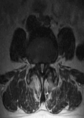
What structure (arrow) is shown in Figure 24?


Explanation

The structure illustrated is the sympathetic chain viewed from an anterolateral view of the lower lumbar spine. It descends along the anterolateral aspect of the spine into the pelvis closely adherent to the vertebral column. The spinal nerves, including L5, can be seen exiting from the foramen. The ureters descend from the kidneys and cross anterior to the iliac vessels to the bladder. Onibokun A, Khoo LT, Holly L: Anterior retroperitoneal approach to the lumbar spine, in Kim DH, Henn JS, Vaccaro AR, et al (eds): Surgical Anatomy and Techniques to the Spine. Philadelphia, PA, Saunders Elsevier, 2006, pp 101-105.


Question 9

The best patient-related outcomes, following the surgical treatment of cauda equina syndrome secondary to a large L5-S1 disk herniation, are most closely related to which of the following?


Explanation

The most predictable positive outcome from spinal surgery due to a cauda equina syndrome is early surgical intervention before any significant neurologic deficit develops. Meta-analysis studies demonstrate that surgical intervention more than 48 hours after the onset of cauda equina syndrome show an increased risk for poor outcomes. Ahn UM, Ahn NU, Buchowski JM, et al: Cauda equina syndrome secondary to lumbar disc herniation: A meta-analysis of surgical outcomes. Spine 2000;25:1515-1522.

Question 10

A 45-year-old man undergoes an anterior cervical diskectomy and fusion at C5-6 and C6-7 with instrumentation. During the first postoperative visit at 1 week, the patient reports difficulty swallowing and mild anterior cervical tightness. The anterior wound is benign and the patient denies any dyspnea or shortness of breath. A postoperative radiograph is seen in Figure 25. What is the most appropriate management at this time?


Explanation

The radiograph shows significant prevertebral soft-tissue swelling following a two-level anterior cervical diskectomy and fusion. The incidence of dysphagia 2 years after anterior cervical spine surgery is 13.6%. Risk factors for long-term dysphagia after anterior cervical spine surgery include gender, revision surgeries, and multilevel surgeries. The use of instrumentation, higher levels, or corpectomy versus diskectomy did not significantly increase the prevalence of dysphagia. Lee and associates demonstrated that while dysphagia after anterior cervical spine surgery is a common early finding, it generally decreases significantly by 6 months with nonsurgical management. A minority of patients experience moderate or severe symptoms by 6 months after the procedure. Female gender and multiple surgical levels have been identified as risk factors for the development of postoperative dysphagia. Lee MJ, Bazaz R, Furey CG, et al: Risk factors for dysphagia after anterior cervical spine surgery: A two-year prospective cohort study. Spine J 2007;7:141-147.


Question 11

Steroids are thought to prevent neurologic deterioration after traumatic spinal cord injury by which of the following mechanisms?


Explanation

The proposed mechanisms by which steroids such as methylprednisolone are thought to prevent neurologic deterioration by limiting secondary insult, include: decreasing the area of ischemia in the cord, reducing TNF-alpha expression and NF-kB binding activity, decreasing free radical oxidation and thus stabilizing cell and lysosomal membranes, and checking the influx of calcium into the injured cells, thus reducing cord edema. Slucky AV: Pathomechanics of spinal cord injury. Spine: State Art Rev 1999;13:409-417.

Question 12

Which of the following mechanisms of inhibition has been linked to cigarette smoking and lumbar spinal fusion?


Explanation

Cigarette smoking has been directly linked to pseudarthrosis in spinal fusions. The direct mechanism of action is diminished revascularization of cancellous bone graft. Additionally, a smaller area of revascularization is seen in these grafts, as well as an increased area of necrosis. Increased activity of osteoblasts would result in more bone production. Increased activity of osteocytes would not affect the fusion because osteocytes are mature bone cells.

Question 13

Which of the following is considered the most effective means of identifying an evolving motor tract injury during cervical spine surgery?


Explanation

In a study of 427 patients undergoing cervical spine surgery, 12 patients demonstrated substantial or complete loss of amplitude of the tceMEPs. Ten of those patients had complete reversal of the loss following prompt intraoperative intervention. SSEP monitoring failed to identify any changes in one of the two patients that awoke with a new motor deficit. SSEP changes lagged behind the tceMEP changes in patients in which major changes were detected by both modalities. TceMEP monitoring was 100% sensitive and 100% specific. SSEP monitoring was only 25% sensitive and 100% specific.

Question 14

A previously healthy 29-year-old man reports a 2-day history of severe atraumatic lower back pain. He denies any bowel or bladder difficulties and no constitutional signs. Examination is consistent with mechanical back pain. No focal neurologic deficits or pathologic reflexes are noted. What is the most appropriate management?


Explanation

In general, a previously healthy patient with an acute onset of nontraumatic lower back pain does not need diagnostic imaging before proceeding with therapeutic treatment. In the absence of any "red flags" during the history and physical examination, such as trauma or constitutional symptoms (ie, fevers, chills, weight loss), the appropriate treatment for acute onset lower back pain is purely symptomatic treatment including limited analgesics and early range of motion. Diagnostic imaging is not necessary unless the initial treatment is unsuccessful and symptoms are prolonged. Miller and associates suggested that the use of radiographs can lead to better patient satisfaction but not necessarily better outcomes. Miller P, Kendrick D, Bentley E, et al: Cost effectiveness of lumbar spine radiographs in primary care patients with low back pain. Spine 2002;27:2291-2297.

Question 15

Sacral fractures are most likely to be associated with neurologic deficits when they involve what portion of the sacrum?


Explanation

Denis divided the sacrum into three zones: zone 1 represents the lateral ala, zone 2 represents the foramina, and zone 3 represents the central canal. A fracture is classified according to its most medial extension. Those in zone 3 are typically bursting-type fractures or fracture-dislocations and are most prone to neurologic sequelae. Denis F, Davis S, Comfort T: Sacral fractures: An important problem. A retrospective analysis of 236 cases. Clin Orthop Relat Res 1988;227:67-81.

Question 16

Which of the following is associated with the use of bisphosphonates in the setting of metastatic breast cancer to the spine?


Explanation

The indications of bisphosphonate therapy in breast cancer patients range from the correction of hypercalcemia to the prevention of cancer treatment-induced bone loss. Bisphosphonates reduce metastatic bone pain in at least 50% of patients and can reduce the frequency of skeletal-related events by 30% to 40%. Osteonecrosis of the jaw could occur in up to 2.5% of breast cancer patients during long-term bisphosphonate therapy.

Question 17

A 67-year-old retired steelworker was involved in a motor vehicle accident and sustained a midcervical spinal cord injury. Radiographs and MRI scans reveal severe cervical stenosis and spondylosis without fractures or dislocations. Neurologic examination reveals an ASIA C spinal cord impairment with greater motor involvement of the upper extremities than the lower extremities. What is the probability that the patient eventually will become ambulatory?


Explanation

The patient sustained an incomplete spinal cord injury known as central cord syndrome. Central cord syndrome characteristically has disproportionate involvement of the upper extremities with the lower extremities being relatively spared. It is most commonly seen after cervical injuries in elderly patients with spondylosis and spinal stenosis, often without fracture. Penrod and associates noted that 23 of 59 patients with central cord syndrome (ASIA C and D) ultimately walked. The poorest prognosis, however, was in ASIA C patients older than age 50, in which only 40% walked. Penrod LE, Hegde SK, Ditunno JF Jr: Age effect on prognosis for functional recovery in acute, traumatic central cord syndrome. Arch Phys Med Rehab 1990;71:963-968.

Question 18

A 20-year-old man involved in a motor vehicle accident is brought to the emergency department with a C6-7 unilateral facet dislocation. His neurologic examination reveals a focal left-sided C7 nerve root palsy. He is awake and cooperative with questioning and has no other obvious traumatic injuries. What is the most appropriate treatment at this time?


Explanation

In the patient who is neurologically intact or has an incomplete injury from a cervical facet dislocation, a closed reduction with weighted tong traction is appropriate when the patient is awake, alert, and cooperative. Although there is a risk that a cervical facet dislocation could occur with an underlying cervical disk herniation, Vaccaro and associates have shown that closed reduction can be safely carried out in the awake, responsive patient. Closed reduction can be performed in the emergency department with traction with skull tongs or a halo ring. A slow stepwise application of weight is added until a reduction is achieved. Any worsening of the neurologic status of the patient requires immediate termination of the closed reduction and further diagnostic imaging before proceeding with further treatment. Vaccaro AR, Falatyn SP, Flanders AE, et al: Magnetic resonance evaluation of the intervertebral disc, spinal ligaments, and spinal cord before and after closed traction reduction of cervical spine dislocations. Spine 1999;24:1210-1217. Hart RA: Cervical facet dislocation: When is magnetic resonance imaging indicated? Spine 2002;27:116-117.

Question 19

A 66-year-old man reports a 2-week history of worsening low back and leg pain. He reports that his pain is aggravated by lying down and relieved by standing and walking. He notes that he has been losing weight recently and that his pain has been awakening him during the night. His medical history is significant for hypertension, coronary artery disease, and prostate cancer. His physical examination is essentially unremarkable. Lumbar radiographs are within normal limits. What is the most appropriate management for this patient?


Explanation

In the initial assessment of acute low back pain in adults, no diagnostic testing is indicated during the first 4 weeks in the absence of "red flags" for a serious underlying condition. The purpose of the initial assessment of acute low back pain in adults is to rule out serious underlying conditions presenting as low back pain. The Agency for Healthcare Policy and Research, in its 1994 clinical practice guideline, identified four serious conditions that may present with low back pain, including fracture, tumor, infection, and cauda equina syndrome. This patient has five "red flags" for a spinal tumor as a possible etiology of his low back pain, including age of older than 50 years, constitutional symptoms (recent weight loss), pain worse when supine, severe nighttime pain, and a history of cancer. Of these, his history of cancer is most significant, as greater than 90% of spinal tumors are metastatic. In order of frequency, breast, prostate, lung, and kidney make up approximately 80% of all secondary spread to the spine. In the presence of "red flags" for tumor or infection, it is recommended that the clinician obtain a CBC count, ESR, and a urinalysis. If these are within normal limits and suspicions still remain, consider consultation or seek further evidence with a bone scan, radiographs, or additional laboratory studies. Negative radiographs alone are insufficient to rule out disease. If radiographs are positive, the anatomy can be better defined with MRI. Agency for Health Care Policy and Research, Bigos SJ (ed): Acute Low Back Problems in Adults. Rockville, MD, US Department of Health and Human Services, AHCPR Publication 95-0642, Clinical Practice Guideline #14, 1994.

Question 20

Which of the following increases radiation exposure to patients and personnel during surgery?


Explanation

Continuous fluoroscopy and cine radiography expose the patient and personnel to markedly increased levels of direct and scatter radiation exposure. Continuous fluoroscopy should be limited to only what is absolutely needed for safe completion of the procedure. By orienting the cathode ray tube beneath the patient and placing the image intensifier as close as clinically possible to the patient, scatter radiation exposure to the personnel is minimized.

Question 21

A 78-year-old woman undergoes her third lumbar decompression and fusion from L3 to L5 without complication. On the morning of postoperative day 3, examination reveals painless, flaccid weakness of both lower extremities. She also has an absent bulbocavernous reflex and a mild saddle paresthesia. MRI scans of the lumbar spine are shown in Figures 26a and 26b. What is the most appropriate management at this time?


Explanation

The MRI scans reveal a large postoperative hematoma causing significant thecal compression. An epidural hematoma with neurologic deficit is a surgical emergency requiring immediate evacuation of the hematoma. Although the incidence of postoperative epidural hematomas is rare, the consequences of a missed diagnosis can be catastrophic. Early recognition and evacuation are essential in preserving or restoring neurologic function. Uribe and associates attributed delayed postoperative hematomas to previous multiple lumbar surgeries as a possible contributing factor. Yi S, Yoon do H, Kim KN, et al: Postoperative spinal epidural hematoma: Risk factor and clinical outcome. Yonsei Med J 2006;47:326-332.


Question 22

Figures 27a through 27c show the radiographs and CT scan of a 27-year-old man who sustained a low-velocity gunshot wound to the neck. He is quadriplegic (ASIA A), hemodynamically stable, and does not have drainage from his wound. After initial resuscitation and stabilization, the cervical spine and spinal cord injuries are best managed by


Explanation

Although the spinal canal has been penetrated, the lateral masses are intact bilaterally with only partial destruction of the vertebral body and penetration of the lamina on one side, thus the cervical spine is not unstable and surgical stabilization is not indicated. Dural repair is not indicated since there is no external cerebrospinal fluid leakage. Surgical treatment should be based on the need to treat extraspinal pathology only. Bono CM, Heary RF: Gunshot wounds to the spine. Spine J 2004;4:230-240.


Question 23

Which of the following is a true statement regarding thoracic disk herniations?


Explanation

Symptomatic herniations of the thoracic spine are much less common than those of the cervical or lumbar region. They tend to occur most commonly during the third to fifth decades of life and although they can be found at all levels, they are most common in the lower third near the thoracolumbar region. Posterior laminectomy and disk excision has the highest rate of neurologic deterioration and is not recommended. Multiple studies have shown that herniated thoracic disks can be found at one or more levels in 40% of asymptomatic individuals. Shah RP, Grauer JN: Thoracoscopic excision of thoracic herniated disc, in Vaccaro AR, Bono CM (eds): Minimally Invasive Spine Surgery. New York, NY, Informa Healthcare, 2007, pp 73-80.

Question 24

A sentinel event is defined as an unexpected occurrence involving death or serious physical or psychological injury, or the risk thereof. What is the most common sentinel event related to spine surgery?


Explanation

Patient safety and prevention of medical errors is a major focus of recent national advocacy groups. Analysis has shown that the most common sentinel event in spine surgery is surgery on the wrong level. Therefore, it is recommended that every patient have the surgical site signed, the level of surgery marked intraoperatively, and a radiograph taken. Surgery on the wrong level is most likely to occur in single-level decompressive procedures. Wong DA, Watters WC III: To err is human: Quality and safety issues in spine care. Spine 2007;32:S2-S8.

Question 25

What structure is most at risk with anterior penetration of C1 lateral mass screws?


Explanation

Posterior screw fixation of the upper cervical spine has gained a great deal of popularity due to its stable fixation, obviating the use of halo vest immobilization, and its high fusion rates. The use of screws in this location, however, has introduced a whole new set of potential complications. Vertebral artery injury is one of the most feared complications associated with screws in the C1/C2 region. This structure, however, is lateral and posterior at the C2 level and then penetrates the foramen transversarium of C1 to lie cephalad to the arch of C1 before entering the foramen magnum. It is the internal carotid artery that lies immediately anterior to the arch of C1 that is particularly at risk by anterior penetration of C1 lateral mass or C1-C2 transarticular screws as demonstrated by Currier and associates. The internal carotid artery lies posterior to the pharynx. The external carotid artery and the glossopharyngeal nerve are not at risk with this method of fixation. Currier BL, Todd LT, Maus TP, et al: Anatomic relationship of the internal carotid artery to the C1 vertebra: A case report of cervical reconstruction for chordoma and pilot study to assess the risk of screw fixation of the atlas. Spine 2003;28:E461-E467. Grant JC: Grant's Atlas of Anatomy, ed 6. Baltimore, MD, Williams & Wilkins, 1972.

Question 26

A 68-year-old man presents with neck pain after a low-speed motor vehicle collision. Radiographs demonstrate a Type II odontoid fracture with 6 mm of posterior displacement. He is neurologically intact. Which of the following factors is most strongly associated with a high risk of nonunion if this injury is treated nonoperatively in a halo vest?


Explanation

Risk factors for nonunion of Type II odontoid fractures include displacement > 5 mm, angulation > 10 degrees, and patient age > 50 years. Posterior displacement is not inherently a higher risk than anterior displacement, provided the magnitude is matched.

Question 27

A 55-year-old man with long-standing ankylosing spondylitis sustains a low-energy mechanical fall. He complains of severe back pain but has a normal neurologic exam. Initial AP and lateral radiographs of the thoracic and lumbar spine show no obvious fracture. What is the most appropriate next step in management?


Explanation

Patients with ankylosing spondylitis are at high risk for highly unstable, occult chalk-stick fractures even from low-energy trauma. A CT scan or MRI of the entire neuroaxis is required when they present with new-onset axial pain after a fall, as these fractures can easily be missed on plain radiographs.

Question 28

A 72-year-old woman with pre-existing cervical spondylosis falls forward, striking her chin. She presents with upper extremity weakness (motor grade 2/5) and lower extremity weakness (motor grade 4/5), along with preserved sacral sensation. Which spinal cord tracts are primarily involved in this classic neurologic pattern?


Explanation

This patient has central cord syndrome, characterized by upper extremity weakness that is more severe than lower extremity weakness. It predominantly affects the central gray matter and the medial portions of the lateral corticospinal tracts, which are somatotopically organized with the cervical tracts located more medially.

Question 29

A 45-year-old man presents with 4 weeks of radiating right arm pain. Examination reveals weakness in wrist extension, a diminished brachioradialis reflex, and altered sensation over the dorsal web space of the hand. Which cervical disc level is most likely herniated?


Explanation

The clinical presentation describes a C6 radiculopathy, which includes weak wrist extension, diminished brachioradialis reflex, and numbness in the thumb, index finger, or dorsal web space. In the cervical spine, the exiting nerve root corresponds to the lower vertebral level, meaning a C5-C6 disc herniation affects the C6 root.

Question 30

A 45-year-old man falls from a roof and sustains an L1 burst fracture. Neurologic examination is normal. Radiographs and CT show 40% loss of anterior vertebral body height and 25% canal compromise. The posterior ligamentous complex is intact on MRI. According to the Thoracolumbar Injury Classification and Severity (TLICS) scale, what is his total score and recommended treatment?


Explanation

The TLICS score assigns 2 points for a burst fracture, 0 points for intact neurologic status, and 0 points for an intact posterior ligamentous complex, totaling 2. A score of 3 or less is an indication for nonoperative management.

Question 31

A 68-year-old man with known cervical spondylosis presents after a hyperextension injury. He has significant upper extremity weakness (motor grade 2/5) but is able to move his lower extremities (grade 4/5). Sensation is variably diminished below the neck. Which of the following is the most likely prognosis for his neurologic recovery?


Explanation

This patient has a central cord syndrome. The typical recovery pattern involves the lower extremities recovering first and most completely, while upper extremity fine motor function often remains impaired.

Question 32

A 13-year-old premenarchal girl (Risser 0) has a right thoracic curve measuring 32 degrees. What is the most appropriate management?


Explanation

Bracing is indicated for skeletally immature patients (Risser 0-2) with curves between 25 and 45 degrees. A dose-response relationship exists, with 16-23 hours per day providing the most effective curve progression prevention.

Question 33

A 65-year-old woman complains of low back pain and neurogenic claudication. Standing radiographs demonstrate a grade I degenerative spondylolisthesis at L4-L5. Dynamic views show 4 mm of translation. She has failed 6 months of conservative treatment. Based on the SPORT trial, what is the most appropriate surgical intervention?


Explanation

The SPORT trial demonstrated that for degenerative spondylolisthesis with spinal stenosis, decompression and arthrodesis yield superior long-term outcomes compared to nonoperative care or decompression alone.

Question 34

A 72-year-old man with cervical spondylotic myelopathy is scheduled for surgery. MRI demonstrates T2 signal changes within the cord at C4-C5. Which MRI finding portends the worst prognosis for neurologic recovery after surgical decompression?


Explanation

While T2 hyperintensity alone indicates edema or myelomalacia, the combination of broad T2 hyperintensity and T1 hypointensity indicates permanent cystic necrosis of the cord, portending a poor prognosis.

Question 35

A 5-year-old child presents to the emergency department after a minor fall. A lateral cervical radiograph shows 3 mm of anterior translation of C2 on C3. Swischuk's line is evaluated. Which of the following findings confirms physiologic pseudosubluxation rather than true injury?


Explanation

Swischuk's line connects the anterior aspects of the posterior arches of C1 and C3. In normal pseudosubluxation, the anterior aspect of the C2 posterior arch should deviate less than 1.5 to 2 mm from this line.

Question 36

A 28-year-old man sustains a Type II odontoid fracture in a motor vehicle collision. The fracture is displaced 6 mm posteriorly with a comminuted base. Which of the following factors is most strongly associated with nonunion if treated nonoperatively in a halo vest?


Explanation

Risk factors for nonunion of Type II odontoid fractures include initial displacement > 5 mm, angulation > 10 degrees, comminution at the fracture base, and advanced age (> 50 years).

Question 37

A 55-year-old man with long-standing ankylosing spondylitis presents with severe neck pain after a minor fall. Neurologic examination is intact. Plain radiographs of the cervical spine appear unchanged from baseline due to marked osteopenia and deformity. What is the most appropriate next step in management?


Explanation

Patients with ankylosing spondylitis are at high risk for highly unstable extension-distraction fractures even with minor trauma. A CT scan of the entire cervical spine is mandatory to rule out occult fractures not visible on plain radiographs.

Question 38

A 15-year-old gymnast presents with a 3-month history of mechanical low back pain. Radiographs are normal. MRI shows bilateral marrow edema in the pars interarticularis of L5 but no obvious fracture line on T1-weighted images. What is the recommended initial management?


Explanation

MRI showing marrow edema without a definitive fracture line indicates an early stress reaction. Management consists of rest, cessation of hyperextension activities, and often a TLSO brace until symptoms resolve to prevent progression to a complete fracture.

Question 39

In planning surgical correction for adult degenerative scoliosis, the surgeon measures the pelvic incidence (PI). If the patient's PI is 55 degrees, what should be the approximate target for lumbar lordosis (LL) to achieve optimal sagittal balance?


Explanation

To achieve harmonious sagittal balance, the lumbar lordosis (LL) should match the pelvic incidence (PI) within 9 degrees (PI - LL < 10 degrees). Therefore, a target LL of 45 to 55 degrees is appropriate.

Question 40

In the Lenke classification for adolescent idiopathic scoliosis, a curve with a structural proximal thoracic curve, a structural main thoracic curve, and a nonstructural thoracolumbar curve is classified as which type?


Explanation

Lenke Type 2 is a Double Thoracic curve pattern, characterized by structural proximal and main thoracic curves, while the thoracolumbar/lumbar curve remains nonstructural.

Question 41

A 16-year-old boy presents with progressive mid-back pain and a cosmetic deformity. Radiographs reveal a thoracic kyphosis of 65 degrees. According to Sorensen's criteria, which radiographic finding is required for the diagnosis of Scheuermann's disease?


Explanation

Sorensen's classic criteria for diagnosing Scheuermann's kyphosis require the presence of anterior wedging of greater than 5 degrees in at least three consecutive apical vertebrae.

Question 42

A 42-year-old man presents with acute back pain, bilateral leg pain, perineal numbness, and urinary retention. MRI reveals a massive L4-L5 central disc herniation. Within what timeframe is surgical decompression generally recommended to optimize bladder function recovery?


Explanation

Cauda equina syndrome with urinary retention is a surgical emergency. Decompression within 48 hours has been shown to significantly improve outcomes regarding motor, sensory, and sphincter function recovery.

Question 43

A 7-year-old girl presents with torticollis 1 week after a pharyngitis infection. CT scan shows C1 rotated on C2 with no anterior displacement. What is the initial treatment of choice for this Fielding Type I rotatory subluxation?


Explanation

For Grisel syndrome (atlantoaxial rotatory subluxation associated with head/neck infections) presenting acutely (less than 1 week) as Fielding Type I, initial management is a soft collar, NSAIDs, and treatment of the underlying infection.

Question 44

A 45-year-old woman complains of right neck pain radiating down the arm. Examination shows weakness of triceps extension and wrist flexion, with an absent triceps reflex. Sensation is decreased over the middle finger. Which cervical nerve root is most likely compressed?


Explanation

The C7 nerve root is responsible for triceps and wrist flexor motor function, the triceps reflex, and sensation over the middle finger. Compression typically occurs at the C6-C7 disc level.

Question 45

During a posterior lumbar decompression for L4-L5 spinal stenosis, a dural tear occurs with CSF leak. It is repaired primarily. What is the recommended postoperative management regarding patient mobilization?


Explanation

Recent studies suggest that prolonged bed rest is unnecessary following a primary repair of an incidental durotomy. Early mobilization does not increase the rate of secondary CSF leak or complications.

Question 46

A 75-year-old woman with severe osteoporosis presents with acute back pain following a coughing fit. Radiographs show an acute L2 compression fracture with 20% height loss. She has intact neurology. What is the recommended first-line treatment?


Explanation

The initial management of an osteoporotic vertebral compression fracture without neurologic deficit is nonoperative, focusing on pain control, early mobilization to prevent deconditioning, and treating the underlying osteoporosis.

Question 47

A 3-year-old boy has a fully segmented hemivertebra at T8. Routine screening should include which of the following imaging modalities?


Explanation

Congenital scoliosis is strongly associated with other VACTERL anomalies. Renal ultrasound is needed to screen for genitourinary abnormalities, and a total spine MRI is critical to rule out intraspinal anomalies like tethered cord or diastematomyelia.

Question 48

A 19-year-old man wearing a lap seatbelt is involved in a high-speed collision. He has a flexion-distraction injury (Chance fracture) at L2. Which of the following concurrent injuries must be highly suspected and investigated?


Explanation

Chance fractures (flexion-distraction injuries) are frequently associated with lap seatbelt use and have a high correlation (up to 50%) with intra-abdominal visceral injuries, particularly bowel perforations.

Question 49

A 62-year-old man undergoes an anterior cervical discectomy and fusion (ACDF) for myelopathy. Postoperatively, he presents with difficulty swallowing solid foods. Which of the following is the most important intraoperative consideration to minimize postoperative dysphagia?


Explanation

Postoperative dysphagia is a common complication of anterior cervical spine surgery. Temporarily deflating the endotracheal tube cuff during retraction and minimizing excessive esophageal retraction pressure can help reduce this risk.

Question 50

A 78-year-old man presents with severe neck pain after a low-energy fall. Radiographs and CT scan reveal a Type II odontoid fracture with 6 mm of posterior displacement and comminution at the fracture base. He is neurologically intact. His medical history includes hypertension and mild osteopenia. What is the most appropriate management for this patient?


Explanation

Posterior C1-C2 instrumented fusion is the most reliable treatment for elderly patients with displaced Type II odontoid fractures. Nonoperative management (halo or collar) has an unacceptably high nonunion rate and morbidity in this age group, while anterior screw fixation is contraindicated given his osteopenia and fracture comminution.

Question 51

A 66-year-old woman presents with progressive neurogenic claudication and bilateral leg pain limiting her walking distance to less than one block. MRI demonstrates severe central canal stenosis at L4-L5 with a Grade I degenerative spondylolisthesis. After 6 months of failed nonoperative management including epidural injections, she elects for surgery. What is the most evidence-based surgical intervention?


Explanation

For patients with lumbar stenosis and concomitant degenerative spondylolisthesis, decompression alone (laminectomy) leads to a higher risk of subsequent instability and reoperation. Laminectomy combined with instrumented fusion provides superior long-term functional outcomes and stability.

Question 52

A 13-year-old premenarchal girl presents for evaluation of a spinal deformity. Examination reveals a right thoracic prominence. Radiographs show a right-sided structural main thoracic curve of 36 degrees. Her Risser stage is 1. What is the most appropriate next step in management?


Explanation

This patient has Adolescent Idiopathic Scoliosis (AIS) with a curve between 25-45 degrees, significant remaining growth (premenarchal, Risser 1), and is at high risk for progression. Full-time bracing (TLSO) with a dose-response goal of >18 hours daily has been shown to effectively decrease the progression of curves to the surgical threshold.

Question 53

A 35-year-old man involved in a high-speed motor vehicle collision as a restrained passenger sustains a flexion-distraction injury (Chance fracture) at L2. He is neurologically intact. Which of the following associated injuries must be most aggressively ruled out during his initial trauma evaluation?


Explanation

Chance fractures (flexion-distraction injuries) are frequently associated with lap-belt use and have a high correlation (up to 40-50%) with intra-abdominal organ injuries. Hollow viscus (bowel) injuries are the most common and must be urgently ruled out via advanced imaging or general surgery consultation.

Question 54

A 68-year-old man falls forward, striking his chin and hyperextending his neck. On examination, he has 3/5 motor strength in his upper extremities and 4+/5 strength in his lower extremities. He has patchy sensory deficits in his arms. What is the most likely diagnosis?


Explanation

Central cord syndrome typically occurs in older individuals with preexisting cervical spondylosis who sustain a hyperextension injury. It is characterized by disproportionately greater motor impairment in the upper extremities compared to the lower extremities.

Question 55

A 24-year-old man is brought to the emergency department after a shallow water diving accident. He is awake, alert, and cooperative, with no other traumatic injuries. Examination reveals full strength and sensation in all extremities. Radiographs and CT scan show a C5-C6 bilateral facet dislocation with 50% translation. What is the most appropriate immediate management?


Explanation

In an awake, alert, and cooperative patient with a cervical facet dislocation, rapid closed reduction using cranial traction is safe and indicated before obtaining an MRI. If the patient has an altered mental status or fails closed reduction, an MRI should be obtained prior to surgical intervention to evaluate for an extruded disc.

Question 56

A 5-year-old boy is evaluated for early-onset spinal deformity. Radiographs demonstrate multiple congenital vertebral anomalies. Which of the following specific anomalies carries the highest risk for rapid curve progression and often requires early prophylactic surgical fusion?


Explanation

A unilateral unsegmented bar with a contralateral hemivertebra represents a combined failure of formation and failure of segmentation. This creates an unopposed, highly asymmetric growth pattern that rapidly progresses, typically requiring early surgical intervention.

Question 57

A 48-year-old man presents with sharp, radiating left lower extremity pain after lifting a heavy box. An MRI of the lumbar spine reveals a large, left-sided far-lateral (extraforaminal) disc herniation at the L4-L5 level. Which nerve root is most likely compressed, and what clinical finding would be expected?


Explanation

A far-lateral (extraforaminal) disc herniation at L4-L5 compresses the exiting L4 nerve root, unlike a paracentral herniation at the same level which compresses the traversing L5 root. L4 radiculopathy clinically presents with anterior thigh pain, weakness in knee extension, and a diminished patellar reflex.

Question 58

A 62-year-old man with a history of long-standing ankylosing spondylitis presents with new-onset mechanical back pain following a minor slip and fall. Plain radiographs of the thoracolumbar spine show bridging syndesmophytes but no obvious fracture. What is the most appropriate next step in management?


Explanation

Patients with ankylosing spondylitis have highly rigid, osteopenic spines and can sustain highly unstable transvertebral or transdiscal fractures from trivial trauma. If clinical suspicion is high and plain films are negative or obscured, advanced imaging (CT or MRI) of the entire spine is mandatory due to the high risk of catastrophic neurologic decline.

Question 59

A 55-year-old woman with advanced rheumatoid arthritis presents for preoperative evaluation of severe cervical myelopathy. Which of the following radiographic parameters best predicts the likelihood of postoperative neurologic recovery following cervical decompression and stabilization?


Explanation

In the rheumatoid cervical spine, the posterior atlanto-dental interval (PADI), also known as the space available for the cord (SAC), is the most critical parameter predicting neurologic recovery. A PADI of less than 14 mm correlates strongly with irreversible cord damage and poor postoperative neurologic recovery.

Question 60

During surgical correction of a complex adult degenerative scoliosis, restoring sagittal balance is a primary goal to optimize postoperative function and pain relief. According to the Schwab criteria, what is the ideal postoperative target relationship between Pelvic Incidence (PI) and Lumbar Lordosis (LL)?


Explanation

In adult spinal deformity surgery, optimal sagittal balance is achieved when the patient's lumbar lordosis matches their innate pelvic incidence. The Schwab classification targets a PI-LL mismatch of less than 10 degrees to improve functional outcomes and minimize the risk of adjacent segment disease.

Question 61

A 16-year-old gymnast presents with persistent, localized low back pain that worsens with extension activities. She has failed 6 months of rest and physical therapy. Radiographs and a CT scan reveal a bilateral pars interarticularis defect at L5 with a Grade II spondylolisthesis. If surgical intervention is pursued, what is the standard treatment of choice?


Explanation

For a symptomatic adolescent with a high-grade (Grade II or above) isthmic spondylolisthesis that fails conservative management, L5-S1 posterolateral in situ fusion (often with instrumentation) is the gold standard. Pars repair is generally reserved for patients with defects but minimal to no slip (Grade 0 or early Grade I) at higher lumbar levels.

Question 62

A 30-year-old male construction worker falls from scaffolding, sustaining a T12 burst fracture. He is neurologically intact. Review of his CT and MRI shows a comminuted burst fracture with 20% canal compromise and an intact posterior ligamentous complex (PLC). According to the Thoracolumbar Injury Classification and Severity (TLICS) score, what is his score and recommended treatment?


Explanation

The TLICS score for this injury is 2: Morphology is burst (2 points), Neurology is intact (0 points), and PLC is intact (0 points). A score of 3 or less indicates nonoperative management, typically with a TLSO brace or early mobilization depending on pain and mechanical stability.

Question 63

A 52-year-old man of Japanese descent presents with clumsiness of the hands, broad-based gait, and hyperreflexia in both upper and lower extremities. Plain radiographs show dense, confluent ossification along the posterior aspect of the cervical vertebral bodies from C3 to C6. What is the primary pathomechanical cause of his symptoms?


Explanation

This presentation is classic for Ossification of the Posterior Longitudinal Ligament (OPLL), which has a higher prevalence in East Asian populations. The ossified ligament compresses the ventral spinal cord, leading to progressive cervical myelopathy.

Question 64

A 22-year-old man sustains a severe fracture-dislocation at T4 with complete spinal cord injury (ASIA A). In the trauma bay, his blood pressure is 80/50 mm Hg and his heart rate is 52 bpm. His extremities are warm and pink. What is the primary etiology of his hemodynamic instability?


Explanation

The patient is in neurogenic shock, characterized by hypotension and bradycardia due to the disruption of descending sympathetic pathways in the cervical or upper thoracic cord (above T6). This loss of sympathetic tone leads to unopposed vagal tone, peripheral vasodilation, and an inability to mount a tachycardic response.

Question 65

A 14-year-old girl is diagnosed with Adolescent Idiopathic Scoliosis. Her standing posteroanterior radiograph shows a 50-degree right thoracic curve and a 35-degree left lumbar curve. On dynamic side-bending radiographs, the thoracic curve corrects to 30 degrees and the lumbar curve corrects to 15 degrees. According to the Lenke Classification system, how is the lumbar curve defined?


Explanation

In the Lenke Classification system, a minor curve is considered non-structural if it bends out to less than 25 degrees on side-bending radiographs. Since her lumbar curve corrects to 15 degrees, it is non-structural, classifying this as a Lenke Type 1 (Main Thoracic) curve.

Question 66

A 45-year-old woman complains of neck and arm pain radiating down to her thumb and index finger. Examination reveals decreased sensation over the radial aspect of the forearm, weakness in wrist extension, and a diminished brachioradialis reflex. An MRI is most likely to show a disc herniation at which cervical level?


Explanation

The patient's findings (thumb/index numbness, weak wrist extension, decreased brachioradialis reflex) correspond to a C6 radiculopathy. In the cervical spine, exiting nerve roots exit above the corresponding pedicle, so a C5-C6 disc herniation compresses the C6 root.

Question 67

A 12-year-old boy with non-ambulatory spastic quadriplegic cerebral palsy presents with a severe, progressive sweeping neuromuscular scoliosis of 85 degrees and a pelvic obliquity of 25 degrees. He is experiencing difficulty sitting in his customized wheelchair. What is the most appropriate definitive surgical intervention?


Explanation

In non-ambulatory patients with severe neuromuscular scoliosis and significant pelvic obliquity, the standard of care to restore sitting balance is a long posterior spinal fusion extending from the upper thoracic spine down to the pelvis. Stopping short of the pelvis frequently leads to recurrent pelvic obliquity and seating difficulties.

Question 68

A 35-year-old woman presents with severe low back pain, bilateral sciatica, and new-onset urinary incontinence. Physical examination reveals saddle anesthesia and decreased anal sphincter tone. To maximize the chance of full bladder function recovery, surgical decompression should ideally be performed within what timeframe from symptom onset?


Explanation

The patient has Cauda Equina Syndrome, a surgical emergency. The literature strongly suggests that surgical decompression performed within 24 to 48 hours of symptom onset provides the best prognosis for the recovery of bladder and bowel function.

Question 69

A 50-year-old man underwent an uncomplicated L4-L5 posterior instrumented fusion three years ago for degenerative spondylolisthesis. He now presents with new-onset severe left thigh pain and weakness in knee extension. Radiographs show solid fusion at L4-L5. What is the most likely diagnosis?


Explanation

The patient is experiencing new L4 radicular symptoms (thigh pain, knee extension weakness) above a solid L4-L5 fusion. This is characteristic of adjacent segment disease at the L3-L4 level, which commonly develops due to increased biomechanical stress adjacent to a rigid construct.

Question 70

A 68-year-old man with a long-standing history of ankylosing spondylitis presents to the emergency department with severe lower neck and upper back pain after a low-speed motor vehicle collision. Neurologic examination is unremarkable. Initial anteroposterior and lateral radiographs of the cervical and thoracic spine show no obvious fracture. What is the most appropriate next step in management?


Explanation

Patients with ankylosing spondylitis are at high risk for highly unstable, occult spinal fractures even after minor trauma. Due to altered bone density and overlapping anatomy, plain radiographs are inadequate; a CT scan is the preferred initial imaging modality to rule out fracture.

Question 71

A 12-year-old girl is evaluated for a spinal deformity. She is premenarchal, has open triradiate cartilages, and is Risser 0. Radiographs reveal a right thoracic adolescent idiopathic scoliosis (AIS) curve of 35 degrees. What is the most appropriate management?


Explanation

In a highly immature patient (Risser 0, open triradiate cartilage, premenarchal) with an AIS curve between 25 and 44 degrees, full-time bracing (16-23 hours/day) is indicated to prevent curve progression. Observation is inappropriate given her extremely high risk of progression.

Question 72

A 75-year-old man presents with a Type II odontoid fracture following a fall. Which of the following fracture characteristics is the strongest independent predictor of nonunion with nonoperative management?


Explanation

Displacement greater than 5 mm is a well-established and strong independent risk factor for nonunion in Type II odontoid fractures treated conservatively. Other relative risk factors include age older than 50 years and initial angulation greater than 10 degrees.

Question 73

A 65-year-old woman with a history of progressive neurogenic claudication over the past 2 years has failed extensive nonoperative management. Imaging shows an L4-L5 grade I degenerative spondylolisthesis with severe central canal and lateral recess stenosis. She undergoes an L4-L5 laminectomy and posterior spinal fusion. Compared to laminectomy alone, the addition of a fusion in this patient primarily decreases the risk of which of the following?


Explanation

Degenerative spondylolisthesis involves dynamic instability. Decompression (laminectomy) alone can destabilize the spine further, leading to progressive slippage and recurrent neurogenic claudication, which is significantly mitigated by adding a fusion.

Question 74

A 28-year-old man presents to the trauma bay after a diving accident. He is awake, alert, and cooperative. Examination reveals 0/5 strength in bilateral triceps, hand intrinsics, and finger flexors, but normal strength in deltoids, biceps, and wrist extensors. Sensation is absent below the C6 dermatome. Lateral cervical radiographs reveal a bilateral C6-C7 facet dislocation. What is the most appropriate next step in management?


Explanation

In an awake, alert, and cooperative patient with a cervical facet dislocation and a neurologic deficit, urgent awake closed reduction with cranial traction is indicated to decompress the spinal cord. MRI is recommended prior to reduction only if the patient is unexaminable (e.g., comatose) to rule out a compressive disc herniation.

Question 75

A 45-year-old man presents with severe right leg pain. Examination reveals a positive femoral nerve stretch test, 4/5 strength in right knee extension, and decreased sensation over the medial aspect of the right lower leg. An MRI of the lumbar spine reveals a far lateral (extraforaminal) disc herniation at the L4-L5 level on the right. Which nerve root is most likely compressed?


Explanation

In the lumbar spine, a far lateral (extraforaminal) disc herniation compresses the exiting nerve root at that level. Therefore, a far lateral disc herniation at L4-L5 compresses the L4 nerve root, whereas a central or paracentral herniation at the same level would compress the traversing L5 nerve root.

Question 76

A 68-year-old woman is planning to undergo posterior spinal instrumentation and fusion for progressive adult spinal deformity and sagittal imbalance. Preoperative radiographic measurements reveal a pelvic incidence (PI) of 55 degrees. To optimize her postoperative sagittal balance and clinical outcomes, the surgical correction should aim for a lumbar lordosis (LL) measurement of approximately:


Explanation

To achieve optimal sagittal balance and reduce the risk of adjacent segment failure in adult spinal deformity, the lumbar lordosis (LL) should be restored to within 10 degrees of the patient's pelvic incidence (PI). Therefore, a goal LL of approximately 55 degrees is ideal.

Question 77

A 14-year-old boy is brought to the emergency department after a high-speed motor vehicle collision in which he was a rear-seat, lap-belted passenger. He complains of severe lower back pain. Radiographs and a CT scan reveal a flexion-distraction injury (Chance fracture) at L2. Given this injury pattern, what additional evaluation is most critical for this patient?


Explanation

Chance fractures (flexion-distraction injuries) are frequently sustained by lap-belted passengers in motor vehicle collisions and are highly associated with concurrent intra-abdominal injuries. Approximately 30% to 50% of these cases present with a concomitant hollow viscus injury.

Question 78

A 13-year-old premenarchal girl presents with a right thoracic curve of 32 degrees. Her Risser stage is 1. What is the most appropriate management?


Explanation

In a growing child (Risser 0-2, premenarchal) with an idiopathic curve between 25 and 45 degrees, bracing is indicated. A rigid TLSO worn for at least 18 hours a day has been shown to significantly decrease the risk of curve progression to surgical magnitude.

Question 79

A 65-year-old man presents with progressive gait instability and poor fine motor skills. Examination shows a positive Hoffmann sign and hyperreflexia. MRI reveals multi-level cervical stenosis from C3 to C6 with cord signal change, and dynamic radiographs show neutral sagittal alignment without instability. What is the most appropriate surgical intervention?


Explanation

Cervical laminoplasty is an ideal motion-preserving option for multi-level cervical myelopathy in the absence of kyphosis or instability. It avoids the morbidity of a multi-level anterior approach and the pseudoarthrosis or adjacent segment risks of a long posterior fusion.

Question 80

A 40-year-old man falls from a height and sustains a T12 burst fracture. He is neurologically intact. CT scan shows 15 degrees of kyphosis, 40% loss of vertebral body height, and an intact posterior ligamentous complex. What is his Thoracolumbar Injury Classification and Severity (TLICS) score and recommended treatment?


Explanation

The TLICS score is calculated by injury morphology (burst = 2), neurologic status (intact = 0), and posterior ligamentous complex integrity (intact = 0). A score of 2 suggests nonoperative management with a brace is appropriate.

Question 81

A 72-year-old man with known cervical spondylosis presents after a hyperextension injury. He has severe weakness in his bilateral hands and arms (1/5 strength) but retains 4/5 strength in his lower extremities. Sensation is intact. What is the most likely diagnosis?


Explanation

Central cord syndrome typically occurs after a hyperextension injury in patients with preexisting cervical spondylosis. It affects the centrally located cervical motor tracts more severely, resulting in disproportionately greater upper extremity weakness compared to the lower extremities.

Question 82

A 62-year-old woman presents with neurogenic claudication and a grade I degenerative spondylolisthesis at L4-L5. Dynamic radiographs demonstrate 4 mm of translation on flexion-extension. She has failed 6 months of conservative care. What is the most appropriate surgical treatment?


Explanation

Laminectomy with instrumented posterolateral fusion is the gold standard for symptomatic degenerative spondylolisthesis with dynamic instability. Decompression alone in the setting of instability leads to a high rate of progressive slip and need for revision surgery.

Question 83

An 80-year-old man sustains a Type II odontoid fracture after a ground-level fall. Which of the following factors most significantly increases his risk of nonunion if treated conservatively with a rigid cervical collar?


Explanation

Risk factors for nonunion in Type II odontoid fractures include age >50 years, displacement >5 mm, posterior displacement, and comminution. Geriatric patients have significantly higher nonunion rates with conservative care, often warranting a discussion on surgical stabilization.

Question 84

A 45-year-old man develops severe, acute right anterior thigh pain and weakness in knee extension. Reflex examination shows a diminished right patellar reflex. MRI shows a far-lateral (extraforaminal) disc herniation at the L4-L5 level. Which nerve root is most likely compressed?


Explanation

A far-lateral (extraforaminal) disc herniation compresses the exiting nerve root at that level. At L4-L5, the exiting nerve root is L4, leading to anterior thigh pain, quadriceps weakness, and a diminished patellar reflex.

Question 85

A 60-year-old man with a long-standing history of ankylosing spondylitis presents with back pain after a minor fall. Radiographs appear unchanged from his baseline, but he reports new-onset leg weakness. What is the most appropriate next step in management?


Explanation

Patients with ankylosing spondylitis are at high risk for unstable, highly displaced fractures even from low-energy trauma. Plain radiographs are notoriously unreliable in these patients, making advanced imaging (CT or MRI of the entire spine) mandatory to rule out occult fractures and epidural hematomas.

Question 86

A 25-year-old man is brought to the ED after a motor vehicle collision. He is awake, alert, and cooperative. Examination reveals intact motor and sensory function in all extremities. Cervical spine imaging demonstrates a right-sided unilateral C5-C6 facet dislocation. What is the recommended initial management?


Explanation

In an awake, cooperative, and neurologically intact patient with a cervical facet dislocation, urgent awake closed reduction via cranial traction is recommended. MRI is generally reserved for patients who fail closed reduction, have an altered mental status, or develop neurologic deficits during traction.

Question 87

A 68-year-old woman with adult degenerative scoliosis is undergoing evaluation for corrective surgery. Her pelvic incidence (PI) is 60 degrees. To achieve optimal sagittal balance and minimize the risk of adjacent segment disease and mechanical failure, her lumbar lordosis (LL) should be reconstructed to approximately what value?


Explanation

Optimal sagittal balance requires the lumbar lordosis (LL) to be within 10 degrees of the pelvic incidence (PI). Therefore, for a PI of 60 degrees, the LL should be reconstructed to approximately 50-60 degrees to prevent flatback deformity.

Question 88

A 16-year-old boy presents with back pain and a rounded back. Radiographs reveal a thoracic kyphosis of 65 degrees. According to Sorensen's criteria, which of the following radiographic findings confirms the diagnosis of Scheuermann's kyphosis?


Explanation

Sorensen's criteria for the diagnosis of Scheuermann's kyphosis require anterior wedging of 5 degrees or more in at least three consecutive vertebrae. It is often accompanied by Schmorl's nodes and endplate irregularities.

Question 89

A 70-year-old man complains of bilateral calf and buttock pain that worsens with walking. He states that leaning forward on a shopping cart completely relieves his symptoms. He has normal lower extremity pulses. Which of the following differentiates neurogenic claudication from vascular claudication?


Explanation

Neurogenic claudication is characteristically relieved by lumbar flexion (e.g., leaning on a shopping cart or sitting), which increases the cross-sectional area of the spinal canal. Vascular claudication is typically relieved simply by resting or standing still and worsens with muscle exertion regardless of posture.

Question 90

A 14-year-old boy with non-ambulatory spastic cerebral palsy presents with a 75-degree thoracolumbar scoliotic curve and severe pelvic obliquity causing skin breakdown over his ischial tuberosity. What is the most appropriate surgical strategy?


Explanation

In non-ambulatory patients with neuromuscular scoliosis and significant pelvic obliquity, long posterior spinal fusion extending from the upper thoracic spine down to the pelvis is required. This extensive fusion corrects the obliquity, restores sitting balance, and prevents pressure ulcers.

Question 91

A newborn is noted to have a hemivertebra at T8 causing a mild congenital scoliosis. Which of the following screening tests is mandatory in the initial evaluation of this patient?


Explanation

Congenital scoliosis is highly associated with VACTERL anomalies, particularly genitourinary and cardiovascular defects. A renal ultrasound and an echocardiogram are mandatory screening tests to rule out associated visceral anomalies.

Question 92

A 22-year-old woman involved in a high-speed motor vehicle collision while wearing a lap belt sustains a flexion-distraction (Chance) injury at L2. She is hemodynamically stable and neurologically intact. What concomitant injury is most highly associated with this fracture pattern?


Explanation

Chance fractures (flexion-distraction injuries) are frequently caused by lap seatbelts acting as a fulcrum. They are highly associated with intra-abdominal injuries, particularly to hollow viscous organs (e.g., bowel perforations), which occur in up to 50% of these patients.

Question 93

A 65-year-old woman presents with right L5 radiculopathy. MRI reveals a cystic structure arising from the L4-L5 facet joint severely compressing the thecal sac and right traversing L5 nerve root. Dynamic radiographs demonstrate grade I degenerative spondylolisthesis at L4-L5. What is the best definitive surgical treatment?


Explanation

Synovial facet cysts are a hallmark of underlying facet joint instability. While excision alone removes the compression, recurrence is high if the underlying instability is not addressed; therefore, decompression with instrumented fusion is the most definitive treatment.

Question 94

A 62-year-old man with a long-standing history of ankylosing spondylitis presents to the emergency department with severe back pain after a mechanical fall from standing height. Neurologic examination is normal. Radiographs and CT scan reveal a displaced extension-type fracture through the C7-T1 disc space. What is the most appropriate management?


Explanation

Fractures in the ankylosed spine are highly unstable and behave biomechanically like long-bone fractures. Rigid internal fixation with long posterior constructs (typically 3 levels above and below) is required to prevent catastrophic neurologic injury and achieve union.

Question 95

A 55-year-old man of East Asian descent presents with progressive clumsiness in his hands, difficulty buttoning his shirts, and a broad-based gait. Examination reveals hyperreflexia, a positive Hoffmann sign, and positive Babinski reflex. Imaging shows a continuous band of ossification along the posterior aspect of the vertebral bodies from C3 to C6, causing spinal cord compression. His cervical spine alignment is lordotic (K-line positive). What is the most appropriate surgical approach?


Explanation

Multi-level OPLL (3 or more levels) with preserved cervical lordosis (K-line positive) is best managed with a posterior decompression such as laminoplasty. This approach decompresses the cord via indirect drift-back, avoiding the high risk of dural tears seen in multi-level anterior OPLL resections and preventing post-laminectomy kyphosis.

Question 96

A 14-year-old Risser 0 female presents with adolescent idiopathic scoliosis. A standing posteroanterior radiograph demonstrates a right thoracic curve measuring 52 degrees and a left lumbar curve measuring 35 degrees. On supine lateral bending films, the thoracic curve reduces to 28 degrees and the lumbar curve reduces to 15 degrees. Sagittal alignment is normal. What is the most appropriate surgical strategy?


Explanation

This patient has a Lenke Type 1 curve, defined by a structural main thoracic curve (residual bend >25 degrees) and a non-structural lumbar curve (bends to <25 degrees). The gold standard surgical treatment is a selective thoracic fusion, which corrects the primary deformity while preserving lumbar motion segments.

None

Clinic OS
Medically Verified Content by
Prof. Clinic OS
Consultant Orthopedic & Spine Surgeon
Chapter Index