العربية
Part of the Master Guide

AAOS Orthopedic MCQs (Set 1): Spine Disorders, Sports Medicine & Arthroplasty | 2026 Board Review

AAOS Spine Surgery MCQs (Set 1): Degenerative, Trauma & Deformity | Board Prep

23 Apr 2026 56 min read 135 Views
Spine 2006 MCQs - Part 1

Key Takeaway

This high-yield question set (Set 1) for the AAOS, ABOS, and OITE exams covers critical spine surgery topics. It includes detailed MCQs on degenerative spine conditions like disc herniation and stenosis, spinal trauma management, and principles of spinal deformity correction. Prepare for your board exams with solved practice questions.

AAOS Spine Surgery MCQs (Set 1): Degenerative, Trauma & Deformity | Board Prep

Comprehensive 100-Question Exam


00:00

Start Quiz

Question 1

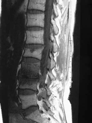
Figures 1a and 1b show the sagittal T2- and T1-weighted MRI scans of a 25-year-old intravenous drug abuser who has low back pain that is increasing in intensity. Laboratory studies show a WBC count of 10,000/mm3 and an erythrocyte sedimentation rate of 80 mm/h. Blood culture is negative. Initial management consist of


Explanation

The MRI scans show vertebral diskitis/osteomyelitis. The treatment of spinal infection in adults should be organism specific; therefore, initial management should consist of CT-guided closed biopsy prior to administration of antibiotic coverage. An open biopsy is indicated for a failed closed biopsy or failure of nonsurgical management. Although Staphylococcus aureus is the most common bacteria, a history of intravenous drug abuse raises suspicion for other organisms, including Pseudomonas. Tay BK, Deckey J, Hu SS: Spinal Infections. J Am Acad Orthop Surg 2002;10:188-197.

Question 2

A 27-year-old man sustained a gunshot wound to the lumbar spine and undergoes an exploratory laparotomy. An injury to the cecum is identified and treated. Management should now include


Explanation

Gunshot wounds to the spine present relatively little risk of infection in most cases. When there has been an injury to the colon, the risk of infection can be minimized with a 7-day course of broad-spectrum antibiotics. Fragment removal is not indicated. Roffi RP, Waters RL, Adkins RH: Gunshot wounds to the spine associated with a perforated viscus. Spine 1989;14:808-811.


Question 3

A 68-year-old man reports a 1-year history of debilitating neck pain without neurologic symptoms. History reveals a C5-6 anterior diskectomy and bone grafting 10 years ago that provided good relief of arm and neck pain. Radiographs show evidence of fibrous union at C5-6, spondylotic disk narrowing at C4-5 and C6-7, and a fixed 2-mm subluxation at C3-4. Examination reveals cervical stiffness and discomfort at the extremes of movement. His neurologic examination is normal. Treatment should now consist of


Explanation

Axial pain can be difficult to manage. Pain management is not always successful, and surgical approaches may provide disappointing results unless there is discrete pathology. Whereas planning of a surgical approach should consider prior approaches and preexisting laryngeal dysfunction, no compelling case for surgical intervention can be made for this patient. Therefore, management should consist of patient education, exercise, and nonnarcotic medication. Ahn NU, Ahn UM, Andersson GB, et al: Operative treatment of the patient with neck pain. Phys Med Rehabil Clin N Am 2003;14:675-692. Algers G, Pettersson K, Hildingsson C, et al: Surgery for chronic symptoms after whiplash injury: Follow-up of 20 cases. Acta Orthop Scand 1993;64:654-656.


Question 4

Which of the following is considered the lowest level that a standard thoracolumbosacral orthosis (TLSO) can immobilize?


Explanation

Without more distal immobilization such as a thigh extension, the lower two lumbar segments generally show the same or even increased mobility with a TLSO. White AA, Panjabi MM: Clinical Biomechanics of the Spine, ed 2. Philadelphia, PA, JB Lippincott, 1990, pp 475-509.


Question 5

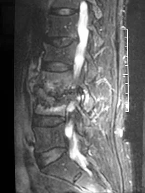
A 65-year-old man with ankylosing spondylitis has neck pain after falling back over his lawnmower, striking his thoracic spine, and forcing his neck into extension. Examination reveals subtle weakness of the intrinsics and finger flexors at approximately 4+/5. Initial management consists of immobilization in a rigid collar, and placing his head in the anatomic position. Radiographs reveal a subtle extension fracture of the lower cervical spine. Approximately 6 hours after the injury, he reports increasing paresthesias in his upper and lower extremities, and examination now shows his intrinsics are 2/5, finger flexors are 3/5, and his triceps are now weak at 4/5 on manual motor testing. In addition, his lower extremities now show weakness in both dorsal and plantar flexion of the ankle in the range of 4/5. Repeat radiographs appear unchanged. An MRI scan is shown in Figure 2. Management should now consist of


Explanation

It is not uncommon for patients with ankylosing spondylitis to sustain extension-type fractures, most typically of the cervicothoracic junction. These fractures can appear nondisplaced or minimally displaced initially, making them difficult to diagnose. Because there is no mobility between vertebrae, fractures tend to occur more like those of a transverse fracture of a long bone. In addition, the vertebral bodies are vascular and their canals are relatively enclosed, making them vulnerable to epidural bleeding. The MRI scan reveals an epidural hematoma located posteriorly on the cord; therefore, the treatment of choice is surgical evacuation and a posterior laminectomy. Because of the intrinsic instability of such fractures at the time of the laminectomy, internal fixation and stabilization with a posterior fusion is warranted. A simple laminectomy will only increase instability, and control is unlikely with halo vest immobilization. An anterior procedure will not effectively treat the problem given the location of the hematoma. Consideration can be given to methylprednisolone and observation; however, this will not eradicate the problem. Bohlman HH: Acute fractures and dislocations of the cervical spine. J Bone Joint Surg Am 1979;61:1119-1142.


Question 6

Figures 3a and 3b show the MRI scans of a patient with neck pain. What is the most likely diagnosis?


Explanation

Muliple neurofibromas result in marked foraminal enlargement as seen on the sagittal MRI scan. Collagen disorders leading to dural ectasia may show similar enlargement, but none of these is listed as a possible answer. Kim HW, Weinstein SL: Spine update: The management of scoliosis in neurofibromatosis. Spine 1997;22:2770-2776.


Question 7

A Trendelenburg gait is most likely to be seen in association with


Explanation

A Trendelenburg gait results from weakness of the gluteus medius, which is innervated by the L5 nerve root. A paracentral disk herniation at L4-L5 most commonly results in an L5 radiculopathy and thus weakness of the gluteus medius. A paracentral herniation at L5-S1 most commonly affects the S1 nerve root. A paracentral herniation at L3-L4, a central herniation at L3-L4, and a far lateral herniation at L4-L5 all affect the L4 root. Fardon DF, Garfin SR, Abitbol J, et al (eds): Orthopedic Knowledge Update: Spine 2. Rosemont, IL, American Academy of Orthopaedic Surgeons, 2002, pp 323-332.


Question 8

An otherwise healthy 70-year-old man has back and bilateral leg pain in an L5 distribution that is aggravated by standing more than 10 minutes or walking more than 100 feet. He has to sit to get relief. Neurologic and pulse examinations are normal. A radiograph and MRI scan are shown in Figures 4a and 4b. Treatment should consist of


Explanation

The patient has a degenerative spondylolisthesis at L4-5 with associated spinal stenosis. His symptoms are consistent with neurogenic claudication. Based on these findings, the surgical treatment of choice is decompression and posterolateral fusion. Use of instrumentation is controversial. Laminectomy alone is reserved for the patient who is frail medically. There is no role for an anterior approach or for fusion alone without decompression. Fischgrund JS, Mackay M, Herkowitz HN, et al: Degenerative lumbar spondylolisthesis with spinal stenosis: A prospective, randomized study comparing decompressive laminectomy and arthrodesis with and without spinal instrumentation. Spine 1997;22:2807-2812.


Question 9

Figures 5a and 5b show the radiograph and MRI scan of a patient who has severe mechanical neck pain but no neurologic problems. Biopsy and work-up show the lesion to be a solitary plasmacytoma. Treatment should consist of


Explanation

Plasmacytoma is very sensitive to radiation therapy and given the complexity of the resection and complications of surgery in the given location, radiation therapy is preferred. However, the patient has clear loss of bony structural integrity, and resultant instability would persist even with tumor irradiation; therefore, posterior stabilization is warranted. Chemotherapy and bone marrow transplant are reserved for systemic disease with multiple myeloma. Corwin J, Lindberg RD: Solitary plasmacytoma of bone vs. extramedullary plasmacytoma and their relationship to multiple myeloma. Cancer 1979;43:1007-1013.


Question 10

A 71-year-old woman undergoes a posterior lumbar decompression and fusion from L4-S1. Thirty-six hours after the procedure, she reports severe right-sided chest pain and shortness of breath. Doppler ultrasound reveals a clot proximal to the knee within the femoral vein. A large pulmonary embolus is confirmed by CT angiography. The next most appropriate step in management should consist of


Explanation

In a review of 13,000 spinal procedures, nine patients were treated with heparin following development of pulmonary emboli. Of these patients, six had serious complications ranging from wound drainage to paralysis. Heparin therapy instituted within 10 days of the surgical procedure resulted in a 100% complication rate. Vena cava filter placement has a complication rate of 0.12% to 10.1%. Removable filters are currently in clinical trials. Cain JE Jr, Major MR, Lauerman WC, et al: The morbidity of heparin therapy after development of pulmonary embolus in patients undergoing thoracolumbar or lumbar spinal fusion. Spine 1995;20:1600-1603. Roberts AC: Venous imaging and inferior vena cava filters. Curr Opin Radiol 1992;4:88-96.


Question 11

Figure 6 shows the lateral radiograph of a 22-year-old woman who has painful Scheuermanns's kyphosis in the middle and lower thoracic spine. When planning surgical correction using instrumentation, the distal aspect of the instrumentation should ideally extend to the


Explanation

Posterior constructs for Scheuermann's kyphosis ideally should extend from the most superior to the most inferior aspect of the Cobb angulation. However, the most distal fusion level must be in a stable or lordotic position to avoid the development of junctional kyphosis. Lowe reported that failure to incorporate the first lordotic segment of the lumbar spine is associated with a higher risk of junctional kyphosis. The first lordotic segment of the lumbar spine is typically at least one level below the distal aspect of the curve as measured by the Cobb technique and most commonly is in the upper part of the lumbar spine. Lowe TG: Scheuermann's disease. Orthop Clin North Am 1999;30:475-487.


Question 12

Figure 7 shows the radiograph of a 64-year-old man who has neck pain and weakness of the upper and lower extremities following a motor vehicle accident. Examination reveals 3/5 quadriceps and 4/5 hip flexors but no ankle dorsiflexion or plantar flexion. His intrinsics are 1/5, with finger flexors of 3/5. He is awake, alert, and cooperative. Management should consist of


Explanation

In patients with facet dislocations and an incomplete neurologic deficit, early decompression of the canal via reduction of the dislocation generally is considered safe if the patient is alert and can cooperate. However, patients who cannot cooperate with serial neurologic examinations during the reduction are at risk for increased deficit secondary to herniated nucleus pulposus, and MRI should be performed prior to either closed or open reduction. Star AM, Jones AA, Cotler JM, et al: Immediate closed reduction of cervical spine dislocations using traction. Spine 1990;15:1068-1072.


Question 13

What is the recommended insertion torque for halo pins in adults?


Explanation

Garfin and associates have shown that halo pins inserted with 8 in-lb of insertion torque results in significantly less loosening with cyclical loading than pins inserted with 6 in-lb of torque. Moreover, Botte and associates reported that 8 in-lb of torque is clinically safe and effective in lowering the incidence of pin loosening and infection. Botte MJ, Byrne TP, Garfin SR: Application of the halo device for immobilization of the cervical spine utilizing an increased torque pressure. J Bone Joint Surg Am 1987;69:750-752. Garfin SR, Lee TO, Roux RD, et al: Structural behavior of the halo orthosis pin-bone interface: Biomechanical evaluation of standard and newly designed stainless steel halo fixation pins. Spine 1986;11:977-981.


Question 14

A 55-year-old woman with a history of untreated idiopathic scoliosis has had neurogenic claudication for the past several months. MRI reveals spinal stenosis at L2-L3, L3-L4, and L4-L5. Radiographs show a 45-degree lumbar curve from T10 to L4, with a degenerative spondylolisthesis at L4-L5. Laminectomy at the stenotic levels and stabilization of the deformity are planned. Which of the following is NOT considered an absolute indication for extending the fusion to the sacrum, rather than stopping at L5?


Explanation

There are several indications for extending adult scoliosis fusions to the sacrum, rather than stopping in the lower lumbar spine. These indications include posterior column deficiencies at L5-S1, such as spondylolysis and laminectomy, and deformities extending to the sacrum, such as fixed tilt of L5-S1 or sagittal imbalance. MRI signal changes in the L5-S1 disk do not preclude stopping the fusion at L5. Some surgeons use diskography or diagnostic facet blocks to evaluate the integrity of the L5-S1 level prior to stopping the fusion at L5. Long scoliosis fusions stopping at L5 have a significant risk of failure, highlighting the importance of careful selection of fusion levels. Bradford DS, Tay BK, Hu SS: Adult scoliosis: Surgical indications, operative management, complications, and outcomes. Spine 1999;24:2617-2629. Bridwell KH: Where to stop the fusion distally in adult scoliosis: L4, L5, or the sacrum? Instr Course Lect 1996;45:101-107.


Question 15

Which of the following findings is considered a contraindication for posterior decompression (with or without fusion) for myelopathy?


Explanation

Although cervical instability is a contraindication to posterior decompression alone, segmental instability in the myelopathic cervical spine can be addressed with concomitant posterior fusion with instrumentation. Cervical lordosis represents the ideal scenario for posterior decompressive procedures for myelopathy (laminectomy and laminoplasty) because compression from anterior osteophytes, if present, is relieved as the spinal cord migrates posteriorly. The anteroposterior diameter of the spinal canal does not have an impact on the selection of surgical approach. Posterior unroofing-type procedures in kyphotic cervical spines, however, are ineffective because anterior impingement on the spinal cord will remain; therefore, kyphosis of more than 10 degrees is considered a contraindication for posterior decompression. Emery SE: Cervical spondylotic myelopathy: Diagnosis and treatment. J Am Acad Orthop Surg 2001;9:376-388.


Question 16

A patient reports progessive bilateral hand clumsiness and ataxia. Examination reveals a positive Hoffmann's sign and intrinsic atrophy. MRI reveals multilevel cervical spondylosis, and lateral flexion and extension radiographs show cervical kyphosis in the neutral position, with restoration of lordosis on extension. Which of the following procedures is most likely to result in poor long-term results?


Explanation

Adequate decompression of the cervical cord can be achieved in a variety of ways depending on the pathoanatomy of the compression, but kyphosis is a relative contraindication to laminectomy alone. For laminectomy to be effective, the lordosis must be maintained so the cord can displace posteriorly away from the anterior structures. In addition, removing the posterior tension band increases the probability that the kyphosis will progress, therefore increasing the force against the front of the cord as it tents across the kyphosis. Albert TJ, Vaccaro A: Postlaminectomy kyphosis. Spine 1998;23:2738-2745. Truumees E, Herkowitz HN: Cervical spondylotic myelopathy and radiculopthy. Instr Course Lect 2000;49:339-360.


Question 17

A 45-year-old man seen in the emergency department reports a 1-week history of worsening low back pain and a progressive neurologic deficit in the S1 distribution. Examination reveals 2/5 strength in the gastrocnemius. Laboratory studies show a WBC count of 13,500/mm3 and an erythrocyte sedimentation rate of 74 mm/h. Radiographs of the lumbosacral spine show narrowing of the L5-S1 disk space, with irregularity of the end plates. A sagittal T2-weighted MRI scan is shown in Figure 8. Definitive management should consist of


Explanation

The history, physical examination, laboratory, and radiographic findings are most consistent with an infectious process. When there are signs of neurologic compromise, surgery is generally recommended. This is an anterior process, and anterior column debridement is necessary, followed by stabilization. Anterior or posterior stabilization is a reasonable option, but posterior decompression alone is unlikely to adequately reverse the process and may lead to segmental kyphosis. Frymoyer JW, Wiesel SW (eds): The Adult and Pediatric Spine, ed 3. Philadelphia, PA, Lippincott Williams and Wilkins, 2003, pp 165-189.


Question 18

In a retroperitoneal approach to the lumbar spine, what structure runs along the medial aspect of the psoas and along the lateral border of the spine?


Explanation

The sympathetic trunk runs longitudinally along the medial border of the psoas. The ilioinguinal nerve emerges along the upper lateral border of the psoas and travels to the quadratus lumborium, and the genitofemoral nerve lies more laterally on the psoas. The ureter is adherent to the posterior peritoneum and falls away from the psoas and the spine in the dissection, as does the aorta. Watkins RG (ed): Surgical Approaches to the Spine. New York, NY, Springer-Verlag, 1983, p 107.


Question 19

What is the most likely primary cause of decreased success rates of bony fusion in smokers undergoing lumbar arthrodesis?


Explanation

A number of studies have shown a lower success rate of arthrodesis in smokers. Animal models also have shown that administration of nicotine can markedly decrease the rate of arthrodesis. Although it may not be possible to completely eliminate some of the other associated factors that contribute to the failure of arthrodesis, it does appear that nicotine is the primary factor. Andersen T, Christensen FB, Laursen M, et al: Smoking as a predictor of negative outcome in lumbar spinal fusion. Spine 2001;26:2623-2628.


Question 20

Flexion-distraction injuries of the thoracolumbar spine are most frequently associated with injury to what organ system?


Explanation

In patients with flexion-distraction injuries of the thoracolumbar spine, 50% have associated, potentially life-threatening, visceral injuries that occasionally are diagnosed hours or even days after admission. Based on these findings, consultation with a general surgeon is recommended. Blunt and penetrating injuries to the cardiopulmonary system or aorta sometimes can be seen with this type of injury, but they are no more common than with other types of thoracolumbar fractures because of the relatively mild bony injury anteriorly. Neurologic trauma with this type of fracture is also somewhat rare. Levine AM (ed): Orthopaedic Knowledge Update: Trauma. Rosemont, IL, American Academy of Orthopaedic Surgeons, 1996, pp 351-360.


Question 21

What is the most common adverse postoperative complication of laminoplasty for multilevel cervical spondylotic myelopathy?


Explanation

A 30% to 50% loss of cervical range of motion is reported postoperatively in most patients following cervical laminoplasty. Inadvertent closure of the laminoplasty does occur but is rare. Laminoplasty is advocated in lieu of laminectomy to prevent progressive kyphosis and can effectively decompress the spinal cord. C5 nerve root palsies are a poorly understood but rare complication of surgical decompression for cervical spondylotic myelopathy. Emery SE: Cervical spondylotic myelopathy: Diagnosis and treatment. J Am Acad Orthop Surg 2001;9:376-388.


Question 22

The thickest bone in the occiput is located


Explanation

Anatomic studies have shown that the thickest bone of the occiput is at the level of the external occipital protuberance. It ranges from 11.5 mm to 15.1 mm in men and from 9.7 mm to 12 mm in women. In general, the bone thins as it extends distally from the external occipital protuberance and it also moves laterally from the midline. The structures at risk during screw placement include the venous sinuses. Nadim Y, Lu J, Sabry FF, et al: Occipital screws in occipitocervical fusion and their relation to the venous sinuses: An anatomic and radiographic study. Orthopedics 2000;23:717-719.


Question 23

A patient who underwent an L5-S1 diskectomy 18 months ago has persistent pain in the left leg. Figures 9a and 9b show postoperative axial T1-weighted MRI scans at the L5-S1 level without and with gadolinium. What is the most likely diagnosis?


Explanation

Persistent or recurrent symptoms after lumbar diskectomy are troublesome and can be difficult to assess. Gadolinium-enhanced MRI scans may be helpful. The images show enhancement about the left S1 root, a finding that is most consistent with perineural (epidural) fibrosis. The root itself does not enhance. Root enhancement has been associated with compressive radicular symptoms. A disk herniation does not enhance with gadolinium. A neurilemmoma enhances with gadolinium, but the involved root would be enlarged. There is no evidence of a fluid collection which would be consistent with an epidural abscess. Babar S, Saifuddin A: MRI of the post-discectomy lumbar spine. Clin Radiol 2002;57:969-981. Kikkawa I, Sugimoto H, Saita K, et al: The role of Gd-enhanced three-dimensional MRI fast low-angle shot (FLASH) in the evaluation of symptomatic lumbosacral nerve roots. J Orthop Sci 2001;6:101-109.


Question 24

Which of the following factors is most closely associated with early postoperative migration of "stand-alone" lumbar interbody fusion cages?


Explanation

Postoperative migration of lumbar interbody fusion cages is a rare complication. It is most commonly seen after placement of the cages through a posterior approach, with instability of the final construct. It is not associated with the design of the cage, the type of graft used, or a resultant pseudarthrosis. McAfee PC: Interbody fusion cages in reconstructive operations on the spine. J Bone Joint Surg Am 1999;81:859-880.


Question 25

If a laminectomy for spinal stenosis is performed, which of the following is an indication for concomitant arthrodesis at that level?


Explanation

A prospective randomized study of patients with degenerative spondylolisthesis and spinal stenosis by Herkowitz and Kurz showed significantly improved clinical outcomes in patients who also received a lumbar arthrodesis. Patients with a laminectomy at an adjacent level do not have improved outcomes with an arthrodesis. Minimal lumbar scoliosis does not require arthrodesis. Arthrodesis is indicated in cases where there is removal of more than 50% of the facets bilaterally but not with an associated foraminal stenosis. Herkowitz HN, Kurz LT: Degenerative lumbar spondylolisthesis with spinal stenosis: A prospective study comparing decompression with decompression and intertransverse process arthrodesis. J Bone Joint Surg Am 1991;73:802-807.


Question 26

A 65-year-old man presents with progressive hand clumsiness and difficulty walking. Examination shows hyperreflexia in the lower extremities, a positive Hoffman's sign, and a wide-based gait. MRI reveals severe cervical stenosis from C3 to C6 with T2 signal change in the spinal cord, and a fixed cervical kyphosis of 15 degrees. What is the most appropriate surgical management?


Explanation

Posterior decompression alone (laminectomy or laminoplasty) is contraindicated in the setting of fixed cervical kyphosis as the cord will not drift backward away from anterior compression. An anterior approach (ACDF or corpectomy) allows for direct decompression of the anterior pathology and correction of the kyphotic deformity.

Question 27

A 45-year-old man falls from a height and sustains an L1 burst fracture. He is neurologically intact. CT and MRI show 15 degrees of kyphosis, 30% canal compromise, and an intact posterior ligamentous complex (PLC). According to the Thoracolumbar Injury Classification and Severity Score (TLICS), what is the most appropriate management?


Explanation

The patient has a TLICS score of 2 (1 point for compression/burst morphology, 0 points for intact neurology, 0 points for intact PLC). A score of 3 or less indicates non-operative management, typically with a TLSO brace and early mobilization.

Question 28

A 72-year-old woman is planning to undergo reconstructive surgery for adult spinal deformity. To achieve optimal sagittal balance and minimize the risk of adjacent segment disease, her postoperative lumbar lordosis (LL) should be matched to which of the following pelvic parameters?


Explanation

Pelvic incidence (PI) is a fixed morphologic parameter that dictates a patient's optimal spino-pelvic alignment. To achieve optimal sagittal balance, the postoperative lumbar lordosis (LL) should typically be within 10 degrees of the patient's pelvic incidence (PI - LL < 10 degrees).

Question 29

A 55-year-old man presents with right leg pain and weakness. Examination reveals 3/5 strength in right knee extension and a diminished right patellar reflex. Sensation is decreased over the medial aspect of the lower leg. An MRI shows a far-lateral disc herniation at the L4-L5 level. Which nerve root is most likely compressed?


Explanation

A far-lateral (extraforaminal) disc herniation at L4-L5 compresses the exiting L4 nerve root, unlike a paracentral herniation which affects the traversing L5 root. An L4 radiculopathy presents with weakness in knee extension (quadriceps) and a diminished patellar reflex.

Question 30

In the treatment of acute traumatic cervical bilateral facet dislocations in an awake, alert, and cooperative patient without distracting injuries, what is the most appropriate initial management step?


Explanation

For awake, alert, and neurologically evaluable patients with cervical facet dislocations, the current standard of care is immediate closed reduction using cranial traction. An MRI prior to reduction delays treatment and is not necessary unless the patient is unexaminable or fails closed reduction.

Question 31

A 25-year-old man sustains a seatbelt-type injury in a high-speed motor vehicle collision. Radiographs demonstrate a flexion-distraction (Chance) fracture of L2. Which of the following associated injuries must be carefully evaluated for?


Explanation

Chance fractures (flexion-distraction injuries) are classically associated with lap seatbelt use in motor vehicle collisions. There is a high incidence (up to 40%) of concomitant intra-abdominal injuries, most commonly hollow viscus perforations.

Question 32

A 68-year-old man with a long-standing history of ankylosing spondylitis presents to the emergency department with severe neck pain after a minor fall from a standing height. Neurologic examination is normal. Initial standard AP and lateral cervical spine radiographs appear unremarkable. What is the next best step in management?


Explanation

Patients with ankylosing spondylitis have highly rigid spines that are susceptible to unstable "chalk-stick" fractures even from trivial trauma. Occult fractures are common on plain radiographs, making a CT or MRI of the entire spine mandatory if there is any clinical suspicion.

Question 33

A 30-year-old man presents with severe central cord syndrome following a hyperextension injury. MRI reveals severe cervical stenosis and cord edema, but no evidence of fracture or instability. Regarding the timing of surgical decompression, what does current literature suggest?


Explanation

Recent evidence supports that early surgical decompression (less than 24 hours) provides better neurologic outcomes and shorter hospital stays compared to delayed surgery in patients with acute central cord syndrome.

Question 34

A 14-year-old girl is diagnosed with Adolescent Idiopathic Scoliosis (AIS). Her standing radiographs show a right thoracic curve of 55 degrees and a left lumbar curve of 35 degrees. On side-bending radiographs, the lumbar curve corrects to 15 degrees. According to the Lenke classification system, what type of curve pattern does she have?


Explanation

The patient has a structural main thoracic curve (greater than 25 degrees on bending) and a non-structural lumbar curve (corrects to less than 25 degrees on bending). This classifies as a Lenke Type 1 curve.

Question 35

A 60-year-old woman presents with classic symptoms of neurogenic claudication. MRI shows severe lumbar spinal stenosis at L4-L5. Which of the following anatomic structures is the primary contributor to dorsal compression of the dural sac in this condition?


Explanation

Degenerative lumbar spinal stenosis is caused by disc space narrowing, facet hypertrophy, and ligamentum flavum hypertrophy. The hypertrophic ligamentum flavum buckles inward, causing the primary dorsal compression of the thecal sac.

Question 36

A 45-year-old diabetic patient presents with 2 weeks of worsening back pain, fever, and new-onset weakness in the lower extremities. Laboratory markers show elevated ESR and CRP. MRI reveals L3-L4 discitis/osteomyelitis with a ventral epidural abscess compressing the cauda equina. What is the most appropriate next step?


Explanation

The patient has a spinal epidural abscess with new-onset neurologic deficits (lower extremity weakness). This is a surgical emergency requiring immediate decompression and debridement to prevent permanent neurologic injury.

Question 37

A 65-year-old man undergoes a multi-level posterior cervical laminectomy and fusion (C3-C7) for cervical spondylotic myelopathy. On postoperative day 2, he develops isolated weakness in right shoulder abduction and external rotation (strength 2/5). His preoperative strength was normal, and long-tract signs are absent. What is the most likely diagnosis?


Explanation

Postoperative C5 palsy is a well-known complication after cervical decompression, presenting as isolated deltoid and/or biceps weakness. It is thought to be caused by posterior drift of the spinal cord resulting in traction on the short, horizontally oriented C5 nerve roots.

Question 38

A 75-year-old woman suffers a Type II odontoid fracture after a ground-level fall. The fracture is displaced 3 mm posteriorly. She has severe medical comorbidities, including advanced COPD and heart failure. What is the most appropriate management?


Explanation

Type II odontoid fractures in elderly patients with significant comorbidities are often best managed non-operatively with a hard cervical collar. Although the nonunion rate is high, stable fibrous nonunions are common and well-tolerated, avoiding the high perioperative morbidity of surgery.

Question 39

A 65-year-old man presents with neurogenic claudication. What is the most common anatomical cause of central canal stenosis in this patient?


Explanation

In degenerative lumbar spinal stenosis, central canal narrowing is primarily caused by ligamentum flavum hypertrophy, facet joint arthropathy, and bulging of the intervertebral disc.

Question 40

A 72-year-old woman presents with dropping objects, a broad-based gait, and hyperreflexia. A positive Hoffmann sign is noted. MRI shows severe C5-C6 stenosis with cord signal changes. What is the best next step in management?


Explanation

The patient has clinical and radiographic signs of cervical spondylotic myelopathy. Given the progressive neurologic deficits and cord signal changes, surgical decompression is indicated to halt progression.

Question 41

A 45-year-old man fell from 10 feet. CT reveals an L1 burst fracture with 40% canal compromise. He is neurologically intact. MRI demonstrates an intact posterior ligamentous complex (PLC). According to the Thoracolumbar Injury Classification and Severity (TLICS) score, what is the recommended treatment?


Explanation

The patient's TLICS score is 2 (burst fracture = 2, intact PLC = 0, neuro intact = 0). A score less than 4 is a strong indication for non-operative management, typically with a TLSO.

Question 42

Which of the following is considered a significant risk factor for nonunion of a Type II odontoid fracture treated nonoperatively with a halo vest?


Explanation

Risk factors for nonunion in Type II odontoid fractures include age > 65 years, displacement > 5 mm, posterior displacement, and angulation > 10 degrees. These patients often require surgical stabilization.

Question 43

A 13-year-old girl presents with adolescent idiopathic scoliosis. She has a right thoracic curve of 55 degrees. She is Risser 0 and premenarchal. What is the most appropriate management?


Explanation

Bracing is typically indicated for curves between 25 and 45 degrees in growing children. For a curve of 55 degrees with significant remaining growth potential (Risser 0), surgical correction with posterior spinal fusion is indicated.

Question 44

A 45-year-old man complains of severe anterior thigh pain and weakness in knee extension. MRI shows a far lateral (extraforaminal) disc herniation at the L3-L4 level. Which nerve root is most likely compressed?


Explanation

In the lumbar spine, a far lateral (extraforaminal) disc herniation impinges upon the exiting nerve root at the same level. Therefore, an L3-L4 far lateral disc compresses the L3 nerve root.

Question 45

A 68-year-old man with a history of cervical spondylosis sustains a hyperextension injury to his neck during a fall. He presents with severe motor weakness in his upper extremities but can move his lower extremities against gravity. What is the most likely diagnosis?


Explanation

Central cord syndrome typically occurs in older patients with preexisting cervical spondylosis who sustain a hyperextension injury. It presents with motor weakness that is proportionally greater in the upper extremities than the lower extremities.

Question 46

A 15-year-old gymnast presents with persistent lower back pain unresponsive to 6 months of conservative management. Radiographs show a grade II isthmic spondylolisthesis at L5-S1. What is the most appropriate surgical treatment?


Explanation

In symptomatic pediatric patients with low-grade isthmic spondylolisthesis who fail conservative care, an in situ single-level (L5-S1) instrumented posterolateral fusion is the surgical treatment of choice.

Question 47

A 55-year-old man with long-standing Ankylosing Spondylitis suffers a minor mechanical fall. He complains of new-onset, severe lower cervical pain. Initial plain radiographs of the cervical spine are obscured by the shoulders and appear inconclusive. What is the mandatory next step?


Explanation

Patients with Ankylosing Spondylitis have rigid, osteopenic spines and are at high risk for unstable occult fractures even from minor trauma. Advanced imaging with CT or MRI is mandatory if plain films are negative or inconclusive.

Question 48

In planning reconstructive surgery for adult spinal deformity, achieving appropriate sagittal balance requires restoring lumbar lordosis (LL) to match the patient's pelvic incidence (PI). What is the widely accepted target formula to minimize the risk of adjacent segment disease and mechanical failure?


Explanation

Sagittal balance is a critical driver of outcomes in adult spinal deformity. The accepted radiographic target is to match the lumbar lordosis to within 10 degrees of the pelvic incidence (PI - LL < 10 degrees).

Question 49

A 62-year-old man presents with progressive gait instability and hand clumsiness over the past 14 months. Examination reveals hyperreflexia, a positive Hoffmann sign, and a positive Romberg test. MRI shows severe stenosis at C5-C6 with T2 hyperintensity and T1 hypointensity within the spinal cord. What is the most significant predictor of poor postoperative neurological recovery in this patient?


Explanation

Prolonged duration of symptoms (typically > 12-18 months) is one of the strongest negative predictors for neurologic recovery following surgery for cervical spondylotic myelopathy. Cord signal changes, particularly T1 hypointensity (more so than T2 hyperintensity alone), also suggest permanent myelomalacia and worse outcomes.

Question 50

A 75-year-old woman sustains a Type II odontoid fracture after a low-energy fall. Surgical stabilization is being considered due to a high risk of nonunion. Which of the following fracture characteristics is an absolute CONTRAINDICATION to anterior odontoid screw fixation?


Explanation

An anterior-superior to posterior-inferior fracture line (barrel vault variant) is an absolute contraindication to anterior screw fixation. Inserting a screw anteriorly in this pattern will cause the fracture to shear and displace rather than compress.

Question 51

A 14-year-old girl with adolescent idiopathic scoliosis (AIS) has a right thoracic curve of 55 degrees and a left lumbar curve of 35 degrees. On lateral bending radiographs, the thoracic curve corrects to 30 degrees, while the lumbar curve corrects to 15 degrees. According to the Lenke classification, what is her curve type?


Explanation

This is a Lenke Type 1 (Main Thoracic) curve. The lumbar curve is non-structural because it bends out to 25 degrees or less (in this case, 15 degrees) on side-bending radiographs.

Question 52

A 30-year-old neurologically intact male sustains an L1 burst fracture. CT shows 30% canal compromise and 15 degrees of local kyphosis. MRI confirms an intact posterior ligamentous complex (PLC). According to the Thoracolumbar Injury Classification and Severity (TLICS) score, what is his total score and the recommended management?


Explanation

The TLICS score assigns 2 points for burst morphology, 0 points for intact neurology, and 0 points for an intact PLC, yielding a total score of 2. A score of 3 or less is an indication for non-operative management.

Question 53

Based on the Spine Patient Outcomes Research Trial (SPORT) data regarding the treatment of degenerative spondylolisthesis, which of the following statements is true?


Explanation

The SPORT trial for degenerative spondylolisthesis showed significant crossover between groups, clouding the intention-to-treat analysis. However, the as-treated analysis demonstrated clear, statistically significant advantages for surgery in pain relief and function at 4 years.

Question 54

A 65-year-old man with long-standing ankylosing spondylitis sustains a low-energy fall. He complains of severe neck pain but remains neurologically intact. Standard anterior-posterior and lateral radiographs of the cervical spine appear normal. What is the most appropriate next step in management?


Explanation

Patients with ankylosing spondylitis are at extremely high risk for unstable cervical spine fractures even after minor trauma. Standard radiographs are notoriously inadequate for visualizing these fractures due to altered bone density and anatomy, making a CT scan mandatory.

Question 55

Which of the following represents the classic Sorensen radiographic criteria required for the diagnosis of typical Scheuermann's kyphosis?


Explanation

The classic Sorensen criteria for Scheuermann's kyphosis require anterior wedging of 5 degrees or more in at least 3 consecutive thoracic vertebrae. Associated findings like Schmorl's nodes and endplate irregularities are common but not the defining strict criteria.

Question 56

A 45-year-old man presents with severe left anterior thigh pain, decreased sensation over the medial leg, and weakness in knee extension. MRI reveals a far lateral (extraforaminal) disc herniation at the L4-L5 level on the left. Which nerve root is most likely compressed?


Explanation

In the lumbar spine, a far lateral (extraforaminal) disc herniation compresses the exiting nerve root at that specific level. Therefore, an L4-L5 far lateral disc herniation compresses the L4 nerve root.

Question 57

A 70-year-old man with preexisting cervical spondylosis presents with upper extremity weakness greater than lower extremity weakness after a hyperextension injury. Which spinal tract's medial fiber topography explains the disproportionate upper extremity involvement in this central cord syndrome?


Explanation

Central cord syndrome preferentially affects the upper extremities because the cervical motor fibers are located medially within the lateral corticospinal tract. The lower extremity and sacral fibers are situated more laterally, sparing them from central damage.

Question 58

In the preoperative surgical planning for an adult spinal deformity, restoring sagittal balance is critical to improving clinical outcomes. The optimal postoperative lumbar lordosis (LL) should be matched to which of the following spinopelvic parameters?


Explanation

Pelvic incidence (PI) is a fixed morphologic parameter. To achieve optimal sagittal balance, the lumbar lordosis (LL) must be matched to the pelvic incidence such that PI minus LL is less than 10 degrees.

Question 59

A 12-year-old boy with Down syndrome is being evaluated for participation in the Special Olympics. Flexion-extension cervical radiographs show an atlantodens interval (ADI) of 6 mm. He is completely asymptomatic and his neurologic examination is normal. What is the most appropriate recommendation?


Explanation

In an asymptomatic patient with Down syndrome, an ADI between 5 and 9 mm indicates instability but does not require immediate fusion. The patient should be restricted from contact sports, gymnastics, and high-risk activities, with continued monitoring.

Question 60

A 65-year-old man presents with progressive clumsiness in his hands, difficulty buttoning his shirts, and an unsteady gait. Examination reveals hyperreflexia in the lower extremities, a positive Hoffmann sign bilaterally, and an inverted brachioradialis reflex. What is the most likely diagnosis?


Explanation

The presentation of upper extremity clumsiness, gait instability, and upper motor neuron signs (hyperreflexia, Hoffmann sign) is classic for cervical spondylotic myelopathy. An inverted brachioradialis reflex specifically localizes the compression to the C5-C6 level.

Question 61

A 72-year-old woman complains of bilateral buttock and posterior thigh pain that worsens after walking for 10 minutes. The pain is rapidly relieved when she sits down or leans forward over a shopping cart. The primary anatomic contributor to her condition is typically hypertrophy of which of the following structures?


Explanation

This patient has classic neurogenic claudication due to lumbar spinal stenosis. The most common primary anatomic contributor to degenerative lumbar stenosis is hypertrophy of the ligamentum flavum, along with facet arthropathy and disc bulging.

Question 62

When evaluating a patient with adult spinal deformity, achieving appropriate sagittal balance is a primary surgical goal. Which of the following spinopelvic parameter relationships correlates most closely with favorable health-related quality of life (HRQOL) scores?


Explanation

A PI-LL mismatch of less than 10 degrees is strongly correlated with improved HRQOL scores in adult spinal deformity patients. Other goals include a sagittal vertical axis (SVA) of < 5 cm and a pelvic tilt (PT) of < 20 degrees.

Question 63

A 12-year-old premenarchal girl presents with a right thoracic curve measuring 32 degrees on standing posteroanterior radiographs. Her Risser stage is 0. What is the most appropriate management?


Explanation

This patient is skeletally immature (premenarchal, Risser 0) and has an adolescent idiopathic scoliosis curve between 25 and 45 degrees. She is at high risk for progression, making her an ideal candidate for full-time TLSO bracing.

Question 64

A 35-year-old man sustains an L1 burst fracture in a motor vehicle collision. He is neurologically intact. Radiographs and CT show 20 degrees of local kyphosis, 40% loss of anterior vertebral body height, and 30% retropulsion of the posterior cortex into the canal. The posterior ligamentous complex is intact on MRI. What is the most appropriate treatment?


Explanation

In a neurologically intact patient with a stable burst fracture (intact posterior ligamentous complex), nonoperative management with a TLSO or hyperextension brace yields clinical outcomes equivalent to surgery. The degree of canal compromise does not strictly correlate with late neurologic deterioration.

Question 65

A 45-year-old man develops severe right lower extremity radiculopathy. MRI reveals a far lateral (extraforaminal) disc herniation at the L4-L5 level on the right side. Which nerve root is most likely compressed?


Explanation

In the lumbar spine, a far lateral (extraforaminal) disc herniation compresses the exiting nerve root at that level. Therefore, an L4-L5 far lateral herniation will compress the L4 nerve root, whereas a paracentral herniation compresses the descending L5 root.

Question 66

An 82-year-old man falls from a standing height and sustains a Type II odontoid fracture with 2 mm of posterior displacement. He is neurologically intact. Given his age and significant medical comorbidities, what is the most appropriate initial management considering the high nonunion rate?


Explanation

Although Type II odontoid fractures in the elderly have a high nonunion rate, halo vest immobilization carries an unacceptably high morbidity and mortality rate in this population. A rigid cervical collar is the preferred initial nonoperative management for a poor surgical candidate.

Question 67

A 24-year-old woman involved in a high-speed motor vehicle collision wearing only a lap belt presents with severe back pain. Radiographs reveal a transverse fracture through the spinous process, pedicles, and vertebral body of L2. What is the primary mechanism of this injury?


Explanation

The injury described is a Chance fracture, classically associated with lap belt injuries in motor vehicle collisions. The mechanism is flexion-distraction, leading to tension failure of the middle and posterior columns.

Question 68

A 68-year-old man with known cervical spondylosis falls forward and strikes his chin, resulting in a hyperextension injury of the neck. Examination reveals motor weakness that is significantly worse in his upper extremities than in his lower extremities, along with patchy sensory loss. What is the most likely diagnosis?


Explanation

Central cord syndrome typically occurs in older patients with underlying cervical spondylosis who sustain a hyperextension injury. The central location of the cervical tracts for the upper extremities causes greater weakness in the arms compared to the legs.

Question 69

A 55-year-old man with a long-standing history of ankylosing spondylitis presents to the emergency department complaining of new-onset neck pain after a minor fall. Plain radiographs show typical osteophyte formation and bamboo spine, but no clear fracture. He has no neurologic deficits. What is the mandatory next step in management?


Explanation

Patients with ankylosing spondylitis have highly rigid spines susceptible to unstable fractures even from minor trauma. Because plain radiographs are notoriously difficult to interpret in these patients, an advanced imaging study (CT or MRI) is mandatory to rule out an occult fracture.

Question 70

A 30-year-old man sustains a traumatic spondylolisthesis of the axis (Hangman's fracture) following a motor vehicle collision. Radiographs show a bilateral fracture of the C2 with 4 mm of anterior displacement of C2 on C3 and severe angulation. This injury involves disruption of which primary anatomic structure?


Explanation

A Hangman's fracture is a traumatic spondylolisthesis of the axis characterized by bilateral fractures through the pars interarticularis (or pedicles) of C2. It is typically caused by hyperextension and axial loading.

Question 71

A 26-year-old man presents after a diving accident. He is awake, alert, and cooperative. He complains of severe neck pain and has right-sided upper extremity weakness (C6 distribution). Radiographs reveal a right unilateral facet dislocation at C5-C6. What is the most appropriate initial management?


Explanation

In an awake, alert, and cooperative patient with a cervical facet dislocation, rapid closed reduction using cranial traction is the recommended initial management. An MRI is not strictly required prior to closed reduction if the patient is fully awake and examinable.

Question 72

A 16-year-old boy presents with progressive mid-back pain and a noticeable rounding of his back. Standing lateral radiographs reveal a thoracic kyphosis of 60 degrees. Which of the following radiographic criteria is required to confirm a diagnosis of classic Scheuermann's disease?


Explanation

The classic Sorensen criteria for Scheuermann's kyphosis require the presence of anterior wedging of 5 degrees or more in at least three consecutive vertebrae. Other common but non-diagnostic findings include Schmorl's nodes and endplate irregularities.

Question 73

A 15-year-old female gymnast complains of 6 months of low back pain that is exacerbated by extension activities. Radiographs demonstrate a pars interarticularis defect at L5 bilaterally and a 15% anterior translation of L5 on S1. Her neurologic examination is normal. What is the most appropriate initial management?


Explanation

This patient has a symptomatic Grade 1 isthmic spondylolisthesis. Initial management for low-grade, symptomatic isthmic spondylolisthesis in adolescents is nonoperative, focusing on activity modification (avoiding hyperextension) and core strengthening physical therapy.

Question 74

A 42-year-old man presents with 24 hours of severe lower back pain, bilateral lower extremity weakness, and new-onset urinary incontinence. Perianal sensation is significantly decreased. MRI confirms a massive extruded L4-L5 disc herniation compressing the thecal sac. What is the most critical prognostic factor for his return of bowel and bladder function following surgical decompression?


Explanation

This patient presents with acute cauda equina syndrome. The most critical prognostic factor for the recovery of bowel, bladder, and sexual function is the timing of surgical decompression, ideally performed within 24 to 48 hours of symptom onset.

Question 75

A 65-year-old woman presents with severe neurogenic claudication and low back pain. Upright lateral flexion-extension radiographs demonstrate a Grade 1 spondylolisthesis at L4-L5 with 4 mm of dynamic translation. After failing 6 months of comprehensive nonoperative management, what is the most appropriate surgical treatment?


Explanation

Laminectomy with instrumented fusion is the gold standard for symptomatic lumbar spinal stenosis with concomitant dynamic instability (degenerative spondylolisthesis). Laminectomy alone in this setting has an unacceptably high risk of progressive instability and clinical failure.

Question 76

A 58-year-old man of East Asian descent presents with progressive clumsiness in his hands, hyperreflexia, and gait instability. Radiographs and MRI show continuous ossification of the posterior longitudinal ligament (OPLL) from C3 to C6, causing severe multilevel cord compression. His cervical alignment is lordotic. What is the most appropriate surgical intervention?


Explanation

Cervical laminoplasty is the ideal procedure for multilevel cord compression due to OPLL in patients with neutral or lordotic alignment. Anterior approaches for multilevel OPLL carry a significantly higher risk of dural tears and neurological injury.

Question 77

A 35-year-old man falls from a height of 10 feet and sustains an L1 burst fracture. He is neurologically intact with normal perianal tone. MRI demonstrates an intact posterior ligamentous complex (PLC). His Thoracolumbar Injury Classification and Severity (TLICS) score is calculated. What is the most appropriate management?


Explanation

This patient has a TLICS score of 2 (1 point for compression morphology, 0 for neurologically intact, 1 for intact PLC). A score of 3 or less is an absolute indication for nonoperative management, typically with a TLSO brace and early mobilization.

Question 78

An 82-year-old man with advanced dementia and severe chronic obstructive pulmonary disease presents after a ground-level fall. CT scan reveals a Type II odontoid fracture with 3 mm of posterior displacement. He is neurologically intact. What is the recommended initial management?


Explanation

In elderly patients with severe medical comorbidities, a rigid cervical collar provides adequate stabilization and has significantly lower morbidity and mortality compared to halo vest immobilization or surgical intervention. Although nonunion rates are higher, clinical outcomes remain acceptable.

Question 79

A 62-year-old woman is planning to undergo reconstructive surgery for adult spinal deformity. Preoperative measurements show her pelvic incidence (PI) is 60 degrees. To achieve optimal sagittal balance postoperatively, what should her target lumbar lordosis (LL) ideally be?


Explanation

To maintain appropriate sagittal balance and prevent flatback deformity, the postoperative lumbar lordosis should ideally be matched within 10 degrees of the patient's pelvic incidence (PI = LL +/- 10 degrees). Therefore, 50 degrees is the optimal target among the choices.

Question 80

A 60-year-old man with a known history of severe cervical spondylosis presents after sustaining a hyperextension injury in a motor vehicle collision. He has severe motor weakness in his hands and arms (1/5) but can still move his lower extremities against gravity (4/5). He retains intact perineal sensation. What is the most likely diagnosis?


Explanation

Central cord syndrome classically occurs following a hyperextension injury in older patients with pre-existing cervical spondylosis. It presents with disproportionately greater motor impairment in the upper extremities compared to the lower extremities.

Question 81

A 24-year-old woman involved in a high-speed motor vehicle collision wearing only a lap belt sustains a flexion-distraction injury (Chance fracture) at L2. She is neurologically intact. What associated injury must be rigorously ruled out before definitive spinal management?


Explanation

Chance fractures (flexion-distraction injuries) are frequently associated with the 'seatbelt syndrome,' which carries a high risk (up to 50%) of concurrent intra-abdominal hollow viscus injuries. Prompt general surgical evaluation is critical.

Question 82

A 55-year-old man with a 20-year history of ankylosing spondylitis presents with acute, severe localized lower back pain after a minor slip and fall. Initial radiographs are equivocal, but a CT scan demonstrates a transdiscal fracture at T11-T12. He is neurologically intact. What is the most appropriate definitive management?


Explanation

Fractures in the ankylosed spine act as highly unstable shear injuries and have a substantial risk of secondary neurological decline or epidural hematoma. Long-segment posterior instrumentation (at least three levels above and below) is required due to the long lever arms of the rigid spine.

Question 83

A 65-year-old man presents with progressive difficulty buttoning his shirts, frequent tripping, and bilateral hand numbness. Physical examination reveals a positive Hoffmann sign bilaterally, hyperreflexia in the lower extremities, and an unsteady, wide-based gait. MRI of the cervical spine shows severe canal stenosis at C4-C5 and C5-C6 with T2 signal changes in the spinal cord. What is the most appropriate next step in management?


Explanation

This patient presents with classic signs of cervical spondylotic myelopathy (CSM). Once myelopathy is clinically apparent and progressive, surgical decompression (anterior, posterior, or combined) is indicated to prevent further neurological decline.

Question 84

When evaluating a 60-year-old female for adult degenerative scoliosis and severe low back pain, standing full-length spine radiographs are obtained. Which of the following radiographic parameters is most closely correlated with improved health-related quality of life (HRQOL) scores following corrective surgery?


Explanation

In adult spinal deformity, restoration of sagittal balance is the primary driver of improved clinical outcomes. An SVA of less than 5 cm and a Pelvic Incidence to Lumbar Lordosis (PI-LL) mismatch of less than 10 degrees are the most critical parameters to achieve.

Question 85

An 82-year-old man falls from a standing height and sustains a Type II odontoid fracture with 3 mm of posterior displacement. He is neurologically intact but has severe neck pain. He has a history of severe COPD, ischemic heart disease, and osteoporosis. What is the most appropriate initial management?


Explanation

In elderly patients with significant comorbidities, rigid cervical collar immobilization is generally preferred for Type II odontoid fractures. Operative morbidity and halo vest complication rates (e.g., pneumonia, aspiration, death) are exceptionally high in this demographic.

Question 86

A 28-year-old construction worker falls from a scaffolding, sustaining an L1 burst fracture. He is neurologically intact. Upright radiographs show 20 degrees of local kyphosis, and CT shows 40% canal compromise. The posterior ligamentous complex is intact on MRI. According to the Thoracolumbar Injury Classification and Severity (TLICS) score, what is the most appropriate treatment?


Explanation

This patient has a TLICS score of 2 (Morphology: burst = 2, PLC: intact = 0, Neurology: intact = 0). A total score of 3 or less typically indicates non-operative management, successfully treated with a TLSO or hyperextension brace.

Question 87

A 45-year-old female presents with persistent lower back pain and bilateral L5 radiculopathy that has failed 6 months of conservative management. Flexion-extension radiographs reveal a Grade II L4-L5 degenerative spondylolisthesis with 5 mm of dynamic instability. MRI shows severe bilateral foraminal stenosis at L4-L5. Which of the following is the most appropriate surgical intervention?


Explanation

In the setting of a symptomatic degenerative spondylolisthesis with dynamic instability, decompression alone has a high failure rate due to progressive slippage. Decompression combined with instrumented fusion is the standard of care to relieve symptoms and stabilize the segment.

Question 88

A 34-year-old male presents to the emergency department with acute onset of severe lower back pain, bilateral sciatica, and perianal numbness after lifting a heavy box. He reports a recent episode of urinary incontinence, and his post-void residual volume is 350 mL. An emergent MRI of the lumbar spine demonstrates a massive central L4-L5 disc herniation. What is the most critical time frame for surgical decompression to optimize the recovery of bladder and bowel function?


Explanation

Cauda equina syndrome is a surgical emergency. Current literature dictates that urgent decompression within 24 to 48 hours of symptom onset maximizes the likelihood of neurological recovery, particularly for bladder, bowel, and sexual function.

None

Clinic OS
Medically Verified Content by
Prof. Clinic OS
Consultant Orthopedic & Spine Surgeon
Chapter Index