AAOS Spine Surgery MCQs (Set 1): Degenerative, Trauma & Deformity | ABOS Board Review 2024
Key Takeaway
This high-yield question set (Set 1) for AAOS/ABOS exams covers essential spine surgery topics. It includes diagnosis and management of degenerative conditions, acute spinal trauma, and complex deformity correction, crucial for board preparation and OITE success.
AAOS Spine Surgery MCQs (Set 1): Degenerative, Trauma & Deformity | ABOS Board Review 2024
Comprehensive 100-Question Exam
00:00
Start Quiz
Question 1
The transverse diameter of the pedicle is most narrow at which of the following levels?
Explanation
Question 2
Subluxation caused by rheumatoid arthritis is most commonly seen at what level of the cervical spine?
Explanation
Question 3
During a transperitoneal approach to the L5-S1 interspace, care must be taken to protect the superior hypogastric plexus from injury. Which of the following techniques reduces the risk of neurologic injury?
Explanation
Question 4
When treating thoracolumbar spine fractures, which of the following is considered the major advantage of using a thoracolumbosacral orthosis (TLSO) when compared to a three-point fixation brace (Jewett)?
Explanation
Question 5
Injury to which of the following structures has been reported following iliac crest bone graft harvest?
Explanation
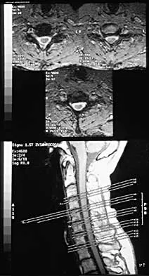
Question 6
A 44-year-old woman has had lower extremity dysesthesias, urinary incontinence, and has been unable to walk for the past 2 days. She reports no pain or history of trauma. She notes that 3 weeks ago she missed work for 2 days because of back pain, but it resolved with rest. Examination shows decreased or absent sensation below the knees, no motor function below the knees, and decreased rectal tone. Catheterization results in a postvoid residual of 2,000 mL. Plain radiographs and MRI scans without contrast are shown in Figures 1a through 1d. What is the next most appropriate step in management?
Explanation
Question 7
During anterior surgery on the cervical spine, at what level would the lateral dissection of the longus coli muscle most likely cause Horner's syndrome?
Explanation
Question 8
When compared with cobalt-chromium and stainless steel implants, a titanium implant has what biomechanical properties?
Explanation
Question 9
A 22-year-old college basketball player who was hit from behind while going up for a rebound is rendered immediately quadraparetic for approximately 10 minutes, followed by complete resolution of motor loss and return of full sensation. The radiograph and MRI scan of the cervical spine shown in Figures 2a and 2b reveal a canal diameter of 13 mm, loss of cerebrospinal fluid space about the spinal cord, and no signal change within the cord. What is the best course of action?
Explanation
Question 10
A 40-year-old woman has local back pain and intense burning pain in her perianal region after being shot twice in the back. Motor and sensory examination of her lower extremities reveals no apparent deficit. She has present but decreased sensation in her perianal region, an intact anal wink, good rectal tone, and an intact bulbocavernosus reflex. Radiographs and CT scans are shown in Figures 3a through 3d. What is the next most appropriate step in management?
Explanation
Question 11
A patient who sustained injuries in a motorcycle accident 30 minutes ago has significant motor and sensory deficits corresponding to a C6 level of injury. A lateral radiograph obtained during the initial on-scene evaluation reveals bilateral jumped facets at C5-C6; this appears to be an isolated injury. The patient is awake and alert. The next step in management of the dislocation should consist of
Explanation
Question 12
Figure 4 shows the MRI scan of a patient who has had bilateral leg pain, weakness, diffuse numbness, and urinary retention for the past week. Examination reveals that motor strength is diffusely decreased, although it may be secondary to pain. The patient is numb throughout both legs, and reflexes in the lower extremities are absent. Rectal examination shows decreased tone, but voluntary tightening is present. Management should consist of
Explanation
Question 13
A 62-year-old man has cervical myelopathy with no evidence of cervical radiculopathy. MRI reveals stenosis at C4-5 and C5-6 with severe cord compression. Examination will most likely reveal which of the following findings?
Explanation
Question 14
A patient who has had neck pain radiating down the arm for the past 4 weeks reports that the pain was excruciating during the first week. Management consisting of anti-inflammatory drugs and physical therapy has decreased the neck and arm symptoms from 10/10 to 3/10. He remains neurologically intact. MRI and CT scans are shown in Figures 5a and 5b. The best course of action should be
Explanation
Question 15
Examination of a supine patient in which the hip is abducted, externally rotated, and flexed is referred to as
Explanation
Question 16
During the evaluation of a patient suspected of having a lumbar disk herniation, T1- and T2-weighted MRI scans reveal a hyperintence lobular, well-defined lesion in the L2 vertebral body. What is the most likely diagnosis?
Explanation
Question 17
A 32-year-old man notes increasing back pain and progressive paraparesis over the past few weeks. He is febrile, and laboratory studies show a WBC of 12,500/mm3. MRI scans are shown in Figures 6a and 6b. Management should consist of
Explanation
Question 18
A 21-year-old woman with Marfan syndrome is seeking evaluation of her scoliosis. She reports no back or leg pain, and the neurologic examination is normal. Lateral and bending radiographs are shown in Figures 7a through 7e. Management should consist of
Explanation
Question 19
Which of the following substances is least likely to affect the success of bone union after lumbar arthrodesis?
Explanation
Question 20
A 33-year-old woman sustains a C6 burst fracture diving into a swimming pool, resulting in a complete spinal cord injury. The canal compromise is shown in Figures 8a and 8b. Functional recovery would be maximized with
Explanation
Question 21
In the upright standing position, approximately what percent of the vertical load is borne by the lumbar spine facet joints?
Explanation
Question 22
A 40-year-old patient who has a type II odontoid fracture is placed in a halo vest for 12 weeks; however, current radiographs show no evidence of healing. The next most appropriate step in management should consist of
Explanation
Question 23
In the initial evaluation of acute low back pain (duration of less than 4 weeks), plain radiographs are recommended in which of the following situations?
Explanation
Question 24
Figure 9 shows a cross-sectional view of the spinal cord at the lower cervical level. Injury to the structure indicated by the black arrow will lead to what neurologic deficit?
Explanation
Question 25
A 26-year-old woman who noted right-sided lumbosacral pain 10 days ago while vacuuming now reports that the pain has intensified. She denies any history of back problems. No radicular component is present, and her neurologic examination is normal. The next most appropriate step in management should consist of
Explanation
Question 26
A 35-year-old male falls from a ladder, sustaining an L1 burst fracture. He is neurologically intact. MRI demonstrates disruption of the posterior ligamentous complex (PLC). According to the Thoracolumbar Injury Classification and Severity Score (TLICS), what is the most appropriate management?
Explanation
Question 27
A 19-year-old male presents with unilateral distal upper extremity weakness and atrophy that distinctly spares the brachioradialis muscle. An MRI of the cervical spine taken in full flexion reveals forward displacement of the posterior dural sac. What is the most likely diagnosis?
Explanation
Question 28
A 6-year-old girl presents with torticollis following an upper respiratory infection. A dynamic CT scan reveals atlantoaxial rotatory subluxation with an anterior displacement of the atlas of 4 mm on the axis. According to the Fielding and Hawkins classification, what type of injury is this?
Explanation
Question 29
A 55-year-old man with advanced ankylosing spondylitis sustains a minor ground-level fall. He complains of back pain but is neurologically intact on presentation. Twelve hours later, he develops progressive lower extremity weakness. CT shows a non-displaced "chalk-stick" fracture at T8. What is the most likely cause of his neurologic deterioration?
Explanation
Question 30
A 72-year-old male sustains a Type II odontoid fracture. He is being evaluated for conservative management in a halo vest. Which of the following factors represents the most significant risk factor for nonunion with nonoperative treatment?
Explanation
Question 31
In a patient presenting with L4-L5 degenerative spondylolisthesis, which of the following MRI findings is most highly predictive of segmental instability and the likelihood of future slip progression?
Explanation
Question 32
To establish a definitive radiographic diagnosis of Scheuermann's kyphosis based on the classic Sorensen criteria, a patient must demonstrate anterior wedging of at least 5 degrees in a minimum of how many consecutive vertebrae?
Explanation
Question 33
In a 3-year-old child diagnosed with congenital scoliosis, which of the following anomalous vertebral patterns carries the highest natural risk of rapid curve progression?
Explanation
Question 34
A 60-year-old diabetic male presents with severe back pain, fever, and progressive lower extremity weakness. MRI reveals a large ventral lumbar epidural abscess. What is the most common causative organism for this condition?
Explanation
Question 35
In preoperative planning for adult spinal deformity correction, achieving optimal global sagittal balance heavily relies on the relationship between pelvic incidence (PI) and lumbar lordosis (LL). What is the generally accepted target formula for a successful correction?
Explanation
Question 36
A 45-year-old patient presents with acute, burning right anterior thigh pain, weakness in right knee extension, and an absent right patellar reflex. MRI reveals a far lateral (extraforaminal) disc herniation. At which lumbar level is this herniation most likely located?
Explanation
Question 37
According to the Denis classification of sacral fractures, fractures extending through which zone are associated with the highest incidence of severe neurologic injury, including bowel and bladder dysfunction?
Explanation
Question 38
A 22-year-old female sustains a severe seatbelt-type flexion-distraction injury (Chance fracture) at the L2 level during a high-speed motor vehicle collision. Which concomitant visceral injury is most classically associated with this specific fracture pattern?
Explanation
Question 39
A 65-year-old Japanese male presents with cervical myelopathy. CT scan shows a dense, continuous calcified mass along the posterior aspect of the C3-C5 vertebral bodies, consistent with Ossification of the Posterior Longitudinal Ligament (OPLL). Which surgical approach carries the highest specific risk of iatrogenic dural tear?
Explanation
Question 40
A Levine-Edwards Type II traumatic spondylolisthesis of the axis (Hangman's fracture) typically demonstrates significant anterior translation and angulation. What is the classic mechanism of injury required to produce this specific Type II pattern?
Explanation
Question 41
A 70-year-old male with pre-existing cervical spondylosis sustains a hyperextension injury, resulting in Central Cord Syndrome. As the patient undergoes rehabilitation, which of the following neurologic functions notoriously has the poorest prognosis for meaningful recovery?
Explanation
Question 42
A 25-year-old male is evaluated after a motor vehicle collision. He complains of right-sided neck pain and a C6 radiculopathy. Lateral cervical radiographs reveal exactly 25% anterior translation of C5 on C6 and a characteristic "bowtie" sign. What is the primary mechanism of this injury?
Explanation
Question 43
Diffuse Idiopathic Skeletal Hyperostosis (DISH) is characterized by flowing ossification along the anterolateral aspect of the spine. According to the Resnick and Niwayama criteria, involvement of how many contiguous vertebral bodies is required to definitively diagnose DISH?
Explanation
Question 44
A 45-year-old male presents with severe myelopathy secondary to a massive, centrally located, calcified T8-T9 disc herniation. The surgeon is contemplating the operative approach. Which of the following approaches is strictly contraindicated due to an unacceptably high risk of catastrophic spinal cord injury?
Explanation
Question 45
Following a severe fracture-dislocation at T4, a patient presents with flaccid paralysis, hypotension (BP 80/40 mmHg), bradycardia (HR 45 bpm), and warm, flushed extremities. Which of the following mechanisms best explains this specific constellation of systemic findings?
Explanation
Question 46
A 65-year-old man presents with progressive gait instability and loss of fine motor skills in his hands. Physical exam reveals hyperreflexia in the lower extremities, a positive Hoffman's sign, and a wide-based gait. MRI of the cervical spine shows severe stenosis at C4-C5 and C5-C6 with T2 signal changes in the spinal cord. Which of the following MRI findings is the most significant predictor of poor neurological recovery after decompressive surgery?
Explanation
Question 47
A 25-year-old man is brought to the trauma bay after a motorcycle accident. He has a T12 burst fracture with 50% loss of vertebral body height and 20 degrees of kyphosis. Neurological examination is completely normal. MRI confirms that the posterior ligamentous complex (PLC) is intact. According to the Thoracolumbar Injury Classification and Severity (TLICS) score, what is the most appropriate management?
Explanation
Question 48
A 78-year-old woman falls from a standing height and sustains a Type II odontoid fracture. She has a history of severe osteoporosis and COPD. Her neurologic exam is normal. What is the most appropriate initial management?
Explanation
Question 49
A 45-year-old male undergoes an L4-L5 microdiscectomy. During the procedure, brisk arterial bleeding is encountered from the anterior aspect of the disc space following the use of pituitary rongeurs, and the patient suddenly becomes hypotensive. Which of the following vascular structures was most likely injured?
Explanation
Question 50
A 62-year-old woman presents with severe low back pain and an inability to stand up straight. Her standing full-length spine radiographs reveal a pelvic incidence (PI) of 60 degrees and a lumbar lordosis (LL) of 30 degrees. Which of the following best describes her spinopelvic parameters?
Explanation
Question 51
A 70-year-old man with a known history of severe cervical stenosis is involved in a rear-end motor vehicle collision, resulting in a hyperextension injury. He presents to the ED with 1/5 motor strength in his bilateral upper extremities and 4/5 strength in his lower extremities. Proprioception and perianal sensation are intact. What is the most likely diagnosis?
Explanation
Question 52
A 42-year-old male with a 15-year history of advanced ankylosing spondylitis presents to the emergency department complaining of severe neck pain after a minor fall at home. Initial cross-table lateral radiographs show extensive syndesmophytes but no obvious fracture line. What is the most appropriate next step in management?
Explanation
Question 53
A 35-year-old construction worker presents with severe left-sided buttock pain radiating down the posterolateral thigh and calf to the dorsum of his foot. On examination, he has decreased sensation over the dorsal web space between his first and second toes and 3/5 weakness in the extensor hallucis longus (EHL). A disc herniation at which level is most likely responsible?
Explanation
Question 54
A 55-year-old woman undergoes an uncomplicated multi-level anterior cervical discectomy and fusion (ACDF) from C3 to C6 for severe myelopathy. On postoperative day 2, she complains of profound inability to abduct her shoulders and flex her elbows, though her hand function and leg strength remain normal. What is the most likely cause of this complication?
Explanation
Question 55
A 16-year-old gymnast presents with chronic, activity-limiting low back pain. Radiographs demonstrate a Grade II isthmic spondylolisthesis at L5-S1 with an intact pars defect. Despite 6 months of comprehensive physical therapy, core strengthening, and bracing, her pain remains severe. What is the most appropriate surgical intervention?
Explanation
Question 56
A 55-year-old male undergoes a multi-level posterior cervical laminectomy and fusion for severe cervical spondylotic myelopathy. On postoperative day 2, he develops profound weakness in bilateral shoulder abduction and elbow flexion, with no sensory deficits or lower extremity symptoms. What is the most likely etiology of this complication?
Explanation
Question 57
A 45-year-old man presents with severe, acute left leg pain. MRI reveals a far-lateral (extraforaminal) disc herniation at the L4-L5 level on the left. Which of the following physical examination findings is most expected?
Explanation
Question 58
A 72-year-old female sustains a Type II odontoid fracture after a ground-level fall. Which of the following radiographic or clinical parameters is the strongest predictor of nonunion if this fracture is treated nonoperatively?
Explanation
Question 59
A 68-year-old man with known cervical spondylosis presents after a hyperextension injury. He exhibits 2/5 motor strength in his bilateral upper extremities and 4/5 motor strength in his bilateral lower extremities. Perianal sensation and proprioception are preserved. What is the most likely diagnosis?
Explanation
Question 60
A 30-year-old male falls from a roof, sustaining an L1 burst fracture. He is neurologically intact. MRI demonstrates an intact posterior ligamentous complex (PLC). According to the Thoracolumbar Injury Classification and Severity (TLICS) score, what is his total score and recommended treatment?
Explanation
Question 61
In the surgical planning for a 65-year-old woman with adult degenerative scoliosis and sagittal imbalance, her pelvic incidence (PI) is measured at 55 degrees. To achieve optimal sagittal alignment and minimize the risk of adjacent segment disease, what should be the target postoperative lumbar lordosis (LL)?
Explanation
Question 62
During an anterior cervical discectomy and fusion (ACDF), a right-sided approach is chosen. Which of the following statements regarding the recurrent laryngeal nerve (RLN) is most accurate?
Explanation
Question 63
A 15-year-old boy presents with progressive thoracic kyphosis. Radiographs reveal anterior wedging of multiple vertebral bodies. What are the classic Sorenson radiographic criteria required to diagnose Scheuermann's kyphosis?
Explanation
Question 64
A 22-year-old female unrestrained backseat passenger is involved in a high-speed motor vehicle collision. Radiographs show a flexion-distraction injury (Chance fracture) at L2. Which of the following injuries is most commonly associated with this spinal fracture pattern?
Explanation
Question 65
A 35-year-old male arrives at the trauma bay with a unilateral C5-C6 facet dislocation following a rugby tackle. He is awake, alert, and complains of C6 radicular pain, but has no myelopathy. What is the most appropriate initial management?
Explanation
Question 66
A 65-year-old man undergoes a C3-C6 posterior laminectomy and instrumented fusion for cervical spondylotic myelopathy. On postoperative day 2, he develops isolated, profound weakness in right shoulder abduction and elbow flexion. His sensation remains intact, and lower extremity function is unchanged. What is the most likely etiology of this complication?
Explanation
Question 67
A 48-year-old man presents with acute onset, severe left-sided anterior thigh pain and knee weakness. Examination reveals weakness in knee extension and a diminished patellar reflex. Sensation is decreased over the medial aspect of the left calf. MRI reveals a far-lateral disc herniation at the L4-L5 level. Which nerve root is most likely compressed?
Explanation
Question 68
A 12-year-old premenarchal female presents for scoliosis evaluation. Radiographs demonstrate a right thoracic curve of 32 degrees. Her Risser stage is 0. What is the most appropriate next step in management?
Explanation
Question 69
A 72-year-old man with a history of cervical stenosis falls forward and strikes his chin. He presents to the emergency department with profound bilateral weakness in his hands and upper extremities (1/5 strength) but retains 4/5 strength in his lower extremities. Sensation is decreased in a cape-like distribution. What is the most likely diagnosis?
Explanation
Question 70
In the surgical planning for adult degenerative scoliosis, sagittal balance is a critical determinant of postoperative outcomes. If a patient has a pelvic incidence (PI) of 55 degrees, what should the target lumbar lordosis (LL) ideally be to minimize the risk of adjacent segment disease?
Explanation
Question 71
A 28-year-old man arrives at the trauma bay after a motor vehicle collision. He is awake, alert, and cooperative (GCS 15). Neurological examination is entirely intact. CT of the cervical spine reveals a unilateral facet dislocation at C5-C6. What is the most appropriate next step in management?
Explanation
Question 72
An 84-year-old woman presents with severe neck pain after a low-energy fall from a standing height. CT imaging reveals a Type II odontoid fracture with 2 mm of posterior displacement. She is neurologically intact. What is the most appropriate management?
Explanation
Question 73
A 16-year-old boy presents with back pain and a prominent thoracic curvature. On physical examination, the kyphosis is rigid and does not correct with hyperextension. Standing lateral radiographs reveal irregular vertebral endplates and Schmorl's nodes. By classic Sørensen criteria, Scheuermann's kyphosis requires anterior wedging of at least 5 degrees in how many consecutive vertebrae?
Explanation
Question 74
During an anterior approach to the lower cervical spine for corpectomy, the surgeon must mobilize the longus colli muscles. Care must be taken laterally to avoid injury to the vertebral artery. In the majority of the population, the vertebral artery enters the transverse foramen at which cervical level?
Explanation
Question 75
A 60-year-old man undergoes a complex T10 to pelvis posterior spinal fusion for severe adult spinal deformity. The surgery lasts 10 hours with an estimated blood loss of 2.5 liters, accompanied by mild intraoperative hypotension. On postoperative day 1, he complains of painless, profound bilateral vision loss. What is the most likely etiology?
Explanation
Question 76
A 35-year-old construction worker falls 10 feet, sustaining an L1 burst fracture. He is neurologically intact. MRI demonstrates an intact posterior ligamentous complex (PLC). According to the Thoracolumbar Injury Classification and Severity (TLICS) score, what is the patient's total score and recommended management?
Explanation
Question 77
A 45-year-old male falls from a height and sustains an L1 burst fracture. Neurological examination is normal. Upright radiographs demonstrate 15 degrees of kyphosis and 30% loss of vertebral body height. CT scan shows 40% canal compromise. What is the most appropriate initial management?
Explanation
Question 78
A 68-year-old female presents with severe neurogenic claudication. Standing radiographs reveal a grade 1 degenerative spondylolisthesis at L4-L5. Dynamic radiographs show 4 mm of translation. She has failed conservative management. What is the most appropriate surgical intervention?
Explanation
Question 79
A 25-year-old male with ankylosing spondylitis sustains a low-energy fall. He complains of severe neck pain but has no neurologic deficits. Plain radiographs of the cervical spine are difficult to interpret due to marked osteopenia and deformity. What is the most appropriate next step in management?
Explanation
Question 80
In evaluating a patient with adult spinal deformity, which of the following spinopelvic parameters is a morphologic constant that does not change with patient positioning?
Explanation
Question 81
A 75-year-old male with a history of rheumatoid arthritis presents with progressive upper extremity weakness and hyperreflexia. Radiographs reveal an atlantodental interval (ADI) of 8 mm and a posterior atlantodental interval (PADI) of 12 mm. Which of the following is the most critical threshold indicating the need for surgical intervention to prevent irreversible neurologic damage?
Explanation
Question 82
A 30-year-old male sustains a C5-C6 bilateral facet dislocation. He is intubated and sedated upon arrival. What is the most appropriate next step in management prior to reduction?
Explanation
Question 83
Which of the following findings is considered the most reliable early indicator of urinary retention in a patient with suspected cauda equina syndrome?
Explanation
Question 84
A 50-year-old female presents with neck pain and right arm radiculopathy in the C6 distribution. She has a positive Spurling's test. Conservative treatment for 6 weeks has failed. MRI shows a right paracentral disc herniation at C5-C6. Which of the following motor deficits is most likely present?
Explanation
Question 85
In the treatment of osteoporotic vertebral compression fractures, which of the following is a known absolute contraindication for balloon kyphoplasty?
Explanation
Question 86
A 22-year-old male dives into shallow water and sustains an isolated Jefferson (C1) burst fracture. An open-mouth odontoid view demonstrates combined lateral overhang of the C1 lateral masses on C2 of 8 mm. What does this radiographic finding indicate?
Explanation
Question 87
A 40-year-old male is involved in a motor vehicle collision and sustains a severe hyperflexion injury to his thoracic spine. Imaging reveals a Chance fracture (flexion-distraction injury) at T12. Which of the following associated injuries is most commonly found with this fracture pattern?
Explanation
Question 88
A 35-year-old male presents with persistent low back pain and stiffness, particularly worse in the morning and improving with activity. He is HLA-B27 positive. Radiographs demonstrate squaring of the vertebral bodies and marginal syndesmophytes. In patients with this condition, which of the following fracture patterns is most likely to result in an epidural hematoma?
Explanation
Question 89
Which of the following is an absolute indication for surgical intervention in a patient with an acute spinal epidural abscess?
Explanation
Question 90
Which of the following is considered the most significant risk factor for nonunion in a patient treated nonoperatively for a Type II odontoid fracture?
Explanation
Question 91
A 13-year-old premenarchal female (Risser 0) presents with a right thoracic adolescent idiopathic scoliosis. Standing radiographs demonstrate a primary thoracic curve of 35 degrees. What is the most appropriate management?
Explanation
Question 92
A 65-year-old man presents with bilateral leg pain and fatigue that worsens after walking two blocks. Which of the following findings is most characteristic of neurogenic claudication secondary to lumbar spinal stenosis, as opposed to vascular claudication?
Explanation
Question 93
A 60-year-old man undergoes a C3-C6 posterior cervical laminectomy and fusion for cervical spondylotic myelopathy. On postoperative day 2, he develops new-onset unilateral weakness in shoulder abduction and elbow flexion. What is the most likely etiology of this complication?
Explanation
None
