العربية
Part of the Master Guide

AAOS Orthopedic MCQs (Set 1): Spine Disorders, Sports Medicine & Arthroplasty | 2026 Board Review

AAOS Pediatrics Board Review MCQs (Set 2): DDH, Fractures & Scoliosis | ABOS 2001

23 Apr 2026 67 min read 89 Views
Pediatrics 2001 MCQs - Part 2

Key Takeaway

This high-yield question set for AAOS/ABOS pediatric exams focuses on key topics like diagnosis & management of Developmental Dysplasia of the Hip (DDH), evaluation & treatment of common pediatric fractures, and principles of scoliosis screening & treatment. Essential for 2001 board preparation.

AAOS Pediatrics Board Review MCQs (Set 2): DDH, Fractures & Scoliosis | ABOS 2001

Comprehensive 100-Question Exam


00:00

Start Quiz

Question 1

Figure 17 shows the radiograph of a 2-year-old girl who sustained a fracture of the femur in a fall while walking with her parents. History reveals that this is her third long bone fracture, having sustained a humerus fracture 1 year ago and a fracture of the opposite femur 9 months ago. There is no family history of any similar problem. Examination reveals distinctly blue sclerae, normal appearing teeth, and no skin lesions. What is the most likely cause of this patient's disorder?


Explanation

Osteogenesis imperfecta (OI) is a genetically determined disorder of type I collagen synthesis that is characterized by bone fragility. This patient has had three fractures of the long bones by age 2 years, with the last one occurring after relatively minor trauma. The patient's history and clinical features are consistent with a diagnosis of Sillence type IA OI. Type I OI is the mildest and most common form. Inheritance is autosomal-dominant; however, as in this patient, new mutations are frequent. Type I is subclassified into the A type (absence of dentinogenesis imperfecta) and B type (presence of dentinogenesis imperfecta). The sclerae are blue, and the first fractures usually occur in the preschool years after walking has begun. Cells from individuals with type I OI largely demonstrate a quantitative defect of type I collagen; they synthesize and secrete about half the normal amount of type I procollagen. Qualitative mutations that lead to an abnormal type I procollagen molecule result in more severe types of the disorder. There are no indications that this child has been abused. Radiographs of the femur show no evidence of rickets, pyknodysostosis, or osteopetrosis. Morquio syndrome, characterized by excess excretion of keratin sulfate in the urine, is not associated with bone fragility. Kocher MS, Shapiro F: Osteogenesis imperfecta. J Am Acad Orthop Surg 1998;6:225-236. Sillence DO, Senn A, Danks DM: Osteogenesis imperfecta: An expanding panorama of variants. Clin Orthop 1981;159:11-25.

Question 2

An 8-year-old boy with severe hemophilia A (factor VIII) and no inhibitor is averaging eight transfusions per month for bleeding into the right ankle. Examination shows synovial hypertrophy; range of motion consists of 0 degrees of dorsiflexion and 20 degrees of plantar flexion. The patient's knees, elbows, and left ankle have no restriction of motion. Standing radiographs of the right ankle are shown in Figure 18. Management should consist of


Explanation

The patient has bilateral hypertrophic synovitis that is causing repeated hemarthroses and progressive arthropathy. Ankle synovectomy in patients with hemophilia is effective in significantly reducing the rate of joint bleeding and in slowing the progression of the arthropathy; therefore, bilateral synovectomies is the treatment of choice. Range of motion can be effectively maintained after ankle synovectomy. Bracing and prophylactic transfusions would be ineffective at this time. Ankle arthrodesis should be reserved for patients with severe pain. Compared with patients who have juvenile rheumatoid arthritis, patients with hemophilia generally do not have involvement of the subtalar joint and rarely require a pantalar arthrodesis. Greene WB: Synovectomy of the ankle for hemophilic arthropathy. J Bone Joint Surg Am 1994;76:812-819.


Question 3

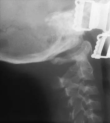
Figures 19a through 19c show radiographs of the cervical spine of an asymptomatic patient with Down syndrome who wants to participate in a Special Olympics running event. The neurologic examination is normal. Management should consist of


Explanation

An atlanto-dens interval (ADI) of up to 4 mm in children is considered normal. Children with Down syndrome have increased ligamentous laxity, with atlantoaxial instability occurring in as many as 15% to 20% of patients. These patients are at risk for catastrophic injury following minor trauma and should be routinely screened for instability, generally beginning when the patient starts to walk. Patients with an ADI of greater than 5 mm should avoid contact sports and high-risk activities such as gymnastics, diving, the high jump, and the butterfly stroke. The American Academy of Pediatrics Committee of Sports Medicine and Fitness guidelines recommend that lateral views of the cervical spine in neutral, flexion, and extension should be obtained in all children with Down syndrome who wish to participate in sports. Patients with normal radiographs and examinations do not need repeat radiographs, although some authors suggest that instability increases with age, and therefore recommend repeat radiographs every 5 years. Cervical spine fusion in patients with Down syndrome has a high rate of complications and should be performed only on patients with symptoms and evidence of myelopathy. American Academy of Pediatrics Committee of Sports Medicine and Fitness: Atlantoaxial instability in Down syndrome. Pediatrics 1995;96:151-154. Copley LA, Dormans JP: Cervical spine disorders in infants and children. J Am Acad Orthop Surg 1998;6:204-214.


Question 4

Compared with amputation, limb salvage for osteosarcoma of the distal end of the femur will result in


Explanation

Major advances in diagnostic imaging, neoadjuvant chemotherapy, and surgical techniques have allowed limb salvage to be performed as an alternative to amputation in children with osteosarcoma. The outcome of treatment of nonmetastatic, high-grade osteosarcoma of the distal femur was studied in 227 patients from 26 institutions. The authors found no difference in the long-term survival or quality of life between patients treated with limb salvage and those treated with amputation. Patients treated with limb salvage had a higher rate of reoperation, but a better functional outcome. Rougraff BT, Simon MA, Kneisl JS, Greenberg DB, Mankin HJ: Limb salvage compared with amputation for osteosarcoma of the distal end of the femur: A long-term oncological, functional, and quality-of-life study. J Bone Joint Surg Am 1994;76:649-656.

Question 5

Examination of a 7-year-old boy reveals 20 degrees of valgus following a lawn mower injury to the lateral femoral epiphysis. Treatment consists of total distal femoral epiphyseodesis and varus osteotomy. Following surgery, he has a limb-length discrepancy of 3 cm and 5 degrees of genu valgum. Assuming that he undergoes no further treatment, the patient's predicted limb-length discrepancy at maturity would be how many centimeters?


Explanation

The distal femoral epiphysis grows approximately 1 cm per year and in boys, growth ceases at approximately age 16 years. Therefore, the patient's limb-length discrepancy at maturity would be 12 cm (9 cm plus the 3-cm discrepancy he has from the previous surgery). Little DG, Nigo L, Aiona MD: Deficiencies of current methods for the timing of epiphyseodesis. J Pediatr Orthop 1996;16:173-179.

Question 6

When the iliac apophysis starts ossifying in the normal adolescent, growth of the sitting height or trunk height is characterized by


Explanation

Studies by Anderson and associates have resulted in a growth-remaining chart for sitting height that shows an increase of 3 to 5 cm in girls and an increase of 5 to 8 cm in boys. Future growth of the spine may impact brace longevity and fit. Anderson M, Hwang SC, Green WT: Growth of the normal trunk in boys and girls during the second decade of life: Related to age, maturity, and ossification of the iliac epiphyses. J Bone Joint Surg Am 1965;47:1554-1564.

Question 7

A 10-year-old girl was thrown over the handlebars of her bicycle and landed directly on her left shoulder. She was treated with a figure-of-8 strap and analgesics. Follow-up examination 2 weeks later reveals that the lateral end of the clavicle is superiorly dislocated relative to the acromion. A radiograph of the shoulder shows calcification lateral to the coracoid process at the level of the acromion, and the clavicle is superiorly displaced. Management should consist of


Explanation

In adults, a direct blow on the acromion usually results in an acromioclavicular dislocation. In children, however, the usual injury from this mechanism is a physeal fracture of the lateral clavicle. The clavicular shaft fragment, analogous to the metaphyseal portion of a physeal fracture, herniates through the periosteum, leaving the distal periosteal sleeve in contact with the lateral (distal) physeal fragment. The treatment of choice is immobilization until the patient is pain-free. Falstie-Jensen S, Mikkelsen P: Pseudodislocation of the acromioclavicular joint. J Bone Joint Surg Br 1982;64:368-369.

Question 8

Figures 20a and 20b show the radiographs of an obese 15-year-old boy who has severe left groin pain and is unable to bear weight following a minor injury. Treatment should consist of


Explanation

The radiographs and history are consistent with an acute unstable slipped capital femoral epiphysis. Aronson and Loder documented an increased rate of osteonecrosis associated with manipulative reduction. They recommended bed rest with skin traction to allow the synovitis to resolve, followed by in situ pinning. They noted, however, that many of these slips reduced with anesthesia and positioning on a fracture table. Biomechanic studies have shown a slight increased resistance to shear stress when two screws are used, but it is unknown if this is significant in the clinical setting. Open epiphyseodesis does not provide postoperative stability; therefore, adjunctive fixation or immobilization is required. Numerous studies have noted the inadvisability of using multiple screws. Casting has a high rate of complications, including chondrolysis and progression of the slip. Aronson DD, Loder RT: Treatment of the unstable (acute) slipped capital femoral epiphysis. Clin Orthop 1996;322:99-110. Karol LA, Doane RM, Cornicelli SF, Zak PA, Haut RC, Manoli A II: Single versus double screw fixation for treatment of slipped capital femoral epiphysis: A biomechanical analysis. J Pediatr Orthop 1992;12:741-745.


Question 9

What is the recommended treatment of a skeletally immature 12-year-old boy who has an anterior cruciate ligament-deficient knee?


Explanation

Traditional surgeries for anterior cruciate ligament-deficient knees carry the potential risk of premature physeal closure in young athletes. Therefore, most surgeons are reluctant to recommend intra-articular reconstruction using bone tunnels with bone-patellar tendon-bone autografts or hamstring tendons. The current recommendation for young athletes is activity modification, rehabilitation, and functional bracing until the patient is near skeletal maturity. At that time, for the very symptomatic patient, the treatment of choice is intra-articular repair of the anterior cruciate ligament. If a skeletally immature patient continues to have instability despite rehabilitation and bracing, a modification of the femoral tunnel to the over-the-top position will not place the lateral femoral physis at risk for premature closure and deformity. A centrally placed tibial tunnel will minimize the risk of angular deformity and minimize limb-length discrepancy if physeal arrest occurs. Barry P: Anterior cruciate ligament injuries, in Andrews JR, Timmerman LA (eds): Diagnostic and Operative Arthroscopy. Philadelphia, Pa, WB Saunders, 1997, p 358. McCarroll JR, Shelbourne KD, Porter DA, Rettig AC, Murray S: Patellar tendon graft reconstruction for midsubstance anterior cruciate ligament rupture in junior high school athletes: An algorithm for management. Am J Sports Med 1994;22:478-484. Nottage WM, Matsuura PA: Management of complete traumatic anterior cruciate ligament tears in the skeletally immature patient: Current concepts and review of the literature. Arthroscopy 1994;10:569-573.

Question 10

Figures 21a and 21b show the radiographs of a 12-year-old patient with an L4-level myelomeningocele who has scoliosis that has been slowly progressing for the past several years. There has been no loss of motor function. An MRI scan shows no syringomyelia or increased hydrocephalus. Management should consist of


Explanation

Scoliosis is a common occurrence in children with myelomeningocele, with the incidence increasing as the neurologic level moves cephalad. The rate of pseudarthrosis for isolated anterior or posterior fusions has been reported as high as 75%. The combination of anterior and posterior fusions with some type of instrumentation has been shown to decrease the rate of pseudarthrosis to 20%. Brace treatment in smaller curves can be used as a temporizing measure to delay surgery, but as with idiopathic scoliosis, the brace is ineffective for larger curves. Observation is not indicated with a curve of this magnitude. Ward WT, Wenger DR, Roach JW: Surgical correction of myelomeningocele scoliosis: A critical appraisal of various spinal instrumentation systems. J Pediatr Orthop 1989;9:262-268.


Question 11

A 3-year-old child is referred for evaluation of bowed legs. History reveals no dietary deficiencies; however, family history is significant for several members with bowed legs. Examination reveals genu varum, and the child is in the 5th percentile for height and weight. Laboratory studies show normal renal function, a normal calcium level, a decreased phosphate level, and an elevated alkaline phosphatase level. A plain radiograph of the lower extremities is shown in Figure 22. What is the most likely diagnosis?


Explanation

The differential diagnosis of genu varum includes physiologic genu varum, Blount's disease, skeletal dysplasia, and metabolic bone disease. Children with Blount's disease are generally in the 95th percentile for height and weight, and usually multiple family members are not affected. The radiographs show widening of the physis and metaphyseal flaring. In Blount's disease, the characteristic radiographic changes involve only the tibia, and at this age, most commonly show beaking of the medial metaphysis. Skeletal dysplasias, such as chondrometaphyseal dysplasia, are associated with short stature, and the radiographic changes are similar to those seen here. However, laboratory studies in these children will be within normal limits. Children with chronic renal disease will often be of short stature, and the radiographic findings are again similar to those shown here. However, BUN and creatinine levels are elevated and phosphate levels are elevated rather than decreased in children with renal disease. The absence of dietary deficiencies and positive family history rules out vitamin D-deficient rickets. There are four types of vitamin D-resistant rickets: failure of production of 1,25-dihydroxy vitamin D, phosphate diabetes (hypophosphatemic rickets), end organ insensitivity to vitamin D, and renal tubular acidosis. All types of vitamin D-resistant rickets are resistant to treatment with physiologic doses of vitamin D. The patient's clinical picture, family history, laboratory studies, and radiographs are most consistent with hypophosphatemic rickets. This entity is inherited as a sex-linked dominant trait. Evans GA, Arulanantham K, Gage JR: Primary hypophosphatemic rickets: Effect of oral phosphate and vitamin D on growth and surgical treatment. J Bone Joint Surg Am 1980;62:1130-1138. Loeffler RD Jr, Sherman FC: The effect of treatment on growth and deformity in hypophosphatemic vitamin D-resistant rickets. Clin Orthop 1982;162:4-10. Loder RT, Johnston CE II: Infantile tibia vara. J Pediatr Orthop 1987;7:639-646.


Question 12

A 14-year-old boy sustained a femoral neck fracture in a fall from a tree and underwent open reduction and internal fixation 6 months ago. Follow-up examination now reveals an antalgic Trendelenburg gait and painful range of motion. A radiograph is shown in Figure 23, and a CT scan shows a nonunion. Treatment should consist of


Explanation

The coxa vara deformity and fracture nonunion should be treated simultaneously; therefore, the treatment of choice is curettage of the nonunion, intertrochanteric valgus osteotomy, and revision internal fixation. In addition, valgus osteotomy will convert the shear forces across the nonunion to compression, aiding in healing of the nonunion. None of the other procedures addresses both issues, and hip fusion is inappropriate under these conditions. Lam SF: Fractures of the neck of the femur in children. J Bone Joint Surg Am 1971;53:1165-1179.


Question 13

A 22-month-old child has scrapes and bruises on his head and a severe deformity of the forearm after being thrown from a car as an unrestrained passenger in a motor vehicle accident. Examination reveals a Glasgow Coma Scale score of 12. Prior to treatment of the forearm, management should include


Explanation

As CT scanning has become available, the use of radiographs of the skull has decreased in importance for evaluation of head trauma. The indications for CT scanning for suspected head trauma include any degree of obtundation, focal neurologic deficit, history of a high-velocity injury, amnesia for the injury, progressive headache, persistent vomiting, children younger than age 2 years, serious facial injury, posttraumatic seizure, skull penetration, or a Glasgow Coma Scale score of 13 or less. Evidence of improved outcome with use of steroids in head trauma is lacking. Steroids are useful for increased intracranial pressure caused by brain tumors or abscesses. High-dose IV methylprednisolone is indicated for spinal cord trauma and improves the ultimate degree of recovery of function. When herniation is suspected in a patient with asymmetric neurologic findings or the patient's condition is deteriorating rapidly, a mannitol infusion may be used. Hall DE: Head injuries, in Hoekelman RA (ed): Primary Pediatric Care. St Louis, Mo, Mosby, 1997, pp 1709-1712. Nelson WE, Behrman RE, Kliegman RM (eds): Nelson Essentials of Pediatrics. Philadelphia, Pa, WB Saunders, 1998, p 712.

Question 14

Examination of a 5-year-old boy with amyoplasia shows a flexion contracture of 70 degrees of the right knee. The active arc of motion is from 70 degrees to 90 degrees, and the opposite knee has a flexion contracture of 10 degrees. Both hips are dislocated with flexion contractures of 10 degrees, passive hip motion is from 10 degrees to 90 degrees of flexion, and the feet are plantigrade and easily braceable. Despite a daily stretching program, the parents and physical therapists note that it is increasingly difficult for him to walk because of the flexion contracture of the right knee. Management of the knee flexion contracture should now include


Explanation

Most children with amyoplasia are ambulatory and when a decrease in function occurs because of a severe contracture, it must be addressed. A radical posterior soft-tissue release, including the posterior knee capsule and often the collateral ligaments and the posterior cruciate ligament, is needed to obtain extension. After the age of 1 year, aggressive physical therapy will do little to correct a contracture. Botulinum toxin A is indicated for spasticity and is contraindicated with severe contractures. Supracondylar femoral extension osteotomy works well, but will remodel at an average rate of 1 degree per month, which is not considered ideal in a young patient. Gradual correction with a circular ring external fixator is an option, but a soft-tissue release will also most likely be needed for a contracture of this severity. Sarwark JF, MacEwen GD, Scott CI Jr: Amyoplasia (a common form of arthrogryposis). J Bone Joint Surg Am 1990;72:465-469. DelBello DA, Watts HG: Distal femoral extension osteotomy for knee flexion contracture in patients with arthrogryposis. J Pediatr Orthop 1996;16:122-126.

Question 15

A 13-year-old girl who is 2 years postmenarche has been referred for management of scoliosis. She denies any history of back pain. Radiographs show a right thoracic curve of 35 degrees. She has a Risser sign of 4 and a bone age of 15.5 years. Management should consist of


Explanation

Because the patient is skeletally mature with a curve of less than 40 degrees, there is no benefit to bracing and surgery is not indicated. Management should consist of observation and follow-up radiographs in 6 months. Lonstein JE, Carlson JM: The prediction of curve progression in untreated idiopathic scoliosis during growth. J Bone Joint Surg Am 1984;66:1061-1071.

Question 16

In children with isolated zone II lacerations of the flexor tendon, poor digital motion is best correlated with


Explanation

In a recent study on restoration of motion following zone I and zone II flexor tendon repairs in children, age was found to have no effect on the results of zone II tendon repairs. Early passive motion offered no better results than immobilization for 3 weeks. Immobilization for more than 4 weeks correlated with poorer results.

Question 17

In a longitudinal study of children with spastic diplegia, analysis of long-term function will most likely reveal


Explanation

In a longitudinal study of 18 patients with spastic diplegia over a period of 32 months, three-dimensional gait analysis revealed a deterioration of gait stability with increases in double support time and decreases in single support time. Kinematic data also identified a loss of excursion about the knee, ankle, and pelvis. Interestingly, the static examination of the children showed a decrease in the popliteal angle over time. The authors concluded that ambulatory ability tends to worsen over time in children with spastic diplegia.

Question 18

Examination of a 7-year-old girl with myelomeningocele reveals calcaneal deformities of both feet. She ambulates on both extremities wearing ankle-foot orthoses and has no upper extremity aids. She has grade 5/5 motor strength to the tibialis anterior muscles and absent motor strength to the triceps surae. There is no varus or valgus deformity of the hindfoot, and the skin over the heels is intact; however, mild callosities are present. Management should consist of


Explanation

A calcaneal deformity of the foot may occur in children who have low lumbar myelomeningocele. Strong dorsiflexors overcome a weak or absent gastrocnemius-soleus complex, leading to downward growth of the calcaneal apophysis. The deformity is usually progressive and does not respond to nonsurgical management. Most authorities recommend transfer of the tibialis anterior muscle through the interosseous membrane to the posterior aspect of the calcaneus. This procedure has been reported to be effective in limiting progression of the deformity. An extra-articular subtalar arthodesis, a treatment option for valgus deformity of the hindfoot, is not indicated. Similarly, Achilles tendon tenodesis to the fibula, an option for managing valgus of the ankle, is not indicated. Calcaneal osteotomy may be used in older children with severe calcaneal deformity. Stott NS, Zionts LE, Gronley JK, Perry J: Tibialis anterior transfer for calcaneal deformity: A postoperative gait analysis. J Pediatr Orthop 1996;16:792-798.

Question 19

Figure 24 shows the radiograph of a 4-year-old girl with spina bifida. Examination reveals an L3 motor level, excellent sitting and standing balance, and satisfactory range of motion at the hips. Management should consist of


Explanation

Children with spina bifida and bilateral symmetrical dislocation of the hips usually do not require treatment. A level pelvis and good range of motion of the hips are more important for ambulation than reduction of bilateral hip dislocations. Because the patient has good sitting and standing balance and good range of motion, maintenance of that range of motion and symmetry is more important than reduction. Surgery is not recommended.


Question 20

Posterior spinal fusion for scoliosis should be performed on a patient with Duchenne muscular dystrophy when


Explanation

Progressive scoliosis develops in most patients with Duchenne muscular dystrophy. The onset of spinal deformity typically follows the cessation of walking, and curves can be expected to progress about 10 degrees per year. Posterior spinal fusion with instrumentation should be performed as soon as a curve of 25 degrees or greater is documented and before deterioration of pulmonary function (a FVC of less than 30%) precludes surgery. Patients with kyphotic posture tend to progress more rapidly than those with lordotic posture. Brace treatment is contraindicated because it is not definitive and it may mask curve progression while pulmonary function is concomitantly worsening. Beaty JH (ed): Orthopaedic Knowledge Update 6. Rosemont, Ill, American Academy of Orthopaedic Surgeons, 1999, pp 635-651.

Question 21

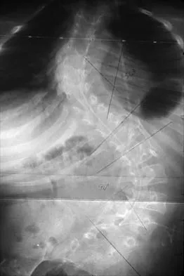
Figures 25a and 25b show the radiograph and MRI scan of a 7 1/2-year-old boy who has been limping for 1 year. His pain has worsened over the past 2 weeks, and his parents note swelling over the dorsum of the foot for the past 4 days. Examination reveals no fever, and laboratory studies show a WBC of 6,700/mm3, an erythrocyte sedimentation rate of 26 mm/h, and a normal C-reactive protein level. What is the most likely diagnosis?


Explanation

The diagnosis of tuberculous osteomyelitis in children is often delayed. In one series of 23 children, the average interval between the onset of symptoms and definite diagnosis was 4.3 months. In these patients, the presenting signs and symptoms were found to be mild, with the most common signs being localized swelling (69.6%) and a painful disability of the involved limbs (65.2%). A mild elevation of the erythrocyte sedimentation rate may be present, but the C-reactive protein level is usually normal. In patients who have osteoarticular tuberculosis, an MRI scan generally shows large intra-articular effusions, periarticular osteoporosis, and gross thickening of the synovial membrane. Differential diagnosis between tuberculosis and pyogenic arthritis is difficult, and an accurate diagnosis usually requires biopsy of synovial tissue. Aspiration of synovial fluid often results in insufficient information to make a diagnosis. Treatment generally consists of surgical debridement and combined antituberculous chemotherapy with isoniazid, ethambutol, and rifampin. Wang MN, Chen WM, Lee KS, Chin LS, Lo WH: Tuberculous osteomyelitis in young children. J Pediatr Orthop 1999;19:151-155.


Question 22

A 10-year-old boy who plays baseball reports acute pain after throwing a softball from the outfield to second base. A radiograph is shown in Figure 26. Management should consist of


Explanation

The patient has a fracture through a unicameral bone cyst, as evidenced by the "falling leaf" sign on the radiograph. Following healing of the fracture, treatment should consist of corticosteroid injection or bone marrow injection. Some cysts heal with the fracture and do not require injections. Biopsy is unnecessary because the radiograph shows that the cyst is benign. Curettage and bone grafting are seldom necessary because these cysts regularly heal with injections. Corticosteroids are useful when injected into the cyst, but are not used systemically. Pulsed electromagnetic fields have not been used therapeutically in this condition.


Question 23

The mother of an otherwise healthy 1-month-old infant reports that he is not moving his left leg after falling from his high chair 2 days ago. He has a temperature of 99.5 degrees F (37.5 degrees C). Examination reveals that the left thigh is moderately tender to palpation. Because the infant is apprehensive, range of motion is difficult to quantify, but appears to be normal at the hips and ankles. Range of motion of the left knee is approximately 25 degrees to 90 degrees. A radiograph of the leg is shown in Figure 27. Management should consist of


Explanation

The patient has a bucket-handle fracture of the distal femur with bilateral corner fractures of the distal femur and a transverse fracture of the proximal tibia. These fractures are virtually pathognomonic of child abuse. The infant should be admitted to the hospital, and child protection services should be notified for investigation of possible abuse. A skeletal survey should be obtained, along with laboratory studies that include a CBC, a platelet count, a prothrombin time, a partial thromboplastin time, and a bleeding time. Akbarnia BA: The role of the orthopaedic surgeon in child abuse, in Morrissy RT, Weinstein SL (eds): Lovell & Winter's Pediatric Orthopaedics, ed 4. Philadelphia, Pa, Lippincott-Raven, 1996, pp 1315-1334.


Question 24

A 12-year-old girl with juvenile rheumatoid arthritis (JRA) has had chronic pain and synovitis about the knee that is now well-controlled medically. Examination reveals 20 degrees of valgus at the knee. Knee range of motion shows 10 degrees to 90 degrees of flexion. Treatment should consist of


Explanation

Children with JRA frequently have valgus in association with hypervascularity because of chronic inflammation. This is normally caused by overgrowth of the medial femoral epiphysis. Staple hemiepiphyseodesis, if done early, can reverse the deformity. Osteotomy is usually unnecessary at this age, and there is a risk of stiffness of the knee following the procedure. Synovectomy may be helpful but will not prevent or correct a deformity.

Question 25

Figure 28 shows the radiograph of a 6-year-old girl who has a right thoracic scoliosis that measures 60 degrees. Examination shows multiple cafe-au-lait spots, and family history reveals that the child's mother has the same disorder. The gene responsible for this disorder codes for


Explanation

The patient has the dystrophic type of scoliosis seen in patients with neurofibromatosis type I (NF-1). The NF-1 gene is located on chromosome 17 and codes for neurofibromin, believed to be a tumor-suppresser gene. Abnormalities in the dystrophin gene are seen in Duchenne muscular dystrophy and Becker muscular dystrophy. A mutation in the frataxin gene is responsible for Friedreich ataxia. The most common type of hereditary motor and sensory neuropathy (Charcot-Marie-Tooth), HMSN type IA is caused by a complete duplication of the peripheral myelin protein gene. A defect in the cellular sulfate transport protein results in undersulfation of proteoglycans seen in diastrophic dysplasia.


Question 26

A 4-week-old female with a history of breech presentation is evaluated for hip instability. Ultrasound reveals an alpha angle of 45 degrees, a beta angle of 77 degrees, and 30% femoral head coverage bilaterally. What is the most appropriate initial management?


Explanation

An alpha angle < 60 degrees with subluxation or dislocation on ultrasound in a 4-week-old is diagnostic of DDH. The first-line treatment is a Pavlik harness to achieve reduction and promote acetabular development.

Question 27

A 6-month-old girl has been treated with a Pavlik harness for 4 weeks for a dislocated left hip. A follow-up ultrasound confirms that the hip remains dislocated within the harness. What is the most appropriate next step in management?


Explanation

Continued use of a Pavlik harness for a dislocated hip beyond 3 to 4 weeks without achieving reduction increases the risk of posterior acetabular wear ("Pavlik harness disease"). The next appropriate step is closed reduction and spica casting under anesthesia.

Question 28

A 6-year-old boy sustains a widely displaced extension-type supracondylar humerus fracture. After closed reduction and percutaneous pinning, the hand is pink and warm, but the radial pulse remains nonpalpable. Capillary refill is less than 2 seconds. What is the most appropriate next step?


Explanation

A "pink, pulseless" hand after anatomic reduction and pinning of a supracondylar humerus fracture typically indicates vascular spasm. It is best managed with close observation and elevation, as collateral circulation is adequate.

Question 29

An 11-year-old girl presents with adolescent idiopathic scoliosis. Standing radiographs reveal a right thoracic curve measuring 35 degrees. She is premenarchal and has a Risser stage of 0. What is the most appropriate management?


Explanation

Bracing is indicated for skeletally immature patients (Risser 0-2, premenarchal) who present with an idiopathic scoliotic curve between 25 and 45 degrees to prevent curve progression.

Question 30

A 14-year-old boy sustains a twisting injury to his right ankle while sliding into a base. Radiographs reveal a Salter-Harris III fracture of the anterolateral aspect of the distal tibial epiphysis (Tillaux fracture). Which ligament is responsible for the avulsion of this fracture fragment?


Explanation

The Tillaux fracture is an avulsion of the anterolateral distal tibial epiphysis caused by an external rotation force. The fragment is avulsed by the anterior inferior tibiofibular ligament (AITFL).

Question 31

A newborn is diagnosed with congenital scoliosis. Radiographs demonstrate a fully segmented unilateral unsegmented bar on the left side with a contralateral fully formed hemivertebra on the right side at the exact same level. What is the anticipated risk of curve progression for this specific deformity pattern?


Explanation

A unilateral unsegmented bar with a contralateral hemivertebra at the same level represents the most severe failure of formation and segmentation. This pattern carries the highest risk of curve progression, approaching 100%, and requires early prophylactic fusion.

Question 32

During the treatment of developmental dysplasia of the hip (DDH) with closed reduction and spica casting, which position places the hip at the greatest risk for avascular necrosis (AVN) of the femoral head?


Explanation

Extreme hip abduction (the "frog-leg" position) forces the cartilaginous femoral head tightly against the acetabulum, compressing the extracapsular epiphyseal vessels and significantly increasing the risk of avascular necrosis.

Question 33

A 3-year-old boy sustains an isolated, closed midshaft femur fracture after tripping. He weighs 14 kg (30 lbs). What is the most appropriate definitive management?


Explanation

For children aged 6 months to 5 years presenting with an isolated femur fracture, early spica casting is the standard of care. Flexible nailing is typically reserved for older children (ages 5 to 11) or those with polytrauma.

Question 34

A 9-year-old girl is evaluated for elbow and forearm pain after a fall. Radiographs reveal a displaced Bado Type I Monteggia fracture-dislocation. What is the characteristic radiographic pattern of this injury?


Explanation

A Bado Type I Monteggia fracture is characterized by an anterior dislocation of the radial head associated with a fracture of the ulnar shaft. It is the most common Monteggia fracture pattern seen in children.

Question 35

A 13-year-old boy with infantile idiopathic scoliosis was treated nonoperatively. Radiographs currently show a 55-degree thoracic curve. Which initial radiographic measurement obtained during infancy was most likely predictive of this curve progression?


Explanation

The rib-vertebra angle difference (RVAD), or Mehta angle, is the most reliable predictor of curve progression in infantile idiopathic scoliosis. An RVAD greater than 20 degrees indicates a high likelihood of relentless progression.

Question 36

An 8-year-old boy presents with a displaced fracture of the lateral condyle of the distal humerus. The fracture fragment is displaced 3 mm on the internal oblique radiograph. What is the most appropriate management?


Explanation

Lateral condyle fractures of the humerus that are displaced greater than 2 mm typically require open reduction and internal fixation. This ensures anatomic restoration of the articular surface and physis to prevent nonunion and cubitus valgus.

Question 37

A 14-year-old female undergoes posterior spinal fusion for adolescent idiopathic scoliosis. Intraoperatively, neuromonitoring demonstrates a sudden, sustained loss of somatosensory evoked potentials (SSEPs) and motor evoked potentials (MEPs) bilaterally during rod derotation. What is the most appropriate immediate first step?


Explanation

In the event of a sudden loss of neuromonitoring signals during deformity correction, the immediate first step is to release the corrective forces (e.g., remove the rods). Subsequent steps include raising mean arterial pressure and optimizing oxygenation.

Question 38

A 4-year-old girl is brought in for a painless limp. Pelvic radiographs show a unilaterally dislocated left hip with a false acetabulum and severe dysplasia of the true acetabulum. She has had no prior treatment. What is the recommended management?


Explanation

In a child older than 3 years presenting with untreated DDH, soft tissue contractures and severe bony dysplasia require a comprehensive approach. This includes open reduction, a femoral shortening derotational osteotomy (to reduce AVN risk), and a pelvic osteotomy to provide adequate coverage.

Question 39

A 7-year-old boy sustains an extension-type supracondylar humerus fracture. Neurologic examination reveals an inability to actively flex the interphalangeal joint of his thumb and the distal interphalangeal joint of his index finger. Which nerve is most likely injured?


Explanation

The anterior interosseous nerve (AIN), a branch of the median nerve, is the most commonly injured nerve in extension-type supracondylar humerus fractures. It provides motor innervation to the flexor pollicis longus and the flexor digitorum profundus to the index and middle fingers.

Question 40

An 8-year-old boy sustains a closed midshaft both-bone forearm fracture. He is being considered for non-operative management with a long arm cast. What is the maximum acceptable angulation for this fracture location in this age group?


Explanation

In an 8-year-old child, the maximum acceptable angulation for a midshaft both-bone forearm fracture is 10 degrees. Distal third fractures may tolerate up to 15 degrees due to greater remodeling potential near the rapidly growing distal physis.

Question 41

A 6-month-old infant is diagnosed with an atypical, left-sided thoracic congenital scoliosis curve. A screening renal ultrasound is normal. What other diagnostic study is mandatory to rule out commonly associated anomalies?


Explanation

Congenital scoliosis is highly associated with VACTERL anomalies, as well as intraspinal abnormalities (e.g., tethered cord, diastematomyelia). Therefore, an echocardiogram to rule out cardiac defects and an MRI of the entire spine to rule out neural axis abnormalities are essential.

Question 42

An infant being treated in a Pavlik harness for developmental dysplasia of the hip (DDH) is noted by the parents to have stopped kicking the knee on the treated side. Physical examination confirms decreased active extension of the knee, though the foot and ankle move symmetrically. What is the most appropriate next step in management?


Explanation

Femoral nerve palsy is a known complication of hyperflexion in a Pavlik harness. The harness should be discontinued until quadriceps function returns, which usually occurs within 1 to 2 weeks.

Question 43

A 6-year-old child sustains a severely displaced type III supracondylar humerus fracture. Upon initial evaluation, the hand is pink but the radial pulse is absent. A satisfactory closed reduction and percutaneous pinning is performed. Following fixation, the hand remains pink but pulseless. What is the most appropriate management?


Explanation

A pink, pulseless hand after satisfactory reduction and pinning indicates adequate collateral perfusion. Observation is recommended, as most radial pulses return within a few days without surgical exploration.

Question 44

Which of the following vertebral anomalies represents the highest risk of curve progression in congenital scoliosis?


Explanation

A unilateral unsegmented bar with a contralateral hemivertebra creates a severe mechanical imbalance and has the highest risk of progression. It almost always requires early prophylactic surgical fusion.

Question 45

During attempted closed reduction of a dislocated hip in a 12-month-old child with DDH, concentric reduction cannot be achieved. Which of the following anatomic structures is NOT a typical obstacle to closed reduction?


Explanation

Common blocks to closed reduction in DDH include an inverted limbus, pulvinar, elongated ligamentum teres, contracted iliopsoas, and tight transverse acetabular ligament. The pectineus muscle does not typically impede reduction.

Question 46

A 5-year-old boy presents with a lateral condyle fracture of the distal humerus displaced by 4 mm. If this fracture is managed nonoperatively in a cast, what is the most likely long-term complication?


Explanation

Displaced lateral condyle fractures treated nonoperatively have a high rate of nonunion. This nonunion can lead to a progressive cubitus valgus deformity, which stretches the ulnar nerve and causes tardy ulnar nerve palsy.

Question 47

In a 6-month-old infant with infantile idiopathic scoliosis, a Mehta rib-vertebra angle difference (RVAD) of 25 degrees at the apical vertebra is most highly predictive of which of the following?


Explanation

In infantile idiopathic scoliosis, a Mehta RVAD greater than 20 degrees is highly predictive of curve progression. An RVAD of less than 20 degrees typically characterizes a resolving curve.

Question 48

A 14-year-old boy sustains a twisting ankle injury. Radiographs reveal a Salter-Harris III avulsion fracture of the anterolateral aspect of the distal tibial epiphysis (Tillaux fracture). Which ligament is responsible for avulsing this fragment?


Explanation

A Tillaux fracture is caused by an external rotation force that creates tension on the anterior inferior tibiofibular ligament (AITFL). This tension avulses the anterolateral distal tibial epiphysis because the medial physis has already fused.

Question 49

A classic triplane fracture of the distal tibia in an adolescent traverses the metaphysis, physis, and epiphysis. Based on the Salter-Harris classification system, what type of physeal injury does this represent?


Explanation

A triplane fracture has a coronal fracture in the metaphysis, an axial fracture through the physis, and a sagittal fracture in the epiphysis. Because the fracture crosses all layers from the metaphysis to the joint surface, it is a Salter-Harris IV equivalent.

Question 50

A 6-week-old female infant born in the breech position presents for a routine evaluation. Her physical examination reveals equal leg lengths and negative Ortolani and Barlow maneuvers. What is the most appropriate imaging modality to evaluate for DDH at this age?


Explanation

For an infant younger than 4 to 6 months with risk factors for DDH, dynamic ultrasound is the screening modality of choice. The femoral head and acetabulum are largely cartilaginous and not well visualized on plain radiographs at this age.

Question 51

Brace treatment for adolescent idiopathic scoliosis is generally most effective and indicated for which of the following patient profiles?


Explanation

Bracing in adolescent idiopathic scoliosis is indicated for skeletally immature patients (Risser 0-2) with progressive curves between 25 and 45 degrees. Curves over 45 degrees usually require surgery, while non-progressive curves under 25 degrees are observed.

Question 52

A 10-year-old boy weighing 38 kg (84 lbs) sustains an isolated, closed, transverse midshaft femur fracture. Which of the following is the most appropriate definitive treatment?


Explanation

For children aged 5 to 11 years weighing less than 50 kg, flexible titanium intramedullary nailing is the standard of care for midshaft femur fractures. Rigid antegrade nailing risks avascular necrosis of the femoral head due to piriformis fossa or greater trochanter entry.

Question 53

A 2-year-old boy is brought to the emergency department with an isolated spiral fracture of the femoral shaft. There are no signs of nonaccidental trauma and shortening is less than 2 cm. What is the most appropriate initial definitive management?


Explanation

In a 2-year-old with an isolated femur fracture and acceptable shortening (<2 cm), early hip spica casting is the most appropriate and effective definitive management. Operative intervention is reserved for older children, multiple trauma, or open fractures.

Question 54

A 13-year-old non-ambulatory boy with Duchenne muscular dystrophy develops a progressive 45-degree neuromuscular scoliosis with pelvic obliquity. What is the most appropriate management?


Explanation

Neuromuscular scoliosis in Duchenne muscular dystrophy is rapidly progressive and poorly controlled by bracing. Surgical stabilization typically involves posterior spinal fusion extending to the pelvis to correct pelvic obliquity and optimize seating balance.

Question 55

A 2-year-old girl recently immigrated to the United States and is noted to have a painless limp. Examination reveals a positive Galeazzi sign and severely limited hip abduction. Radiographs show a dislocated left hip with a dysplastic acetabulum. What is the most appropriate treatment?


Explanation

In a child older than 18-24 months presenting with untreated DDH, closed reduction carries a very high risk of AVN and is rarely successful. Treatment requires open reduction, often combined with femoral shortening to reduce joint tension and a pelvic osteotomy for acetabular coverage.

Question 56

A 7-year-old child presents with a Bado type I Monteggia fracture-dislocation (ulnar shaft fracture with anterior radial head dislocation). Closed reduction of the ulna is performed, but the radial head remains subluxated. What is the most critical technical factor to ensure stable reduction of the radial head?


Explanation

In a pediatric Monteggia fracture, the radial head dislocation is driven by ulnar deformity. Restoring the anatomic length and perfect alignment of the ulna is the most critical step to achieve and maintain spontaneous reduction of the radial head.

Question 57

A 3-week-old female infant is evaluated for a suspected hip abnormality. Examination reveals a palpable clunk when her hips are abducted with anteriorly directed pressure on the greater trochanter. What is the most appropriate initial management?


Explanation

The palpable clunk on abduction describes a positive Ortolani test, indicating a dislocated but reducible hip. Immediate treatment with a Pavlik harness is the gold standard for reducible DDH in infants younger than 6 months.

Question 58

A 12-year-old premenarchal girl (Risser 0) presents with adolescent idiopathic scoliosis. Radiographs demonstrate a progressive right thoracic curve of 52 degrees. What is the most appropriate definitive management?


Explanation

Curves greater than 50 degrees in skeletally immature patients have a high risk of progression into adulthood and generally require surgical intervention with a posterior spinal fusion.

Question 59

A 7-month-old infant is referred for evaluation of a developmental dysplasia of the hip (DDH) that was missed at birth. Ultrasound confirms a completely dislocated right hip. What is the most appropriate initial management?


Explanation

The Pavlik harness is most effective in infants under 6 months of age. For an infant 6 to 18 months old, closed reduction and spica casting is the preferred initial treatment for DDH.

Question 60

A 6-year-old boy sustains a completely displaced supracondylar humerus fracture. Upon arrival, his hand is pink but the radial pulse is absent. Following closed reduction and percutaneous pinning, the hand remains pink but the pulse is still not palpable. What is the next best step in management?


Explanation

In a "pink, pulseless" hand after anatomical reduction and pinning of a supracondylar humerus fracture, observation is recommended as collateral circulation is adequate. Routine exploration is not indicated unless the hand becomes cool and pale.

Question 61

A 3-year-old child is found to have congenital scoliosis due to a fully segmented hemivertebra at T8. Which of the following imaging modalities is most essential to evaluate for the most common associated non-spinal anomalies?


Explanation

Congenital scoliosis is highly associated with VACTERL anomalies. Up to 30% of patients have genitourinary abnormalities and 10% have congenital heart defects, making renal ultrasound and echocardiography essential.

Question 62

During attempted closed reduction of a dysplastic hip in a 1-year-old child, the surgeon notes that the hip reduces but is highly unstable in extension. What is the most common extra-articular anatomical block to concentric reduction that may necessitate an open approach?


Explanation

The iliopsoas tendon is the most common extra-articular block to reduction, often compressing the capsule in an hourglass configuration. Intra-articular blocks include the inverted limbus, pulvinar, and transverse acetabular ligament.

Question 63

A 13-year-old boy presents with right knee pain and a noticeable limp for 3 weeks. Radiographs reveal an unstable slipped capital femoral epiphysis (SCFE) of the right hip. He is unable to bear weight, even with crutches. What is the most severe potential complication associated with this condition and its surgical fixation?


Explanation

Avascular necrosis (AVN) is the most devastating complication following SCFE and occurs at a much higher rate in unstable slips (up to 47%) compared to stable slips.

Question 64

In the initial radiographic evaluation of infantile idiopathic scoliosis, which of the following measurements is the most reliable predictor of curve progression?


Explanation

Mehta's rib-vertebra angle difference (RVAD) is the most prognostic factor for infantile idiopathic scoliosis. An RVAD greater than 20 degrees strongly predicts progressive disease requiring intervention.

Question 65

A 2-year-old child presents with an isolated closed diaphyseal fracture of the left femur after a reported fall from a low bed. There are no signs of child abuse. What is the standard of care for definitive management?


Explanation

Early spica casting is the standard of care for isolated diaphyseal femur fractures in children aged 6 months to 5 years with less than 2 cm of shortening. Flexible nailing is typically reserved for children over age 5.

Question 66

A newborn girl with a positive Ortolani sign on the left hip is fitted with a Pavlik harness. Which of the following complications is most likely to occur if the anterior straps of the harness are adjusted to place the hips in excessive flexion (>120 degrees)?


Explanation

Excessive flexion in a Pavlik harness causes compression of the femoral nerve against the inguinal ligament, leading to transient femoral nerve palsy. Excessive abduction (tight posterior straps) increases the risk of avascular necrosis.

Question 67

A 5-year-old girl falls on her outstretched hand and sustains a displaced lateral condyle fracture of the humerus. Radiographs show 4 mm of displacement. If this fracture progresses to a symptomatic nonunion, which of the following long-term complications is most characteristic?


Explanation

Nonunion of a lateral condyle fracture typically leads to progressive cubitus valgus deformity. Over time, this stretches the ulnar nerve, resulting in a tardy ulnar nerve palsy.

Question 68

A 14-year-old boy with Duchenne muscular dystrophy presents with a progressive thoracolumbar scoliosis measuring 45 degrees. His forced vital capacity (FVC) is 45% of predicted. What is the recommended treatment?


Explanation

In Duchenne muscular dystrophy, spinal fusion to the pelvis is indicated when curves exceed 20-30 degrees and the patient is non-ambulatory, provided FVC >30%. This improves sitting balance and slows pulmonary decline; bracing is ineffective.

Question 69

A 2-year-old boy undergoes open reduction for a late-presenting DDH. During a medial approach, which structure must be carefully protected as it passes posterior to the iliopsoas tendon to avoid vascular compromise to the femoral head?


Explanation

The medial femoral circumflex artery (MFCA) provides the primary blood supply to the femoral head in this age group. It is at significant risk during a medial approach for DDH as it courses posterior to the iliopsoas tendon.

Question 70

A 13-year-old boy presents with an ankle fracture characterized by a sagittal fracture through the epiphysis, a transverse fracture through the physis, and a coronal fracture through the posterior metaphysis. What is the primary anatomical mechanism responsible for this specific fracture pattern?


Explanation

This describes a triplane fracture, which occurs due to the asymmetric closure of the distal tibial physis. The physis typically closes first centrally, then medially, and finally laterally, making the lateral aspect vulnerable to this injury pattern.

Question 71

According to the Bracing in Adolescent Idiopathic Scoliosis Trial (BRAIST), a rigid thoracolumbosacral orthosis is most effective at preventing curve progression to surgical thresholds when worn for a minimum of how many hours per day?


Explanation

The BRAIST trial established a clear dose-response relationship for bracing in adolescent idiopathic scoliosis, demonstrating highly significant clinical success in preventing surgery when braces are worn for 18 or more hours daily.

Question 72

A 10-year-old boy presents with an elbow dislocation and an associated displaced fracture of the medial epicondyle of the humerus. Which nerve is most commonly injured in association with this specific injury pattern?


Explanation

The ulnar nerve passes directly posterior to the medial epicondyle in the cubital tunnel. It is highly susceptible to traction or direct impingement in medial epicondyle fractures and elbow dislocations.

Question 73

A 6-week-old female infant born breech presents for evaluation. Ultrasound demonstrates an alpha angle of 43 degrees and a beta angle of 78 degrees on the left hip. The right hip is normal. What is the most appropriate initial management?


Explanation

An alpha angle less than 60 degrees and a beta angle greater than 55 degrees (Graf Type III/IV) indicates developmental dysplasia of the hip (DDH). The Pavlik harness is the gold standard initial treatment for DDH in infants under 6 months of age.

Question 74

A 12-month-old child undergoes closed reduction and spica casting for developmental dysplasia of the hip. Which of the following positions during casting is the most significant risk factor for the development of iatrogenic avascular necrosis of the femoral head?


Explanation

Extreme abduction during spica casting forces the femoral head against the acetabulum and stretches the medial circumflex femoral artery, significantly increasing the risk of avascular necrosis (AVN). The hip should ideally be immobilized in the 'human position' of moderate flexion and mild abduction.

Question 75

A 6-year-old boy sustains a closed, isolated midshaft fracture of the right femur after a fall from a playground structure. He has no other associated injuries. What is the current standard of care for definitive management?


Explanation

For children aged 5 to 11 years with length-stable diaphyseal femur fractures, flexible intramedullary nailing (e.g., titanium elastic nails) is the standard of care. Rigid intramedullary nailing is contraindicated in this age group due to the risk of avascular necrosis and proximal femoral physeal arrest.

Question 76

A 9-month-old boy presents with an infantile idiopathic scoliosis. Radiographs demonstrate a 30-degree left thoracic curve. The rib-vertebral angle difference (RVAD) is calculated to be 25 degrees. What is the most appropriate management?


Explanation

An RVAD (Mehta's angle) greater than 20 degrees in infantile idiopathic scoliosis strongly predicts curve progression. Serial casting (Mehta casting) is indicated to control and potentially correct the deformity in young infants with progressive curves.

Question 77

A 12-year-old obese boy presents with sudden inability to bear weight on his left leg. He reports a 2-month history of vague left knee pain. Radiographs reveal a severe, posterior translation of the proximal femoral epiphysis. He cannot bear weight even with crutches. What intervention best minimizes the risk of osteonecrosis?


Explanation

This is an unstable slipped capital femoral epiphysis (SCFE), defined by the inability to bear weight. Urgent/emergent capsulotomy to decompress the intracapsular hematoma combined with in situ single-screw fixation is recommended to reduce the high risk of osteonecrosis associated with unstable SCFE.

Question 78

A 14-year-old boy with Duchenne muscular dystrophy who is wheelchair-bound develops a 45-degree progressive thoracolumbar scoliosis. His forced vital capacity (FVC) is 40% of predicted. What is the most appropriate management of his spinal deformity?


Explanation

In Duchenne muscular dystrophy, scoliosis progresses rapidly once the patient becomes wheelchair-bound, and bracing is ineffective. Posterior spinal fusion to the pelvis is indicated to maintain sitting balance and comfort before pulmonary function deteriorates too severely.

Question 79

A 5-year-old girl falls from monkey bars and sustains a widely displaced, extension-type supracondylar humerus fracture. On examination, the hand is pink and well-perfused, but the radial pulse is absent. What is the most appropriate next step in management?


Explanation

A 'pink, pulseless' hand after a supracondylar fracture indicates adequate collateral circulation. The next step is urgent closed reduction and percutaneous pinning. The pulse often returns after reduction; vascular exploration is only indicated if the hand becomes or remains dysvascular (white and pulseless) after reduction.

Question 80

A 3-year-old girl presents with a painless waddling gait. Radiographs show a completely dislocated left hip with a false acetabulum and a dysplastic true acetabulum. She has had no prior treatment. What is the most appropriate surgical management?


Explanation

In children over 2 to 3 years of age with an untreated completely dislocated hip, the soft tissues are contracted and the acetabulum is dysplastic. Successful management typically requires an open reduction, a femoral shortening osteotomy (to relieve tension and prevent AVN), and a pelvic osteotomy (e.g., Salter or Dega) to provide anterior/lateral coverage.

Question 81

A 6-year-old boy is evaluated for a displaced lateral condyle fracture of the humerus. If this fracture is managed non-operatively and progresses to nonunion, what is the most likely late clinical complication?


Explanation

Lateral condyle fractures are prone to nonunion because they are intra-articular and bathed in synovial fluid. A nonunion leads to progressive lateral growth arrest, resulting in cubitus valgus. This stretch on the medial side frequently causes a tardy ulnar nerve palsy years later.

Question 82

A 13-year-old premenarcheal female presents for scoliosis screening. Radiographs demonstrate a 32-degree right thoracic curve. Her Risser stage is 0. What is the most appropriate management?


Explanation

This patient has adolescent idiopathic scoliosis (AIS) with a curve between 25 and 45 degrees and significant growth remaining (premenarcheal, Risser 0). The standard of care to prevent curve progression is full-time bracing (TLSO) for 16 to 23 hours a day.

Question 83

A 7-year-old boy is diagnosed with Legg-Calve-Perthes disease. Which of the following is considered the most important prognostic factor for long-term hip joint congruency?


Explanation

Age at the onset of Legg-Calve-Perthes disease is the most critical prognostic factor. Children younger than 6 years generally have a favorable outcome due to greater remodeling potential, whereas older children (especially >8 years) have a higher risk of developing a permanently deformed, incongruous femoral head.

Question 84

A 13-year-old boy sustains an ankle injury while playing soccer. Radiographs reveal a distal tibia fracture that appears as a Salter-Harris III pattern on the AP view and a Salter-Harris II pattern on the lateral view. What is the classic mechanism of injury for this fracture?


Explanation

The described injury is a triplane fracture, which occurs in early adolescence as the distal tibial physis closes (central first, then medial, then lateral). It is characteristically caused by an external rotation force, leading to a multiplanar fracture.

Question 85

A 2-year-old child presents with a congenital spinal deformity. Radiographs show multiple vertebral anomalies. Which of the following anomaly patterns is associated with the highest risk of rapid curve progression?


Explanation

A unilateral unsegmented bar combined with a contralateral fully segmented hemivertebra produces maximal asymmetric growth. This specific congenital pattern has a nearly 100% chance of relentless, rapid progression and mandates early surgical fusion.

Question 86

A 5-month-old infant with developmental dysplasia of the hip has been treated in a Pavlik harness for 4 weeks. Serial ultrasounds show that the hip remains persistently dislocated and cannot be reduced in the harness. What is the most appropriate next step?


Explanation

If a hip remains dislocated after 3 to 4 weeks of Pavlik harness treatment, the harness must be discontinued. Prolonged use of the harness on a dislocated hip causes posterior acetabular wear ('Pavlik harness disease'). The next step is a closed reduction with an arthrogram and spica casting.

Question 87

A 14-year-old boy sustains a Salter-Harris II fracture of the distal femur with 30% posterior translation. Which of the following best describes the preferred definitive management and rationale?


Explanation

Distal femur physeal fractures have a high risk of growth arrest and displacement due to strong muscle pull (gastrocnemius). Because of this instability and the devastating consequences of displacement, they require anatomic reduction and secure fixation (typically crossed smooth pins or cannulated screws), avoiding simple casting.

Question 88

During a posterior spinal fusion for adolescent idiopathic scoliosis in a 15-year-old female, the intraoperative neuromonitoring demonstrates a sudden loss of transcranial motor evoked potentials (MEPs) bilaterally, while somatosensory evoked potentials (SSEPs) remain intact. What is the most likely neurologic event?


Explanation

MEPs monitor the anterior (motor) pathways of the spinal cord, which are supplied by the anterior spinal artery. SSEPs monitor the posterior columns (sensory). Isolated loss of MEPs with intact SSEPs strongly suggests ischemia of the anterior spinal cord, which is highly sensitive to hypotension or over-distraction.

Question 89

An 8-year-old boy falls from a tree and sustains a Delbet Type II (transcervical) femoral neck fracture. Following prompt open reduction and internal fixation, what is the most significant complication he is at risk of developing?


Explanation

Pediatric femoral neck fractures, particularly transcervical (Delbet Type II) and transepiphyseal (Delbet Type I) fractures, carry a high risk of osteonecrosis (avascular necrosis) of the femoral head due to the disruption of the tenuous retinacular blood supply.

Question 90

A 14-year-old girl sustains a juvenile Tillaux fracture. Which ligament is responsible for the avulsion of the anterolateral fragment of the distal tibial epiphysis?


Explanation

A juvenile Tillaux fracture is a Salter-Harris III fracture of the anterolateral distal tibia. It occurs via external rotation forces where the intact anterior inferior tibiofibular ligament (AITFL) avulses the anterolateral epiphysis, which is the last portion of the distal tibial physis to close.

Question 91

A 13-year-old gymnast presents with chronic lower back pain. Radiographs reveal an isthmic spondylolisthesis at L5-S1 with 60% forward translation (Meyerding Grade III). She has failed 6 months of physical therapy. What is the most appropriate surgical management?


Explanation

For high-grade isthmic spondylolisthesis (>50% slip, Grade III-V) in adolescents that is symptomatic or progressive, in situ posterior or posterolateral spinal fusion (typically extending from L4 to S1) is the standard treatment. Attempting complete reduction carries a high risk of L5 nerve root injury.

Question 92


A 10-year-old boy with neurofibromatosis type 1 (NF-1) develops a 45-degree, sharp, short angular thoracic kyphoscoliosis. Rib penciling and dural ectasia are noted on advanced imaging. What is the recommended surgical management for this patient?


Explanation

Dystrophic curves in neurofibromatosis type 1 are notorious for relentless progression and an exceptionally high rate of pseudarthrosis if treated with posterior fusion alone. The standard of care for dystrophic kyphoscoliosis in NF-1 is a combined anterior and posterior spinal fusion.

Question 93

A 4-year-old girl presents with an untreated, completely dislocated left hip. Radiographs confirm developmental dysplasia of the hip (DDH) with a false acetabulum and significant superior migration of the femoral head. What is the most appropriate surgical management?


Explanation

In a 4-year-old with an untreated dislocated hip, open reduction is required. Femoral shortening is necessary to reduce joint reaction forces and minimize the risk of osteonecrosis, while a pelvic osteotomy addresses the secondary acetabular dysplasia.

Question 94

A 6-year-old boy falls from monkey bars and sustains a widely displaced extension-type supracondylar humerus fracture. Examination reveals an absent radial pulse but a warm, pink hand. After closed reduction and percutaneous pinning, the hand remains warm and pink, but the pulse remains absent. What is the next best step in management?


Explanation

A "pink, pulseless" hand after anatomical reduction and pinning of a supracondylar humerus fracture typically indicates adequate collateral circulation. Observation is indicated as the pulse often returns within a few days; vascular exploration is reserved for a pale, pulseless hand.

Question 95

A 3-year-old boy presents with a 35-degree right thoracic curve. The rib-vertebral angle difference (RVAD) of Mehta is calculated at 25 degrees on the AP radiograph. What is the most appropriate management for this condition?


Explanation

Infantile idiopathic scoliosis with a curve greater than 30 degrees and an RVAD greater than 20 degrees has a very high risk of progression. Early intervention with serial elongation-derotation-flexion (EDF) casting (Mehta casting) is the gold standard to control or resolve the deformity.

Question 96

A 3-month-old infant is being treated with a Pavlik harness for a reducible, dislocated right hip. During the follow-up visit, you notice the infant lacks active knee extension on the right side. What complication is most likely occurring?


Explanation

Femoral nerve palsy in a Pavlik harness is typically caused by excessive hip flexion. The harness must be adjusted to reduce flexion or temporarily removed to allow nerve recovery, as persistent hyperflexion can lead to permanent palsy or inferior hip dislocation.

Question 97

A 13-year-old boy presents with a painful, swollen ankle after a skateboarding injury. Radiographs show a Salter-Harris III fracture of the anterolateral distal tibia. What is the primary pathomechanical force and structure responsible for this specific fracture pattern?


Explanation

A juvenile Tillaux fracture is a Salter-Harris III fracture of the anterolateral distal tibia epiphysis. It occurs secondary to an external rotation force where the AITFL avulses the anterolateral fragment due to the asymmetric closure pattern of the distal tibial physis.

Question 98

A 14-year-old girl with adolescent idiopathic scoliosis (AIS) has a right thoracic curve of 55 degrees and a left lumbar curve of 30 degrees. On side-bending radiographs, the lumbar curve reduces to 10 degrees. According to the Lenke classification, what type of curve pattern is this, and what is the recommended surgical approach?


Explanation

This is a Lenke 1 (main thoracic) curve pattern, because the lumbar curve is nonstructural (bends out to less than 25 degrees). The standard surgical management is a selective thoracic fusion, which spares the lumbar spine to preserve spinal mobility.

Question 99

A 5-year-old girl sustains a displaced lateral condyle fracture of the distal humerus. If left untreated and progressing to nonunion, which of the following is the most likely long-term complication?


Explanation

Nonunion of a pediatric lateral condyle fracture typically leads to a progressive cubitus valgus deformity due to the proximal migration of the un-united lateral fragment. Over time, this valgus deformity stretches the ulnar nerve, resulting in a tardy ulnar nerve palsy.

Question 100

A 6-week-old female infant, born breech at 39 weeks gestation, presents for a routine check-up. Clinical examination of the hips reveals symmetric thigh folds and negative Barlow and Ortolani maneuvers bilaterally. What is the most appropriate next step in hip screening for this patient?


Explanation

Breech presentation and female sex are major risk factors for DDH. Current clinical practice guidelines strongly recommend screening ultrasound at 4 to 6 weeks of age for infants with these risk factors, regardless of a normal clinical examination.

None

Clinic OS
Medically Verified Content by
Prof. Clinic OS
Consultant Orthopedic & Spine Surgeon
Chapter Index