العربية
Part of the Master Guide

AAOS Orthopedic MCQs (Set 1): Spine Disorders, Sports Medicine & Arthroplasty | 2026 Board Review

Orthopedic Spine 2026 MCQs: Board Review Questions & Answers (Part 1)

23 Apr 2026 87 min read 71 Views
Figure for Spine 2006 MCQs - Part 1 - Question 1

Key Takeaway

In this comprehensive guide, we discuss everything you need to know about Orthopedic Spine 2026 MCQs: Board Review Questions & Answers (Part 1). Top-rated Orthopedic Spine 2026 MCQs bank. Practice with clinical case questions, orthopedic surgery board review, and evidence-based answers updated for 2026.

Orthopedic Spine 2026 MCQs: Board Review Questions & Answers (Part 1)

Comprehensive 100-Question Exam


00:00

Start Quiz

Question 1

Figures 1a and 1b show the sagittal T2- and T1-weighted MRI scans of a 25-year-old intravenous drug abuser who has low back pain that is increasing in intensity. Laboratory studies show a WBC count of 10,000/mm3 and an erythrocyte sedimentation rate of 80 mm/h. Blood culture is negative. Initial management consist of


Explanation

1b The MRI scans show vertebral diskitis/osteomyelitis. The treatment of spinal infection in adults should be organism specific; therefore, initial management should consist of CT-guided closed biopsy prior to administration of antibiotic coverage. An open biopsy is indicated for a failed closed biopsy or failure of nonsurgical management. Although Staphylococcus aureus is the most common bacteria, a history of intravenous drug abuse raises suspicion for other organisms, including Pseudomonas. Tay BK, Deckey J, Hu SS: Spinal Infections. J Am Acad Orthop Surg 2002;10:188-197.

Question 2

A 27-year-old man sustained a gunshot wound to the lumbar spine and undergoes an exploratory laparotomy. An injury to the cecum is identified and treated. Management should now include


Explanation

Gunshot wounds to the spine present relatively little risk of infection in most cases. When there has been an injury to the colon, the risk of infection can be minimized with a 7-day course of broad-spectrum antibiotics. Fragment removal is not indicated. Roffi RP, Waters RL, Adkins RH: Gunshot wounds to the spine associated with a perforated viscus. Spine 1989;14:808-811.

Question 3

A 68-year-old man reports a 1-year history of debilitating neck pain without neurologic symptoms. History reveals a C5-6 anterior diskectomy and bone grafting 10 years ago that provided good relief of arm and neck pain. Radiographs show evidence of fibrous union at C5-6, spondylotic disk narrowing at C4-5 and C6-7, and a fixed 2-mm subluxation at C3-4. Examination reveals cervical stiffness and discomfort at the extremes of movement. His neurologic examination is normal. Treatment should now consist of


Explanation

Axial pain can be difficult to manage. Pain management is not always successful, and surgical approaches may provide disappointing results unless there is discrete pathology. Whereas planning of a surgical approach should consider prior approaches and preexisting laryngeal dysfunction, no compelling case for surgical intervention can be made for this patient. Therefore, management should consist of patient education, exercise, and nonnarcotic medication. Ahn NU, Ahn UM, Andersson GB, et al: Operative treatment of the patient with neck pain. Phys Med Rehabil Clin N Am 2003;14:675-692. Algers G, Pettersson K, Hildingsson C, et al: Surgery for chronic symptoms after whiplash injury: Follow-up of 20 cases. Acta Orthop Scand 1993;64:654-656.

Question 4

Which of the following is considered the lowest level that a standard thoracolumbosacral orthosis (TLSO) can immobilize?


Explanation

Without more distal immobilization such as a thigh extension, the lower two lumbar segments generally show the same or even increased mobility with a TLSO. White AA, Panjabi MM: Clinical Biomechanics of the Spine, ed 2. Philadelphia, PA, JB Lippincott, 1990, pp 475-509.

Question 5

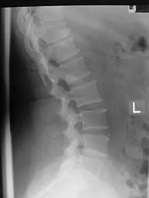
A 65-year-old man with ankylosing spondylitis has neck pain after falling back over his lawnmower, striking his thoracic spine, and forcing his neck into extension. Examination reveals subtle weakness of the intrinsics and finger flexors at approximately 4+/5. Initial management consists of immobilization in a rigid collar, and placing his head in the anatomic position. Radiographs reveal a subtle extension fracture of the lower cervical spine. Approximately 6 hours after the injury, he reports increasing paresthesias in his upper and lower extremities, and examination now shows his intrinsics are 2/5, finger flexors are 3/5, and his triceps are now weak at 4/5 on manual motor testing. In addition, his lower extremities now show weakness in both dorsal and plantar flexion of the ankle in the range of 4/5. Repeat radiographs appear unchanged. An MRI scan is shown in Figure 2. Management should now consist of


Explanation

It is not uncommon for patients with ankylosing spondylitis to sustain extension-type fractures, most typically of the cervicothoracic junction. These fractures can appear nondisplaced or minimally displaced initially, making them difficult to diagnose. Because there is no mobility between vertebrae, fractures tend to occur more like those of a transverse fracture of a long bone. In addition, the vertebral bodies are vascular and their canals are relatively enclosed, making them vulnerable to epidural bleeding. The MRI scan reveals an epidural hematoma located posteriorly on the cord; therefore, the treatment of choice is surgical evacuation and a posterior laminectomy. Because of the intrinsic instability of such fractures at the time of the laminectomy, internal fixation and stabilization with a posterior fusion is warranted. A simple laminectomy will only increase instability, and control is unlikely with halo vest immobilization. An anterior procedure will not effectively treat the problem given the location of the hematoma. Consideration can be given to methylprednisolone and observation; however, this will not eradicate the problem. Bohlman HH: Acute fractures and dislocations of the cervical spine. J Bone Joint Surg Am 1979;61:1119-1142.

Question 6

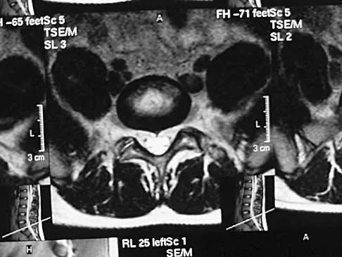
Figures 3a and 3b show the MRI scans of a patient with neck pain. What is the most likely diagnosis?


Explanation

3b Muliple neurofibromas result in marked foraminal enlargement as seen on the sagittal MRI scan. Collagen disorders leading to dural ectasia may show similar enlargement, but none of these is listed as a possible answer. Kim HW, Weinstein SL: Spine update: The management of scoliosis in neurofibromatosis. Spine 1997;22:2770-2776.

Question 7

A Trendelenburg gait is most likely to be seen in association with


Explanation

A Trendelenburg gait results from weakness of the gluteus medius, which is innervated by the L5 nerve root. A paracentral disk herniation at L4-L5 most commonly results in an L5 radiculopathy and thus weakness of the gluteus medius. A paracentral herniation at L5-S1 most commonly affects the S1 nerve root. A paracentral herniation at L3-L4, a central herniation at L3-L4, and a far lateral herniation at L4-L5 all affect the L4 root. Fardon DF, Garfin SR, Abitbol J, et al (eds): Orthopedic Knowledge Update: Spine 2. Rosemont, IL, American Academy of Orthopaedic Surgeons, 2002, pp 323-332.

Question 8

An otherwise healthy 70-year-old man has back and bilateral leg pain in an L5 distribution that is aggravated by standing more than 10 minutes or walking more than 100 feet. He has to sit to get relief. Neurologic and pulse examinations are normal. A radiograph and MRI scan are shown in Figures 4a and 4b. Treatment should consist of


Explanation

4b The patient has a degenerative spondylolisthesis at L4-5 with associated spinal stenosis. His symptoms are consistent with neurogenic claudication. Based on these findings, the surgical treatment of choice is decompression and posterolateral fusion. Use of instrumentation is controversial. Laminectomy alone is reserved for the patient who is frail medically. There is no role for an anterior approach or for fusion alone without decompression. Fischgrund JS, Mackay M, Herkowitz HN, et al: Degenerative lumbar spondylolisthesis with spinal stenosis: A prospective, randomized study comparing decompressive laminectomy and arthrodesis with and without spinal instrumentation. Spine 1997;22:2807-2812.

Question 9

Figures 5a and 5b show the radiograph and MRI scan of a patient who has severe mechanical neck pain but no neurologic problems. Biopsy and work-up show the lesion to be a solitary plasmacytoma. Treatment should consist of


Explanation

5b Plasmacytoma is very sensitive to radiation therapy and given the complexity of the resection and complications of surgery in the given location, radiation therapy is preferred. However, the patient has clear loss of bony structural integrity, and resultant instability would persist even with tumor irradiation; therefore, posterior stabilization is warranted. Chemotherapy and bone marrow transplant are reserved for systemic disease with multiple myeloma. Corwin J, Lindberg RD: Solitary plasmacytoma of bone vs. extramedullary plasmacytoma and their relationship to multiple myeloma. Cancer 1979;43:1007-1013.

Question 10

A 71-year-old woman undergoes a posterior lumbar decompression and fusion from L4-S1. Thirty-six hours after the procedure, she reports severe right-sided chest pain and shortness of breath. Doppler ultrasound reveals a clot proximal to the knee within the femoral vein. A large pulmonary embolus is confirmed by CT angiography. The next most appropriate step in management should consist of


Explanation

In a review of 13,000 spinal procedures, nine patients were treated with heparin following development of pulmonary emboli. Of these patients, six had serious complications ranging from wound drainage to paralysis. Heparin therapy instituted within 10 days of the surgical procedure resulted in a 100% complication rate. Vena cava filter placement has a complication rate of 0.12% to 10.1%. Removable filters are currently in clinical trials. Cain JE Jr, Major MR, Lauerman WC, et al: The morbidity of heparin therapy after development of pulmonary embolus in patients undergoing thoracolumbar or lumbar spinal fusion. Spine 1995;20:1600-1603. Roberts AC: Venous imaging and inferior vena cava filters. Curr Opin Radiol 1992;4:88-96.

Question 11

Figure 6 shows the lateral radiograph of a 22-year-old woman who has painful Scheuermanns's kyphosis in the middle and lower thoracic spine. When planning surgical correction using instrumentation, the distal aspect of the instrumentation should ideally extend to the


Explanation

Posterior constructs for Scheuermann's kyphosis ideally should extend from the most superior to the most inferior aspect of the Cobb angulation. However, the most distal fusion level must be in a stable or lordotic position to avoid the development of junctional kyphosis. Lowe reported that failure to incorporate the first lordotic segment of the lumbar spine is associated with a higher risk of junctional kyphosis. The first lordotic segment of the lumbar spine is typically at least one level below the distal aspect of the curve as measured by the Cobb technique and most commonly is in the upper part of the lumbar spine. Lowe TG: Scheuermann's disease. Orthop Clin North Am 1999;30:475-487.

Question 12

Figure 7 shows the radiograph of a 64-year-old man who has neck pain and weakness of the upper and lower extremities following a motor vehicle accident. Examination reveals 3/5 quadriceps and 4/5 hip flexors but no ankle dorsiflexion or plantar flexion. His intrinsics are 1/5, with finger flexors of 3/5. He is awake, alert, and cooperative. Management should consist of


Explanation

In patients with facet dislocations and an incomplete neurologic deficit, early decompression of the canal via reduction of the dislocation generally is considered safe if the patient is alert and can cooperate. However, patients who cannot cooperate with serial neurologic examinations during the reduction are at risk for increased deficit secondary to herniated nucleus pulposus, and MRI should be performed prior to either closed or open reduction. Star AM, Jones AA, Cotler JM, et al: Immediate closed reduction of cervical spine dislocations using traction. Spine 1990;15:1068-1072.

Question 13

What is the recommended insertion torque for halo pins in adults?


Explanation

Garfin and associates have shown that halo pins inserted with 8 in-lb of insertion torque results in significantly less loosening with cyclical loading than pins inserted with 6 in-lb of torque. Moreover, Botte and associates reported that 8 in-lb of torque is clinically safe and effective in lowering the incidence of pin loosening and infection. Botte MJ, Byrne TP, Garfin SR: Application of the halo device for immobilization of the cervical spine utilizing an increased torque pressure. J Bone Joint Surg Am 1987;69:750-752. Garfin SR, Lee TO, Roux RD, et al: Structural behavior of the halo orthosis pin-bone interface: Biomechanical evaluation of standard and newly designed stainless steel halo fixation pins. Spine 1986;11:977-981.

Question 14

A 55-year-old woman with a history of untreated idiopathic scoliosis has had neurogenic claudication for the past several months. MRI reveals spinal stenosis at L2-L3, L3-L4, and L4-L5. Radiographs show a 45-degree lumbar curve from T10 to L4, with a degenerative spondylolisthesis at L4-L5. Laminectomy at the stenotic levels and stabilization of the deformity are planned. Which of the following is NOT considered an absolute indication for extending the fusion to the sacrum, rather than stopping at L5?


Explanation

There are several indications for extending adult scoliosis fusions to the sacrum, rather than stopping in the lower lumbar spine. These indications include posterior column deficiencies at L5-S1, such as spondylolysis and laminectomy, and deformities extending to the sacrum, such as fixed tilt of L5-S1 or sagittal imbalance. MRI signal changes in the L5-S1 disk do not preclude stopping the fusion at L5. Some surgeons use diskography or diagnostic facet blocks to evaluate the integrity of the L5-S1 level prior to stopping the fusion at L5. Long scoliosis fusions stopping at L5 have a significant risk of failure, highlighting the importance of careful selection of fusion levels. Bradford DS, Tay BK, Hu SS: Adult scoliosis: Surgical indications, operative management, complications, and outcomes. Spine 1999;24:2617-2629. Bridwell KH: Where to stop the fusion distally in adult scoliosis: L4, L5, or the sacrum? Instr Course Lect 1996;45:101-107.

Question 15

Which of the following findings is considered a contraindication for posterior decompression (with or without fusion) for myelopathy?


Explanation

Although cervical instability is a contraindication to posterior decompression alone, segmental instability in the myelopathic cervical spine can be addressed with concomitant posterior fusion with instrumentation. Cervical lordosis represents the ideal scenario for posterior decompressive procedures for myelopathy (laminectomy and laminoplasty) because compression from anterior osteophytes, if present, is relieved as the spinal cord migrates posteriorly. The anteroposterior diameter of the spinal canal does not have an impact on the selection of surgical approach. Posterior unroofing-type procedures in kyphotic cervical spines, however, are ineffective because anterior impingement on the spinal cord will remain; therefore, kyphosis of more than 10 degrees is considered a contraindication for posterior decompression. Emery SE: Cervical spondylotic myelopathy: Diagnosis and treatment. J Am Acad Orthop Surg 2001;9:376-388.

Question 16

A patient reports progessive bilateral hand clumsiness and ataxia. Examination reveals a positive Hoffmann's sign and intrinsic atrophy. MRI reveals multilevel cervical spondylosis, and lateral flexion and extension radiographs show cervical kyphosis in the neutral position, with restoration of lordosis on extension. Which of the following procedures is most likely to result in poor long-term results?


Explanation

Adequate decompression of the cervical cord can be achieved in a variety of ways depending on the pathoanatomy of the compression, but kyphosis is a relative contraindication to laminectomy alone. For laminectomy to be effective, the lordosis must be maintained so the cord can displace posteriorly away from the anterior structures. In addition, removing the posterior tension band increases the probability that the kyphosis will progress, therefore increasing the force against the front of the cord as it tents across the kyphosis. Albert TJ, Vaccaro A: Postlaminectomy kyphosis. Spine 1998;23:2738-2745. Truumees E, Herkowitz HN: Cervical spondylotic myelopathy and radiculopthy. Instr Course Lect 2000;49:339-360.

Question 17

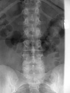
A 45-year-old man seen in the emergency department reports a 1-week history of worsening low back pain and a progressive neurologic deficit in the S1 distribution. Examination reveals 2/5 strength in the gastrocnemius. Laboratory studies show a WBC count of 13,500/mm3 and an erythrocyte sedimentation rate of 74 mm/h. Radiographs of the lumbosacral spine show narrowing of the L5-S1 disk space, with irregularity of the end plates. A sagittal T2-weighted MRI scan is shown in Figure 8. Definitive management should consist of


Explanation

The history, physical examination, laboratory, and radiographic findings are most consistent with an infectious process. When there are signs of neurologic compromise, surgery is generally recommended. This is an anterior process, and anterior column debridement is necessary, followed by stabilization. Anterior or posterior stabilization is a reasonable option, but posterior decompression alone is unlikely to adequately reverse the process and may lead to segmental kyphosis. Frymoyer JW, Wiesel SW (eds): The Adult and Pediatric Spine, ed 3. Philadelphia, PA, Lippincott Williams and Wilkins, 2003, pp 165-189.

Question 18

In a retroperitoneal approach to the lumbar spine, what structure runs along the medial aspect of the psoas and along the lateral border of the spine?


Explanation

The sympathetic trunk runs longitudinally along the medial border of the psoas. The ilioinguinal nerve emerges along the upper lateral border of the psoas and travels to the quadratus lumborium, and the genitofemoral nerve lies more laterally on the psoas. The ureter is adherent to the posterior peritoneum and falls away from the psoas and the spine in the dissection, as does the aorta. Watkins RG (ed): Surgical Approaches to the Spine. New York, NY, Springer-Verlag, 1983, p 107.

Question 19

What is the most likely primary cause of decreased success rates of bony fusion in smokers undergoing lumbar arthrodesis?


Explanation

A number of studies have shown a lower success rate of arthrodesis in smokers. Animal models also have shown that administration of nicotine can markedly decrease the rate of arthrodesis. Although it may not be possible to completely eliminate some of the other associated factors that contribute to the failure of arthrodesis, it does appear that nicotine is the primary factor. Andersen T, Christensen FB, Laursen M, et al: Smoking as a predictor of negative outcome in lumbar spinal fusion. Spine 2001;26:2623-2628.

Question 20

Flexion-distraction injuries of the thoracolumbar spine are most frequently associated with injury to what organ system?


Explanation

In patients with flexion-distraction injuries of the thoracolumbar spine, 50% have associated, potentially life-threatening, visceral injuries that occasionally are diagnosed hours or even days after admission. Based on these findings, consultation with a general surgeon is recommended. Blunt and penetrating injuries to the cardiopulmonary system or aorta sometimes can be seen with this type of injury, but they are no more common than with other types of thoracolumbar fractures because of the relatively mild bony injury anteriorly. Neurologic trauma with this type of fracture is also somewhat rare. Levine AM (ed): Orthopaedic Knowledge Update: Trauma. Rosemont, IL, American Academy of Orthopaedic Surgeons, 1996, pp 351-360.

Question 21

What is the most common adverse postoperative complication of laminoplasty for multilevel cervical spondylotic myelopathy?


Explanation

A 30% to 50% loss of cervical range of motion is reported postoperatively in most patients following cervical laminoplasty. Inadvertent closure of the laminoplasty does occur but is rare. Laminoplasty is advocated in lieu of laminectomy to prevent progressive kyphosis and can effectively decompress the spinal cord. C5 nerve root palsies are a poorly understood but rare complication of surgical decompression for cervical spondylotic myelopathy. Emery SE: Cervical spondylotic myelopathy: Diagnosis and treatment. J Am Acad Orthop Surg 2001;9:376-388.

Question 22

The thickest bone in the occiput is located


Explanation

Anatomic studies have shown that the thickest bone of the occiput is at the level of the external occipital protuberance. It ranges from 11.5 mm to 15.1 mm in men and from 9.7 mm to 12 mm in women. In general, the bone thins as it extends distally from the external occipital protuberance and it also moves laterally from the midline. The structures at risk during screw placement include the venous sinuses. Nadim Y, Lu J, Sabry FF, et al: Occipital screws in occipitocervical fusion and their relation to the venous sinuses: An anatomic and radiographic study. Orthopedics 2000;23:717-719.

Question 23

A patient who underwent an L5-S1 diskectomy 18 months ago has persistent pain in the left leg. Figures 9a and 9b show postoperative axial T1-weighted MRI scans at the L5-S1 level without and with gadolinium. What is the most likely diagnosis?


Explanation

9b Persistent or recurrent symptoms after lumbar diskectomy are troublesome and can be difficult to assess. Gadolinium-enhanced MRI scans may be helpful. The images show enhancement about the left S1 root, a finding that is most consistent with perineural (epidural) fibrosis. The root itself does not enhance. Root enhancement has been associated with compressive radicular symptoms. A disk herniation does not enhance with gadolinium. A neurilemmoma enhances with gadolinium, but the involved root would be enlarged. There is no evidence of a fluid collection which would be consistent with an epidural abscess. Babar S, Saifuddin A: MRI of the post-discectomy lumbar spine. Clin Radiol 2002;57:969-981. Kikkawa I, Sugimoto H, Saita K, et al: The role of Gd-enhanced three-dimensional MRI fast low-angle shot (FLASH) in the evaluation of symptomatic lumbosacral nerve roots. J Orthop Sci 2001;6:101-109.

Question 24

Which of the following factors is most closely associated with early postoperative migration of "stand-alone" lumbar interbody fusion cages?


Explanation

Postoperative migration of lumbar interbody fusion cages is a rare complication. It is most commonly seen after placement of the cages through a posterior approach, with instability of the final construct. It is not associated with the design of the cage, the type of graft used, or a resultant pseudarthrosis. McAfee PC: Interbody fusion cages in reconstructive operations on the spine. J Bone Joint Surg Am 1999;81:859-880.

Question 25

If a laminectomy for spinal stenosis is performed, which of the following is an indication for concomitant arthrodesis at that level?


Explanation

A prospective randomized study of patients with degenerative spondylolisthesis and spinal stenosis by Herkowitz and Kurz showed significantly improved clinical outcomes in patients who also received a lumbar arthrodesis. Patients with a laminectomy at an adjacent level do not have improved outcomes with an arthrodesis. Minimal lumbar scoliosis does not require arthrodesis. Arthrodesis is indicated in cases where there is removal of more than 50% of the facets bilaterally but not with an associated foraminal stenosis. Herkowitz HN, Kurz LT: Degenerative lumbar spondylolisthesis with spinal stenosis: A prospective study comparing decompression with decompression and intertransverse process arthrodesis. J Bone Joint Surg Am 1991;73:802-807.

Question 26

A 45-year-old man presents with acute right anterior thigh pain and weakness in knee extension following a lifting injury. Physical examination reveals a diminished right patellar reflex and a positive femoral nerve stretch test. MRI of the lumbar spine reveals a large, far lateral (extraforaminal) disc herniation at the L4-L5 level on the right. Which of the following nerve roots is most likely being compressed?


Explanation

In the lumbar spine, a far lateral (extraforaminal) disc herniation compresses the exiting nerve root at that specific level. Therefore, an L4-L5 far lateral disc herniation will compress the exiting L4 nerve root, leading to L4 radiculopathy (anterior thigh pain, weak quadriceps, diminished patellar reflex). Conversely, a classic paracentral disc herniation at the L4-L5 level typically spares the exiting L4 root and compresses the traversing L5 nerve root.

Question 27

A 62-year-old man with a 30-year history of ankylosing spondylitis presents to the emergency department with severe back pain after a mechanical fall from standing height. Neurologic examination is unremarkable. Radiographs and CT scans reveal a transverse fracture through the T10-T11 disc space extending through the fused posterior elements.

What is the most appropriate management for this patient?


Explanation

Patients with ankylosing spondylitis have rigidly fused spines that act as long lever arms. Even low-energy trauma can cause highly unstable, three-column 'chalk stick' fractures. Because of the altered biomechanics and tremendous shear forces across the fracture site, conservative management or short-segment fixation carries an unacceptably high risk of nonunion, displacement, and secondary neurologic injury. The standard of care is long-segment posterior spinal instrumentation and fusion (typically 2 to 3 levels above and below the fracture).

Question 28

A 58-year-old woman with a history of metastatic renal cell carcinoma presents with progressive back pain and mild bilateral lower extremity weakness (motor strength 4/5). MRI demonstrates an isolated L2 vertebral body metastasis with significant epidural extension and spinal cord compression. The patient has a life expectancy of greater than 12 months. What is the most appropriate treatment paradigm?


Explanation

Renal cell carcinoma, along with melanoma, thyroid cancer, and gastrointestinal malignancies, is known to be highly radioresistant to conventional external beam radiation. For patients with epidural spinal cord compression from a radioresistant tumor and a reasonable life expectancy, the modern gold standard is 'separation surgery' (posterolateral decompression to separate the tumor from the dura) coupled with spinal stabilization. This creates a safe margin for the neural elements so that high-dose, tumor-ablative stereotactic body radiation therapy (SBRT) can be safely administered postoperatively.

Question 29

A 75-year-old woman sustains a Type II odontoid fracture after a fall. She is neurologically intact. Nonoperative management with a rigid cervical collar is being considered. Which of the following factors is most strongly associated with an increased rate of nonunion in this scenario?


Explanation

Type II odontoid fractures occur at the base of the dens, a region with a watershed blood supply, predisposing them to nonunion. Established risk factors for nonunion when treated nonoperatively include patient age greater than 50 years, initial displacement greater than 5 mm, posterior displacement, and angulation greater than 10 degrees. Age is one of the most critical determinants, with significantly higher nonunion rates seen in the elderly population.

Question 30

An 11-year-old girl is evaluated for scoliosis. She is premenarchal. Radiographs demonstrate a right thoracic curve with a Cobb angle of 32 degrees. Her Risser stage is 0. What is the most appropriate recommendation?


Explanation

This patient has adolescent idiopathic scoliosis (AIS) with a high risk of curve progression due to her significant skeletal immaturity (premenarchal, Risser 0) and curve magnitude (32 degrees). The BrAIST (Bracing in Adolescent Idiopathic Scoliosis Trial) study definitively showed that full-time bracing (at least 16-18 hours per day) significantly decreases the progression of high-risk curves to the surgical threshold (curves between 25 and 45 degrees in skeletally immature patients).

Question 31

A 16-year-old male gymnast has a 9-month history of localized lower back pain that worsens with extension. He has no radicular symptoms and his neurologic examination is normal. He has failed a comprehensive regimen of rest, NSAIDs, physical therapy, and a trial of bracing. Radiographs reveal a Grade 1 isthmic spondylolisthesis at L5-S1. What is the most appropriate surgical intervention?


Explanation

In a skeletally immature or young adult patient with symptomatic low-grade (Grade 1 or 2) isthmic spondylolisthesis at L5-S1 that has failed nonoperative management, the gold standard surgical treatment is an uninstrumented or instrumented L5-S1 posterolateral fusion in situ. Direct pars repair is generally contraindicated at L5-S1 due to high sheer forces and poor biomechanics, leading to high failure rates (it is more appropriate for L4 and above). Laminectomy alone without fusion is contraindicated as it exacerbates instability.

Question 32

A 65-year-old man presents with deteriorating handwriting, frequent dropping of objects, and a broad-based, unsteady gait. Physical examination reveals an inverted brachioradialis reflex. This highly specific finding is indicative of pathology at which of the following spinal levels?


Explanation

The patient's symptoms and examination are classic for cervical spondylotic myelopathy (CSM). The inverted brachioradialis reflex is elicited by tapping the brachioradialis tendon; a positive sign is the absence of the normal response (elbow flexion/radial deviation) combined with paradoxical spontaneous flexion of the fingers. It is a highly specific upper motor neuron sign indicative of spinal cord compression at the C5-C6 level, as it demonstrates lower motor neuron dysfunction at C5/C6 and upper motor neuron hyperreflexia below this level.

Question 33

A 54-year-old man with poorly controlled diabetes mellitus presents with severe, unrelenting mid-back pain, fevers, and new-onset bilateral leg weakness that has progressed over the last 12 hours. MRI with contrast reveals a large, dorsally located epidural collection from T6 to T9 with significant cord compression. What is the most appropriate next step in management?


Explanation

The patient has a spinal epidural abscess (SEA) causing progressive neurologic deficit (bilateral leg weakness). This is an orthopedic/neurosurgical emergency. Medical management (antibiotics alone) is only indicated in patients without neurologic deficits, those who are prohibitively high risk for surgery, or those with complete paralysis lasting >48-72 hours. Because the abscess is dorsally located, an emergent posterior decompressive laminectomy with debridement and culture is the procedure of choice.

Question 34

An 8-year-old boy with Down syndrome is brought in by his parents for medical clearance to participate in gymnastics. He is completely asymptomatic and his neurologic exam is normal. Flexion-extension radiographs of the cervical spine demonstrate an atlantodens interval (ADI) of 7 mm. What is the most appropriate recommendation?


Explanation

In children, an atlantodens interval (ADI) up to 4-5 mm can be considered normal. In patients with Down syndrome, an ADI between 5 mm and 9 mm in the absence of neurologic symptoms indicates mild to moderate atlantoaxial instability. These patients do not require prophylactic surgery but should be restricted from contact sports and activities that place the cervical spine at risk (like gymnastics, diving, or rugby) and monitored closely. Surgical fusion is indicated if the patient develops neurologic symptoms, the ADI exceeds 10 mm, or the space available for the cord (SAC) is less than 14 mm.

Question 35

A 40-year-old man presents to the emergency department with acute onset of bilateral lower extremity weakness, perineal numbness, and sexual impotence following a heavy lifting incident. Examination reveals symmetrical 3/5 weakness in bilateral hip flexors, knee extensors, and ankle dorsiflexors. Deep tendon reflexes at the knees and ankles are hyperactive (3+). Anal sphincter tone is absent. Which of the following is the most likely diagnosis?


Explanation

The presentation of sudden, symmetrical, bilateral lower extremity weakness, early sphincter dysfunction, impotence, and mixed upper motor neuron (hyperreflexia) and lower motor neuron signs (absent anal tone) points to Conus medullaris syndrome. The conus medullaris is the terminal end of the spinal cord (usually L1-L2) and contains both UMNs and LMNs. Cauda equina syndrome, in contrast, involves only lower motor nerve roots and classically presents with asymmetrical, unilateral or bilateral radicular pain, hyporeflexia, and a later onset of bowel/bladder dysfunction.

Question 36

A 65-year-old female presents with progressive low back pain and difficulty standing upright. She has to bend her knees to look straight ahead. Radiographic analysis reveals a pelvic incidence (PI) of 60 degrees, a lumbar lordosis (LL) of 25 degrees, and a sagittal vertical axis (SVA) of +12 cm. She has failed all conservative management. What is the most critical radiographic goal of her surgical correction?


Explanation

In adult spinal deformity, restoring sagittal balance is highly correlated with improved patient outcomes (HRQOL scores). The accepted radiographic goals defined by the Scoliosis Research Society (SRS) include maintaining a Sagittal Vertical Axis (SVA) < 5 cm, a Pelvic Tilt (PT) < 20 degrees, and a PI-LL mismatch within +/- 9 degrees. Achieving a PI-LL mismatch of less than 10 degrees is crucial to restoring global sagittal alignment and reducing compensatory mechanisms like knee flexion.

Question 37

A 70-year-old male with progressive gait clumsiness and hand dexterity issues undergoes a C3-C6 cervical laminectomy and instrumented fusion. On postoperative day 2, he reports isolated profound bilateral deltoid and biceps weakness (Medical Research Council grade 2/5). Sensation and lower extremity function remain completely unchanged. Which of the following is the most likely etiology of this patient's new neurologic deficit?


Explanation

C5 palsy is a known complication following cervical decompression, classically seen after a posterior laminectomy and fusion due to the posterior drift of the spinal cord. The C5 nerve root has a short, horizontal course and can undergo stretch/traction when the spinal cord shifts posteriorly following decompression (the 'tethering effect'). Presentation is typically isolated deltoid and/or biceps weakness postoperatively without long tract signs. An epidural hematoma would typically present with a broader deterioration including global myelopathy.

Question 38

A 35-year-old construction worker falls from a 15-foot scaffolding, sustaining severe middle back pain. Neurological examination is intact bilaterally (ASIA E). A CT scan of the thoracic spine demonstrates a T12 burst fracture with 40% loss of anterior vertebral body height, 15 degrees of focal kyphosis, and a vertical splitting fracture of the lamina. An MRI reveals fluid signal and disruption of the posterior ligamentous complex (PLC). Based on the Thoracolumbar Injury Classification and Severity (TLICS) score, what is the most appropriate management?


Explanation

The Thoracolumbar Injury Classification and Severity (TLICS) score evaluates three parameters: fracture morphology, neurologic status, and posterior ligamentous complex (PLC) integrity. Burst fracture = 2 points. Intact neurologic status = 0 points. Disrupted PLC = 3 points. The total score is 5. A TLICS score of > 4 is an indication for surgical stabilization. A score of 3 or less is typically treated non-surgically, while 4 is indeterminate.

Question 39

A 58-year-old male with a history of renal cell carcinoma presents with acute-onset bilateral lower extremity weakness (3/5 hip flexion, 4/5 knee extension), saddle anesthesia, and urinary retention. MRI reveals an extensive T8 metastatic lesion with high-grade epidural spinal cord compression (ESCC). What is the most appropriate next step in management?


Explanation

Renal cell carcinoma is a classically radioresistant tumor. For radioresistant tumors causing high-grade epidural spinal cord compression (ESCC) and acute neurologic deficits, the NOMS (Neurologic, Oncologic, Mechanical, Systemic) framework favors urgent 'separation surgery' (posterolateral decompression to create a gap between the spinal cord and tumor) followed by Stereotactic Body Radiation Therapy (SBRT). SBRT delivers high-dose radiation effectively to radioresistant tumors but requires a safe margin from the spinal cord to prevent radiation myelopathy.

Question 40

A 14-year-old female gymnast presents with a 6-month history of mechanical low back pain. Radiographs demonstrate a Grade II L5-S1 spondylolisthesis. Advanced imaging confirms bilateral pars interarticularis defects at L5. She has failed 6 months of physical therapy, bracing, and NSAIDs. Her pain restricts her activities of daily living and sports. What is the most appropriate surgical treatment?


Explanation

In symptomatic adolescent isthmic spondylolisthesis (Grade I or II) that has failed extensive conservative management, L5-S1 fusion (posterolateral with or without interbody) is the gold standard. Direct pars repair (e.g., Buck's or Scott's wiring) is generally reserved for patients with pars defects without significant slip, and is more commonly performed at L4 or above, as L5 is technically difficult and less reliable for direct repair. Artificial disc replacement is contraindicated in the presence of instability and pars defects.

Question 41

An 82-year-old male falls from a standing height. He has severe neck pain but a normal neurologic examination. CT scan reveals a Type II odontoid fracture with 2 mm of posterior displacement. Comorbidities include severe COPD, diabetes mellitus, and severe osteoporosis. Which of the following treatments provides the best balance of safety and efficacy for this specific patient?


Explanation

Type II odontoid fractures in the elderly have a high nonunion rate, but surgical intervention carries significant morbidity and mortality, especially with severe comorbidities like COPD. Halo vest immobilization in the elderly is associated with unacceptably high morbidity and mortality (e.g., respiratory complications, pin site infections). Studies (including the AOSpine North America Geriatric Odontoid Fracture Initiative) have shown that rigid cervical collar immobilization is a viable, safe option, often leading to a stable fibrous nonunion with satisfactory clinical outcomes. Anterior screw fixation is contraindicated in the presence of severe osteoporosis.

Question 42

A 45-year-old male with a long-standing history of ankylosing spondylitis presents to the emergency department after a low-speed motor vehicle collision. He complains of new-onset neck pain. Neurologic examination is unremarkable. Standard AP, lateral, and open-mouth odontoid plain radiographs are negative for fracture or dislocation. What is the most appropriate next step in the management of this patient?


Explanation

Patients with ankylosing spondylitis (AS) have a rigid, osteopenic spine that acts as a long lever arm. They are highly susceptible to unstable fractures even from minor trauma. Plain radiographs are notoriously unreliable in AS due to altered anatomy, osteopenia, and superimposition of the shoulders. A CT scan of the entire cervical (or whole) spine is mandatory in any AS patient presenting with neck or back pain following trauma, even if plain films are interpreted as negative. Flexion-extension views are contraindicated in the acute trauma setting with potential occult unstable fractures.

Question 43

A 52-year-old female presents with right-sided neck pain radiating down her arm. On examination, she has profound weakness in elbow extension and wrist flexion on the right side. She also exhibits diminished pinprick sensation over her middle finger and an absent triceps reflex. Which cervical nerve root is most likely compressed?


Explanation

The patient's clinical presentation is classic for a C7 radiculopathy. The C7 nerve root supplies the triceps (elbow extension) and flexor carpi radialis (wrist flexion). Sensory innervation covers the middle finger. The triceps reflex evaluates the C7 nerve root. In contrast, C5 affects the deltoid/biceps; C6 affects wrist extension and brachioradialis reflex with thumb sensation; and C8 affects finger flexors and hand intrinsics.

Question 44

A 60-year-old diabetic patient presents with acute worsening back pain, low-grade fevers, and progressive bilateral lower extremity weakness over the last 48 hours. ESR and CRP are markedly elevated. MRI with contrast shows L3-L4 discitis/osteomyelitis with an associated anterior epidural abscess causing severe thecal sac compression. Blood cultures are pending. Examination reveals 3/5 strength in hip flexion and knee extension bilaterally. What is the most appropriate immediate management?


Explanation

This patient has a spinal epidural abscess presenting with an acute, progressive neurologic deficit (3/5 strength). While broad-spectrum antibiotics and CT-guided biopsy are appropriate for uncomplicated discitis/osteomyelitis without neurologic compromise, the presence of an active neurologic deficit and significant epidural compression on MRI mandates emergent surgical decompression and debridement. Medical management alone in the face of progressive neurologic deficits carries an unacceptably high risk of irreversible paralysis.

Question 45

A 72-year-old male presents with bilateral leg pain and cramping that worsens with walking. The pain reliably improves when he sits down or leans over a shopping cart. To distinguish between neurogenic claudication and vascular claudication, the physician performs a stationary bicycle test (van Gelderen bicycle test). Which of the following findings during the test is most characteristic of neurogenic claudication?


Explanation

The stationary bicycle test (van Gelderen bicycle test) differentiates neurogenic claudication (from lumbar spinal stenosis) from vascular claudication. Patients with neurogenic claudication typically experience relief or can pedal substantially further when leaning forward in a flexed posture. Spinal flexion increases the cross-sectional area of the spinal canal and neural foramina, relieving compression. In contrast, patients with vascular claudication experience ischemic pain related to muscle workload, and the onset of pain will occur at a consistent workload/distance regardless of spinal posture.

Question 46

A 65-year-old woman presents with severe low back pain and leaning forward when walking. Standing lateral radiographs show a Pelvic Incidence (PI) of 60 degrees, Pelvic Tilt (PT) of 35 degrees, and Lumbar Lordosis (LL) of 20 degrees. Her Sagittal Vertical Axis (SVA) is +12 cm. What is the goal of surgical correction for this patient based on the SRS-Schwab classification?


Explanation

Based on the SRS-Schwab classification for adult spinal deformity, surgical correction goals to achieve ideal sagittal alignment include matching the Lumbar Lordosis (LL) to the Pelvic Incidence (PI) within 9 degrees (PI-LL < +/- 9 degrees). For a PI of 60 degrees, the target LL should be approximately 60 degrees. Additional goals include restoring Pelvic Tilt (PT) to < 20 degrees and Sagittal Vertical Axis (SVA) to < 5 cm. Option B accurately reflects all these goals.

Question 47

A 78-year-old man presents with neck pain after a ground-level fall. CT scan reveals a Type II odontoid fracture with 3 mm of posterior displacement. He is neurologically intact. He has a history of severe COPD, congestive heart failure, and coronary artery disease. What is the most appropriate management?


Explanation

In the elderly population, particularly octogenarians or those with severe comorbidities, Type II odontoid fractures are associated with high morbidity and mortality regardless of treatment. Rigid immobilization with a halo vest is poorly tolerated and associated with life-threatening complications (e.g., aspiration, pin site infection, pneumonia) without a significant improvement in union rates. While posterior C1-C2 fusion provides high union rates, this patient is a poor surgical candidate due to his severe comorbidities. Nonoperative management with a hard cervical collar aims for fibrous nonunion and symptom control while minimizing morbidity, making it the most appropriate choice.

Question 48

A 55-year-old male with diabetes presents with a 2-week history of worsening severe midthoracic back pain, low-grade fevers, and new-onset bilateral lower extremity weakness (motor strength 3/5). MRI with gadolinium reveals a posterior epidural fluid collection with rim enhancement at T7-T9 causing severe spinal cord compression. What is the most appropriate next step in management?


Explanation

The patient presents with a thoracic spinal epidural abscess causing acute neurologic deficit (paraparesis). The presence of a progressing neurologic deficit or profound weakness is an absolute indication for urgent surgical decompression and evacuation of the abscess. Since the abscess is located posteriorly, a posterior decompression (laminectomy) is appropriate. Medical management or percutaneous aspiration alone is reserved for neurologically intact patients, those with prohibitive surgical risks, or those with prolonged (>48-72 hours) complete paralysis.

Question 49

A 62-year-old man of East Asian descent presents with progressive clumsiness in his hands and an unsteady, broad-based gait. Examination shows a positive Hoffmann's sign bilaterally. Cervical spine CT demonstrates continuous ossification of the posterior longitudinal ligament (OPLL) from C3 to C6, causing severe canal stenosis. The K-line on a neutral lateral radiograph is plotted, and the OPLL mass anteriorly crosses the K-line (K-line negative). Which of the following is true regarding this finding?


Explanation

The K-line is defined as a straight line connecting the midpoints of the spinal canal at C2 and C7 on a neutral lateral radiograph. In patients with OPLL, if the ossified mass crosses this line posteriorly (K-line negative), it indicates massive OPLL or cervical kyphosis. In this scenario, posterior decompression alone (such as laminoplasty) will not allow the spinal cord to drift backward sufficiently, leading to inadequate decompression. Such patients require an anterior decompression or a posterior decompression combined with instrumented fusion to correct alignment.

Question 50

A 22-year-old restrained driver is involved in a high-speed motor vehicle collision. He complains of severe back pain. CT scans of the thoracolumbar spine show a transverse fracture through the L1 spinous process, pedicles, and posterior vertebral body, with widening of the posterior elements. There is no translation. What associated intra-abdominal injury is most likely to be present?


Explanation

The imaging describes a flexion-distraction injury, classically known as a Chance fracture. This type of injury is commonly sustained in high-energy deceleration accidents where a lap belt acts as a fulcrum. These fractures have a high association (up to 40-50%) with intra-abdominal injuries, most notably hollow viscus injuries (e.g., bowel perforations or mesenteric tears). Therefore, a high index of suspicion and appropriate workup (e.g., abdominal CT) are mandatory.

Question 51

A 50-year-old man with a long-standing history of ankylosing spondylitis presents to the emergency department after a low-speed rear-end motor vehicle collision. He reports new-onset neck pain but no neurologic deficits. Initial plain radiographs of the cervical spine are reported as 'normal'. What is the most appropriate next step in management?


Explanation

Patients with ankylosing spondylitis have highly brittle, osteopenic spines that are highly susceptible to unstable fractures even from minor trauma, most commonly extension-distraction injuries. Plain radiographs are notoriously difficult to interpret in these patients due to altered anatomy, osteopenia, and deformity, frequently missing fractures. Therefore, any patient with ankylosing spondylitis who experiences trauma and presents with new spinal pain must undergo a CT scan of the spine to rule out a fracture. Flexion-extension views are contraindicated due to the risk of displacing an occult fracture.

Question 52

A 16-year-old female gymnast presents with a 6-month history of low back pain and left leg pain that worsens with activity. Examination reveals hamstring tightness and an L5 step-off. Radiographs show a grade II L5-S1 isthmic spondylolisthesis. She is scheduled for L5-S1 decompression and instrumented fusion. During reduction of the L5 vertebra, which nerve root is at highest risk for iatrogenic injury?


Explanation

In L5-S1 isthmic spondylolisthesis, the L5 nerve root exits through the L5-S1 foramen. As the L5 vertebral body slips anteriorly, the L5 root can be stretched or compressed by the pars defect fibrocartilaginous tissue or the pedicle of L5. During surgical reduction of the slip, pulling the L5 vertebra posteriorly places significant traction on the L5 nerve root, making it the most vulnerable to iatrogenic stretch injury.

Question 53

A 45-year-old man presents with acute onset severe low back pain, bilateral lower extremity radicular pain, and numbness in his perineal region. He reports difficulty urinating for the past 12 hours. Bladder scan reveals a post-void residual (PVR) of 450 mL. An MRI of the lumbar spine shows a massive L4-L5 central disc herniation. What is the most significant prognostic factor for recovery of bladder function following surgical decompression?


Explanation

The patient is presenting with acute Cauda Equina Syndrome (CES). The most significant prognostic factor for the return of normal bladder, bowel, and sexual function is the time to surgical decompression. Decompression within 24 to 48 hours of symptom onset maximizes the chances of full neurologic recovery. Delays beyond this window are associated with a significantly higher risk of permanent sphincter dysfunction.

Question 54

A 35-year-old male undergoes an anterior lumbar interbody fusion (ALIF) at L5-S1 for degenerative disc disease. Postoperatively, he notes normal erectile function but reports a lack of seminal emission during orgasm. This complication is most likely due to injury of which of the following structures?


Explanation

The patient is experiencing retrograde ejaculation, a known complication of the anterior approach to the lower lumbar spine, particularly at L5-S1. This condition occurs due to injury to the superior hypogastric plexus, which lies over the bifurcation of the aorta and the anterior aspect of the L5 vertebral body. It carries sympathetic nerve fibers that control the closure of the internal urethral sphincter during ejaculation. Erectile function, mediated by parasympathetic fibers (pelvic splanchnic nerves), is typically preserved.

Question 55

A 60-year-old woman with a history of breast cancer presents with progressive mechanical back pain. Standing radiographs and MRI reveal a lytic metastatic lesion at T10 involving the vertebral body and left pedicle. The lesion occupies 60% of the vertebral body height. She has kyphosis of 15 degrees at this level, and pain is reproducible with loading and relieved by lying down. According to the Spinal Instability Neoplastic Score (SINS), which of the following features contributes most to classifying this lesion as unstable?


Explanation

The Spinal Instability Neoplastic Score (SINS) incorporates six variables: location of the lesion, character of pain, bone lesion quality (lytic vs. blastic), spinal alignment, extent of vertebral body collapse, and posterolateral involvement. Mechanical pain (pain that worsens with movement/loading and improves with recumbency) is awarded the highest number of points (3 points) for any single criterion in the SINS system and is a strong clinical indicator of spinal instability. Tumor histology and age are not components of the SINS score.

Question 56

A 65-year-old man presents with a 6-month history of progressive difficulty buttoning his shirts and an unsteady gait. On examination, he demonstrates bilateral positive Hoffmann signs, an inverted brachioradialis reflex, and lower extremity hyperreflexia.

The lateral cervical radiograph demonstrates a fixed 15-degree cervical kyphosis centered at C4-C6. MRI reveals severe central canal stenosis with anterior cord compression and T2 hyperintensity at C4-C5 and C5-C6. Which of the following is the most appropriate surgical intervention?


Explanation

In patients with cervical spondylotic myelopathy and fixed cervical kyphosis, anterior decompression and fusion (ACDF or corpectomy) is preferred. Posterior procedures like laminectomy or laminoplasty rely on the spinal cord drifting posteriorly away from anterior compressive lesions, which does not effectively occur in rigid kyphotic deformities. Furthermore, laminectomy alone in a kyphotic spine can exacerbate the deformity.

Question 57

A 55-year-old diabetic man presents to the emergency department with severe midthoracic back pain, a fever of 39.0°C (102.2°F), and rapidly progressive paraparesis over the last 24 hours. A STAT thoracic spine MRI with contrast demonstrates a large dorsal epidural abscess spanning from T8 to T10, causing severe spinal cord compression. There is no evidence of anterior column instability, discitis, or osteomyelitis. What is the most appropriate immediate management?


Explanation

This patient has a rapidly progressive neurologic deficit in the setting of a dorsal spinal epidural abscess. Emergent surgical decompression is indicated. Given the dorsal (posterior) location of the abscess without associated anterior vertebral osteomyelitis or instability, a posterior decompressive laminectomy with evacuation and irrigation of the abscess is the most direct and appropriate surgical approach.

Question 58

A 30-year-old man presents to the trauma bay after falling 10 feet from a ladder. He complains of moderate low back pain. He is neurologically intact with 5/5 strength in all myotomes and normal bowel/bladder function.

A CT scan shows an L2 burst fracture with 20% loss of anterior vertebral body height, 10 degrees of regional kyphosis, 30% canal compromise, and an intact posterior osseous-ligamentous complex. What is the recommended treatment?


Explanation

Thoracolumbar burst fractures with a Thoracolumbar Injury Classification and Severity (TLICS) score of less than 4 (neurologically intact, intact posterior ligamentous complex, stable height loss/kyphosis) are generally considered stable. Multiple randomized controlled trials have demonstrated that stable thoracolumbar burst fractures in neurologically intact patients have equivalent long-term functional outcomes when treated non-operatively with bracing (TLSO) or early mobilization compared to surgical fixation.

Question 59

An 82-year-old woman with a history of severe osteoporosis and multiple medical comorbidities presents after a low-speed motor vehicle collision. She reports severe upper neck pain. She is neurologically intact.

A cervical CT scan reveals a Type II odontoid fracture with 3 mm of posterior displacement. What is the most appropriate initial management for this patient?


Explanation

In elderly patients (typically >80 years) with Type II odontoid fractures, morbidity and mortality are significantly higher than in younger cohorts. Halo vest immobilization in the elderly is poorly tolerated and associated with alarmingly high mortality and complication rates (e.g., respiratory distress, aspiration, falls). A rigid cervical collar is generally preferred as the safest initial non-operative management for symptomatic relief in elderly, infirm patients, despite accepting a higher rate of fibrous nonunion, which often proves clinically stable. Surgical stabilization (posterior C1-2 fusion) may be considered if patients fail conservative care or have progressive instability/neurologic deficits, but odontoid screws are heavily contraindicated in severe osteoporosis.

Question 60

A 45-year-old man with known ankylosing spondylitis presents to the emergency department after a seemingly minor mechanical fall from standing height. He complains of severe, localized, and new-onset mid-back pain. Anteroposterior and lateral thoracic spine radiographs demonstrate extensive syndesmophyte formation but are otherwise read as 'unremarkable' by the radiologist. His neurologic examination is perfectly normal. What is the most appropriate next step in management?


Explanation

Patients with ankylosing spondylitis possess rigid, osteopenic spines that act as long lever arms, making them highly susceptible to unstable fractures (often transdiscal or transvertebral extension/shear injuries) even from very low-energy trauma. These fractures are notoriously difficult to visualize on plain radiographs due to the altered anatomy and extensive ossification. Any patient with ankylosing spondylitis who presents with new-onset back pain after trauma mandates advanced imaging—preferably a CT scan or MRI of the entire spine—to rule out a potentially disastrous occult fracture.

Question 61

A 60-year-old woman presents with a 4-month history of right-sided neck and arm pain radiating into her middle finger, consistent with a C7 radiculopathy. Five years ago, she underwent a C5-C6 anterior cervical diskectomy and fusion (ACDF). Radiographs show a solid fusion at C5-C6 and new, severe disc space narrowing and foraminal osteophytosis at C6-C7.

She has failed 6 months of comprehensive non-operative management. What is the most appropriate surgical option?


Explanation

Symptomatic adjacent segment disease (ASD) requiring surgery after a previous ACDF is most reliably treated with an adjacent level ACDF. Cervical disc arthroplasty (CDA) is generally not indicated (and often explicitly contraindicated in FDA labeling) adjacent to a prior fusion due to altered biomechanics, although some off-label use is being studied. A corpectomy or an extensive posterior laminectomy/fusion is excessively morbid for isolated single-level adjacent segment radiculopathy.

Question 62

A 14-year-old female gymnast presents with persistent lower back pain that has prevented her from participating in sports for the past 8 months. She has undergone extensive physical therapy, bracing, and activity modification without relief. Radiographs reveal a Grade II isthmic spondylolisthesis at L5-S1 with 35% translation. Her neurologic examination is intact. What is the recommended surgical procedure for this patient?


Explanation

In pediatric patients with a symptomatic, high-grade isthmic spondylolisthesis (or a symptomatic low-grade slip that has completely failed comprehensive non-operative management), an L5-S1 posterior instrumented fusion with bone grafting is the standard of care. Direct pars repairs are generally reserved for young patients with early-stage spondylolysis (defect only) or very minimal Grade I slips, typically at the L1-L4 levels rather than L5-S1 due to biomechanical stresses. A laminectomy alone is strictly contraindicated as it will aggressively destabilize the segment and dramatically worsen the slip.

Question 63

A 40-year-old man is transported to the trauma center after a high-speed motorcycle collision. On physical examination, he has dense bilateral loss of motor function, as well as loss of pain and temperature sensation below the T10 dermatomal level. However, his proprioception, vibratory sense, and fine touch sensation remain intact in his lower extremities. Which of the following spinal cord injury syndromes does this presentation most accurately describe?


Explanation

This classic presentation describes Anterior Cord Syndrome, which results from injury to the anterior two-thirds of the spinal cord (frequently secondary to direct compression or anterior spinal artery territory ischemia). It is characterized by the bilateral loss of motor function (corticospinal tracts) and pain/temperature sensation (spinothalamic tracts) below the level of the injury, with the complete preservation of the dorsal columns (proprioception, vibration, and light touch).

Question 64

A 68-year-old woman presents with progressively worsening lower back pain and an inability to stand completely upright. She notes that she must consciously bend her knees to maintain a forward gaze. Standing full-length scoliosis radiographs reveal a Pelvic Incidence (PI) of 60 degrees, a Lumbar Lordosis (LL) of 20 degrees, and a Sagittal Vertical Axis (SVA) of +12 cm. What is the primary radiographic goal in the surgical correction of her adult spinal deformity?


Explanation

In the surgical management of adult spinal deformity, restoring regional and global sagittal balance is paramount for favorable clinical outcomes. The widely accepted goal is to restore the patient's Lumbar Lordosis (LL) to within 10 degrees of their fixed Pelvic Incidence (PI) (i.e., PI - LL < 10 degrees). The Sagittal Vertical Axis (SVA) should ideally be corrected to less than 5 cm. Pelvic Incidence is a fixed, innate morphologic parameter of the pelvis and cannot be surgically altered or 'corrected.'

Question 65

A 72-year-old man presents with a 2-year history of worsening bilateral buttock and posterior thigh pain when walking. His symptoms are consistently relieved by sitting down or leaning forward over a shopping cart. Bilateral lower extremity pulses are palpable and bounding, and his neurologic exam is unremarkable. An MRI reveals severe central canal stenosis at L3-L4 and L4-L5 secondary to significant ligamentum flavum hypertrophy and facet arthropathy. Flexion-extension radiographs show no evidence of spondylolisthesis or dynamic instability. After failing 6 months of physical therapy and epidural steroid injections, what is the most appropriate surgical management?


Explanation

In older patients with symptomatic lumbar spinal stenosis (neurogenic claudication) who have failed conservative management and have absolutely no evidence of preoperative instability (such as a degenerative spondylolisthesis) or deformity, a decompressive laminectomy alone is the gold standard surgical intervention. Major prospective trials, including the SPORT trial, have shown that adding a fusion to a decompression in patients without instability provides no additional clinical benefit but significantly increases surgical time, blood loss, complication rates, and healthcare costs.

Question 66

A 32-year-old man is evaluated in the emergency department after a shallow water diving accident. He is awake, alert, and fully cooperative. Neurologic examination demonstrates 0/5 strength in the bilateral triceps, finger flexors, and hand intrinsics, but 5/5 strength in the deltoids and biceps. Sensation is decreased in the C7, C8, and T1 dermatomes bilaterally. Lateral radiographs reveal a bilateral C6-C7 facet dislocation. Which of the following is the most appropriate initial management?


Explanation

In an awake, alert, and cooperative patient with a cervical spine facet dislocation and a neurologic deficit, urgent closed reduction using cranial traction is indicated prior to an MRI. The awake patient can provide real-time neurologic feedback during the reduction process. If the patient were obtunded or uncooperative, an MRI would be required prior to any closed or open reduction maneuver to rule out a herniated disc that could cause secondary neurologic injury during the reduction.

Question 67

A 62-year-old woman presents with severe, unrelenting mid-back pain and progressive lower extremity weakness. She has a history of renal cell carcinoma. Imaging reveals a solitary lytic lesion at T12 with severe collapse of the vertebral body and bony retropulsion causing severe spinal cord compression. Oncology determines her expected survival is greater than 12 months. Which of the following treatment strategies is most appropriate?


Explanation

Renal cell carcinoma (RCC) metastases to the spine are highly vascular. In a patient with mechanical instability, spinal cord compression, a solitary lesion, and expected survival >12 months, surgical decompression and stabilization are indicated (NOMS framework). Because RCC and thyroid metastases are hypervascular, pre-operative selective arterial embolization is strongly recommended to minimize catastrophic intraoperative blood loss.

Question 68

A 68-year-old woman with a prior L3-S1 fusion presents with severe back pain, a stooped posture, and an inability to stand up straight. Standing full-length radiographs show a pelvic incidence (PI) of 60 degrees, lumbar lordosis (LL) of 20 degrees, pelvic tilt (PT) of 35 degrees, and a sagittal vertical axis (SVA) of +12 cm. Revision corrective spinal osteotomy is planned. To optimize sagittal balance, what is the minimum lumbar lordosis (LL) that should be targeted during the reconstruction?


Explanation

For optimal sagittal alignment in adult spinal deformity, the lumbar lordosis (LL) should be matched to the patient's fixed pelvic incidence (PI). The recognized formula is PI - LL ≤ 10 degrees. With a PI of 60 degrees, the targeted LL should be at least 50 degrees (preferably closer to 60) to restore sagittal balance, reduce the compensatory high pelvic tilt, and bring the SVA under 5 cm.

Question 69

A 14-year-old girl presents with severe low back pain and significant hamstring tightness. She stands with a characteristic 'pelvic waddle' gait. Lateral radiographs demonstrate a Grade 4 dysplastic isthmic spondylolisthesis at L5-S1 with a high slip angle. Nonoperative management has failed. Surgical planning includes an L4-to-pelvis posterior instrumented fusion with partial reduction of the L5 vertebral body. Which nerve root is at the highest risk of injury during the reduction maneuver?


Explanation

Reduction of a high-grade dysplastic or isthmic spondylolisthesis at L5-S1 carries a well-documented risk of neurologic injury. The L5 exiting nerve root is at the highest risk. As the displaced L5 vertebral body is reduced (pulled posteriorly and translated cranially), the L5 nerve root is stretched tightly across the anterior sacral ala.

Question 70

A 55-year-old Asian man presents with an 8-month history of progressive clumsy hands, gait instability, and hyperreflexia. A positive Hoffman's sign is present bilaterally. CT of the cervical spine shows continuous ossification of the posterior longitudinal ligament (OPLL) from C3 to C6. The sagittal MRI shows cervical kyphosis, and the ossified mass crosses the K-line (K-line negative). Which surgical approach is most appropriate to halt the progression of his myelopathy?


Explanation

The K-line is a line drawn from the midpoints of the spinal canal at C2 and C7 on a sagittal radiograph. When the OPLL mass crosses or exceeds the K-line (K-line negative), or in the presence of cervical kyphosis, posterior decompression alone (laminectomy or laminoplasty) will fail because the spinal cord cannot shift backward and remains tethered over the anterior compressive mass. Therefore, an anterior approach (such as corpectomy) or a combined approach is required.

Question 71

A 65-year-old woman is evaluated for neurogenic claudication secondary to a symptomatic Grade 1 degenerative spondylolisthesis at L4-L5. She is deciding between operative and nonoperative management. Based on the 8-year long-term results of the Spine Patient Outcomes Research Trial (SPORT) for degenerative spondylolisthesis, what should she be counseled regarding her treatment options?


Explanation

The Spine Patient Outcomes Research Trial (SPORT) evaluated surgical versus nonoperative treatment for degenerative spondylolisthesis. The long-term (8-year) results demonstrated that the surgical group maintained significantly greater improvement in pain, function, and satisfaction compared to patients treated nonoperatively. Progressive paralysis in the nonoperative group is exceedingly rare.

Question 72

A 28-year-old male is intubated following a high-speed motor vehicle collision. A lateral cervical spine radiograph reveals a basion-dental interval (BDI) of 14 mm and a basion-posterior axial line interval (BBAI) of 15 mm. A subsequent MRI demonstrates complete disruption of the tectorial membrane and alar ligaments. What is the definitive management for this injury?


Explanation

The patient has an occipitocervical dissociation (craniocervical dislocation), indicated by the 'Rule of 12s' (BDI > 12 mm or BBAI > 12 mm on plain radiographs) and MRI confirmation of craniocervical ligamentous disruption (tectorial membrane, alar ligaments). This is a highly unstable injury that does not heal reliably with external immobilization. Definitive treatment requires rigid internal fixation via an occipitocervical fusion.

Question 73

A 72-year-old woman undergoes a T10 to pelvis posterior instrumented fusion for adult spinal deformity. Six months later, she returns with localized mechanical back pain at the thoracolumbar junction. Radiographs demonstrate an abnormal kyphotic angulation measuring 18 degrees between the upper instrumented vertebra (UIV) and the UIV+2 (compared to 4 degrees postoperatively). She is neurologically intact. Which of the following is the most significant modifiable surgical risk factor for this specific complication?


Explanation

The clinical scenario describes Proximal Junctional Kyphosis (PJK), defined as a proximal junctional sagittal Cobb angle >10 degrees and at least 10 degrees greater than the immediate postoperative measurement. A major modifiable intraoperative risk factor is the iatrogenic disruption of the posterior ligamentous complex (supraspinous/interspinous ligaments and facet capsules) at the UIV and adjacent proximal segments. Preserving these structures helps mitigate the risk of PJK.

Question 74

A 54-year-old man with uncontrolled diabetes presents with severe mid-back pain, fevers, and acute lower extremity weakness evolving over 24 hours. His temperature is 38.8°C. ESR is 95 mm/hr and CRP is 120 mg/L. Gadolinium-enhanced MRI reveals a T8-T9 discitis/osteomyelitis with a large ventral epidural abscess causing severe anterior compression of the thoracic spinal cord. What is the most appropriate initial surgical approach?


Explanation

The patient has acute neurologic compromise secondary to a ventral epidural abscess and thoracic osteomyelitis, necessitating emergent surgical decompression. Because the pathology (infected bone/disc and abscess) is entirely ventral to the spinal cord, an anterior approach (corpectomy, debridement, and stabilization) provides direct decompression and eradicates the infected nidus. A posterior laminectomy for a ventral thoracic lesion is contraindicated as it fails to address the anterior pathology and often leads to catastrophic spinal destabilization and progressive kyphosis.

Question 75

A 42-year-old man presents with an acute onset of severe bilateral sciatica, saddle anesthesia, and urinary retention. Post-void residual volume is 600 mL. MRI demonstrates a massive central disc herniation at L4-L5 completely obliterating the thecal sac. Symptoms began 12 hours ago, and urgent surgical decompression is planned. What is the predominant pathophysiological mechanism causing nerve root injury in this syndrome?


Explanation

Cauda Equina Syndrome occurs when massive compression of the lumbosacral nerve roots occurs. The cauda equina nerve roots have a poorly developed epineurium and rely on cerebrospinal fluid and a fine radicular vascular network for nutrition. Massive mechanical compression primarily leads to venous congestion, which causes interstitial edema, increased intraneural pressure, and subsequent ischemic injury to the nerve roots. Urgent decompression aims to restore perfusion and prevent irreversible ischemic necrosis.

Question 76

A 55-year-old man presents with progressive clumsiness in his hands and difficulty walking. He has a positive Hoffman's sign and hyperreflexia in the lower extremities. MRI shows multilevel cervical stenosis from C3 to C6. He has neutral sagittal alignment but prominent retrovertebral osteophytes. He undergoes a multilevel posterior cervical laminectomy and fusion. What is the most common postoperative neurologic complication specific to this posterior approach?


Explanation

C5 palsy is a well-known complication after cervical decompression, particularly following posterior laminectomy and fusion. It is thought to occur due to the posterior shift of the spinal cord and subsequent traction on the tethered C5 nerve roots, or due to direct intraoperative trauma. Rates typically range from 5% to 15%. Recurrent laryngeal nerve injury, esophageal perforation, and Horner syndrome are common complications of the anterior approach to the cervical spine.

Question 77

A 35-year-old construction worker falls from a height of 15 feet and sustains a T12 burst fracture. On examination, he is neurologically intact with 5/5 motor strength and normal bowel/bladder function. Upright radiographs show 25 degrees of local kyphosis, and CT shows 40% canal compromise. An MRI reveals that the posterior ligamentous complex (PLC) is completely intact. What is the most appropriate management?


Explanation

For neurologically intact patients with a thoracolumbar burst fracture and an intact posterior ligamentous complex (TLICS score <= 3), nonoperative management with a TLSO or hyperextension brace is the standard of care. Surgical stabilization is typically indicated if there is a progressive neurologic deficit, definite PLC injury (indicating instability), or severe progressive kyphotic collapse. Laminectomy alone is strictly contraindicated as it further destabilizes the fracture.

Question 78

A 65-year-old woman presents with severe low back pain and an inability to stand up straight, reporting progressive fatigue when walking. Radiographs reveal a degenerative lumbar scoliosis with marked sagittal imbalance. Her measured pelvic incidence (PI) is 65 degrees. For an optimal postoperative functional outcome in sagittal alignment, her lumbar lordosis (LL) should be surgically restored to approximately:


Explanation

The primary goal of sagittal realignment in adult spinal deformity surgery is to achieve a lumbar lordosis (LL) that is proportional to the patient's fixed pelvic incidence (PI). The widely accepted Schwab-SRS classification dictates that the target LL should be within 10 degrees of the PI (PI - LL = <10 degrees). Therefore, for a PI of 65 degrees, restoring the LL to approximately 65 degrees provides the most physiologic standing posture and minimizes adjacent segment stress.

Question 79

A 15-year-old male gymnast presents with a 3-week history of worsening low back pain that is significantly exacerbated by extension activities. Plain radiographs, including oblique views, show no definitive fracture lines. An MRI of the lumbar spine without contrast reveals increased T2/STIR signal (marrow edema) in the L5 pars interarticularis bilaterally, without a visible fracture gap on T1-weighted sequences. What is the most appropriate initial management?


Explanation

The clinical presentation and MRI findings of isolated marrow edema in the pars interarticularis without a clear fracture defect are highly consistent with an acute pars stress reaction (early spondylolysis). The treatment of choice for acute/early pars stress reactions is nonoperative, consisting of strict activity modification (cessation of extension-heavy sports), rest, and frequently an anti-lordotic brace to allow the stress reaction to heal and prevent progression to a complete nonunion. Surgery is reserved for patients failing 6 months of conservative care.

Question 80

An 82-year-old woman with a medical history of severe severe chronic obstructive pulmonary disease and recent myocardial infarction falls from standing height. She complains of isolated neck pain. A CT scan of the cervical spine reveals a Type II odontoid fracture with 2 mm of posterior displacement. She is neurologically intact. What is the most appropriate definitive management?


Explanation

In elderly patients with severe medical comorbidities, the morbidity and mortality associated with surgical intervention or halo vest immobilization are unacceptably high. Halo vest placement in the elderly carries a mortality rate of up to 40% due to respiratory restrictions, aspiration, and pin site infections. The contemporary treatment of choice for an elderly, high-risk patient with a Type II odontoid fracture is a hard cervical collar. Even if a stable fibrous nonunion develops, it is typically well-tolerated and preferable to the risks of surgery or rigid external fixation.

Question 81

A 45-year-old man underwent an L4-L5 posterior lumbar interbody fusion (PLIF) 10 days ago for degenerative spondylolisthesis. He now presents to the emergency department with worsening incisional back pain, fever to 38.8°C, and new purulent drainage from the wound. Laboratory tests reveal a CRP of 150 mg/L and an ESR of 85 mm/h. MRI with gadolinium demonstrates an enhancing fluid collection deep to the fascia, adjacent to the spinal instrumentation. What is the next best step in management?


Explanation

The patient presents with an acute postoperative deep surgical site infection (SSI). The gold standard for an acute, deep postoperative spine infection is a prompt return to the operating room for aggressive irrigation and debridement (I&D). Spinal instrumentation that is well-fixed should be retained during the early postoperative period, as its removal can lead to catastrophic spinal instability and nonunion. I&D must be followed by targeted intravenous antibiotics based on intraoperative cultures.

Question 82

A 42-year-old man with a 15-year history of ankylosing spondylitis presents to the trauma bay after a minor, low-speed motor vehicle collision. He complains of moderate lower neck pain. Neurological examination is unremarkable. Standard anteroposterior, lateral, and open-mouth odontoid radiographs are obtained and read as normal. What is the most appropriate next step in his evaluation?


Explanation

Patients with ankylosing spondylitis have fused, highly brittle, and biomechanically altered spines. They are at an extremely high risk for highly unstable extension-distraction fractures even after trivial trauma. These fractures are notoriously difficult to visualize on plain radiographs, especially at the cervicothoracic junction due to overlapping shoulder anatomy. A CT scan of the entire cervical and thoracic spine is mandatory to definitively rule out an occult fracture. Delayed diagnosis can result in catastrophic neurologic deterioration.

Question 83

A 60-year-old man with an established history of metastatic prostate cancer presents with rapidly progressive bilateral lower extremity weakness, sensory deficits, and urinary retention over the last 48 hours. MRI reveals an anterior metastatic lesion at T8 causing severe anterior spinal cord compression. He has a Karnofsky Performance Status of 80 and an expected survival of greater than 6 months. What is the most appropriate initial surgical management?


Explanation

According to the landmark trial by Patchell et al., for patients with high-grade malignant spinal cord compression who have a good functional status and an expected survival of > 3 months, surgical decompression and stabilization followed by radiation is superior to radiation alone in preserving ambulatory capability. Because the lesion is anterior, a laminectomy alone is strictly contraindicated; it removes the only remaining stable column (posterior) and exacerbates instability. Circumferential stabilization combined with decompression is required.

Question 84

A 40-year-old construction worker presents with a 2-year history of debilitating low back pain and bilateral L5 radiculopathy that has failed comprehensive conservative management. Upright radiographs demonstrate a Grade II L5-S1 isthmic spondylolisthesis. MRI confirms bilateral severe L5 foraminal stenosis. What is the most appropriate surgical treatment?


Explanation

In adult patients with symptomatic isthmic spondylolisthesis (pars defect) that has failed conservative treatment, surgical decompression of the exiting nerve roots combined with an instrumented fusion is the standard of care. Decompression alone (Gill procedure) increases spinal instability and often leads to progressive slip and recurrent pain. The addition of pedicle screw instrumentation significantly improves fusion rates and clinical outcomes compared to non-instrumented (in situ) posterolateral fusion.

Question 85

A 6-year-old girl is brought to the clinic by her parents. She is holding her head tilted to the right and rotated to the left. Her parents report this deformity occurred suddenly following a mild upper respiratory tract infection 2 weeks ago. She has tenderness over the upper cervical spine. An open-mouth odontoid view and dynamic CT scan confirm atlantoaxial rotatory subluxation (AARS) Fielding Type I. What is the initial treatment of choice?


Explanation

The patient is presenting with Grisel syndrome, which is a non-traumatic atlantoaxial rotatory subluxation (AARS) secondary to an inflammatory process in the head and neck. For AARS present for less than 1 month, conservative management with halter cervical traction, muscle relaxants, and analgesics is the initial treatment of choice. Surgical fusion is indicated only for chronic subluxations (>3 months), intractable pain failing conservative measures, or the presence of a neurologic deficit.

Question 86

A 24-year-old male arrives at the trauma bay after a high-speed motor vehicle collision. He has 0/5 strength in his lower extremities, 0/5 in wrist flexion and finger extension, but 3/5 in bilateral elbow flexion. He is awake, alert, and fully cooperative. Lateral cervical radiographs demonstrate a bilateral C5-C6 facet dislocation. What is the most appropriate next step in his management?


Explanation

For an awake, cooperative patient with a cervical facet dislocation and a neurologic deficit, immediate closed reduction with skeletal traction is indicated to decompress the spinal cord as quickly as possible. Time is spine. MRI is indicated before reduction only if the patient is unexaminable (e.g., comatose, heavily intoxicated) to rule out a massive anterior disc herniation that could cause a secondary spinal cord injury during reduction.

Question 87

A 65-year-old male with long-standing ankylosing spondylitis presents to the emergency department after a ground-level fall. He complains of severe neck pain. He is neurologically intact. Initial plain radiographs of the cervical spine are obscured by the shoulders but read by the resident as showing no acute fracture.

What is the most appropriate next step in his management?


Explanation

Patients with ankylosing spondylitis (AS) have a highly rigid, osteoporotic spine and are at high risk for highly unstable extension-type fractures even after low-energy trauma (e.g., ground-level falls). Due to altered bone density and distorted anatomy, plain radiographs frequently miss these fractures. A CT scan of the cervical spine is mandatory to rule out a fracture in an AS patient presenting with neck pain after trauma.

Question 88

A 62-year-old female presents with severe low back pain and difficulty standing upright. She constantly leans forward to walk. Standing full-length scoliosis radiographs show a pelvic incidence (PI) of 65 degrees, lumbar lordosis (LL) of 30 degrees, and a sagittal vertical axis (SVA) of +12 cm. If surgical intervention is planned, which of the following sagittal alignment goals is most critical to achieve optimal clinical outcomes and reduce the risk of adjacent segment disease?


Explanation

In adult spinal deformity, restoring sagittal balance is the most critical factor for a good clinical outcome. A key parameter is the mismatch between pelvic incidence (PI) and lumbar lordosis (LL). The goal of surgical correction is to restore the LL to within 10 degrees of the PI (PI - LL < 10 degrees). Pelvic incidence is a fixed morphological parameter and cannot be changed surgically.

Question 89

An 82-year-old male with a history of severe COPD and ischemic heart disease presents with neck pain after a low-speed motor vehicle collision. CT scan demonstrates a Type II odontoid fracture with 2 mm of posterior displacement. He is neurologically intact. Which of the following is the most appropriate initial management for this patient?


Explanation

Type II odontoid fractures in the elderly (especially >80 years) present a difficult challenge. Halo vest immobilization is associated with unacceptably high morbidity and mortality (e.g., pneumonia, respiratory failure) and is contraindicated. Anterior screw fixation has a high failure rate in osteoporotic bone. For minimally displaced fractures in a frail elderly patient with severe comorbidities, a rigid cervical collar is increasingly recommended as initial management, accepting a high rate of fibrous nonunion which is generally well-tolerated and avoids perioperative risks.

Question 90

A 65-year-old female presents with a 2-year history of bilateral lower extremity heaviness and cramping that worsens with walking and improves when leaning over a shopping cart. She has failed 6 months of conservative management. MRI reveals severe L4-L5 spinal stenosis with a stable Grade I degenerative spondylolisthesis. According to the Spine Patient Outcomes Research Trial (SPORT), which of the following statements is true regarding her treatment options?


Explanation

The SPORT trial for degenerative spondylolisthesis demonstrated that patients treated surgically (decompression and fusion) had significantly greater improvements in pain and function at 4 years (sustained at 8 years) compared to those treated nonoperatively. Historically, decompression alone in the setting of degenerative spondylolisthesis is associated with a higher risk of progressive instability and reoperation compared to decompression with fusion.

Question 91

A 58-year-old male presents with deteriorating handwriting, difficulty buttoning his shirts, and frequent tripping over the last six months. On physical examination, flicking the nail of his middle finger results in reflexive flexion of the thumb and index finger. This specific clinical sign indicates pathology in which of the following anatomic locations?


Explanation

The scenario describes the Hoffmann sign. A positive Hoffmann sign indicates an upper motor neuron lesion, characteristic of cervical myelopathy (compression of the cervical spinal cord). It does not indicate lower motor neuron pathology such as radiculopathy, brachial plexopathy, or peripheral nerve entrapment (cubital or carpal tunnel syndromes).

Question 92

A 54-year-old male with a history of intravenous drug use presents with severe mid-back pain, fevers, and new-onset lower extremity weakness (motor strength 3/5 bilaterally). His temperature is 38.9°C (102.0°F), ESR is 110 mm/hr, and CRP is 85 mg/L. MRI of the thoracic spine with gadolinium shows a posterior epidural fluid collection at T6-T8 compressing the spinal cord.

What is the most appropriate next step in management?


Explanation

This patient presents with a spinal epidural abscess and progressive neurologic deficits (lower extremity weakness). Medical management (IV antibiotics alone) is reserved for neurologically intact patients or those definitively unfit for surgery. In the presence of a progressive neurologic deficit and focal cord compression, urgent surgical decompression (via posterior laminectomy for a posterior abscess) and debridement is the standard of care.

Question 93

A 60-year-old male undergoes a multi-level posterior cervical laminectomy and instrumented fusion (C3-C7) for severe cervical spondylotic myelopathy. On postoperative day 2, he is noted to have new profound weakness in right shoulder abduction and external rotation (strength 1/5). His grip strength, wrist extension, and lower extremity strength remain intact. What is the most likely etiology of this new deficit?


Explanation

C5 nerve root palsy is a known complication following cervical decompression surgery, particularly extensive posterior laminectomies. It typically presents as deltoid and/or biceps weakness 24 to 48 hours postoperatively, without worsening of myelopathic symptoms or long-tract signs. The widely accepted etiology is the traction or tethering effect on the relatively short and horizontal C5 nerve root as the spinal cord shifts posteriorly following decompression.

Question 94

A 15-year-old boy is brought by his parents for evaluation of a 'hunchback' posture. He reports mild achy pain in the mid-back after playing sports. Standing lateral radiographs demonstrate a thoracic kyphosis of 65 degrees. According to Sorensen's criteria, which of the following radiographic findings is required to confirm the diagnosis of Scheuermann's disease?


Explanation

Sorensen's criteria define classic Scheuermann's disease radiographically as a structural thoracic kyphosis > 40 degrees with anterior wedging of at least 5 degrees in 3 or more consecutive vertebral bodies. While Schmorl's nodes and irregular endplates are often present, sequential wedging is the definitive diagnostic criterion.

Question 95

A 42-year-old male presents to the emergency department with severe lower back pain and bilateral sciatica. He reports new-onset perineal numbness and difficulty initiating urination for the past 12 hours. On physical examination, he has decreased perianal sensation and decreased rectal tone.

The urinary retention seen in this syndrome is primarily due to dysfunction of which of the following nerve roots?


Explanation

The patient presents with cauda equina syndrome. The parasympathetic innervation to the detrusor muscle, which is responsible for bladder contraction and emptying, originates from the S2, S3, and S4 spinal nerve roots (pelvic splanchnic nerves). Compression of these roots in the cauda equina leads to an areflexic bladder and subsequent urinary retention with overflow incontinence.

Question 96

An 84-year-old man is brought to the emergency department after a ground-level fall. He complains of severe neck pain but denies any numbness, tingling, or weakness in his extremities. His medical history is significant for severe chronic obstructive pulmonary disease (COPD), coronary artery disease with a previous myocardial infarction, and poorly controlled diabetes mellitus. Neurological examination is completely intact. A CT scan of the cervical spine reveals a Type II odontoid fracture with 2 mm of posterior displacement. What is the most appropriate management for this patient?


Explanation

In octogenarians with significant medical comorbidities, non-operative management with a rigid cervical collar is favored for isolated, minimally displaced Type II odontoid fractures. Halo vest immobilization carries an unacceptably high morbidity and mortality rate in the elderly (up to 20-40%) due to restrictive respiratory mechanics, pin site infections, and increased fall risk. Operative intervention (anterior screw or posterior fusion) also carries a high perioperative risk in patients with severe cardiopulmonary disease. While the non-union rate of Type II fractures treated with a collar is high, the vast majority of these patients develop a stable fibrous non-union that is clinically asymptomatic and does not compromise neurological function or longevity.

Question 97

A 62-year-old man presents with progressive hand clumsiness, difficulty walking, and frequent tripping over the past 8 months. Physical examination demonstrates hyperreflexia in the bilateral lower extremities, a positive Hoffmann sign bilaterally, and an inverted brachioradialis reflex. Imaging shows multi-level cervical spondylosis from C3 to C6. A sagittal MRI reveals severe spinal cord compression from large anterior diskosteophyte complexes. Standing lateral radiographs demonstrate a rigid 15-degree kyphotic deformity of the cervical spine from C3 to C6. Which of the following surgical approaches is most appropriate?


Explanation

In the setting of cervical spondylotic myelopathy (CSM) accompanied by a rigid kyphotic deformity, an anterior approach (such as multi-level ACDF or corpectomy) is indicated. Posterior decompression alone (laminectomy or laminoplasty) is strictly contraindicated in a rigid kyphotic spine because the spinal cord will remain draped over the anterior compressive pathology (loss of the 'bowstring' effect). Therefore, the cord will not translate posteriorly, and the neural decompression will be inadequate. Furthermore, laminectomy without fusion in this setting would further destabilize the spine and exacerbate the kyphotic deformity. An anterior approach directly addresses the anterior compressive pathology while allowing for the correction of sagittal alignment.

Question 98

A 65-year-old woman is being evaluated for progressive low back pain and an inability to stand up straight, which severely limits her daily activities. Standing full-length lateral spinopelvic radiographs are obtained to plan a multi-level corrective spinal fusion. Measurement of her spinopelvic parameters demonstrates a pelvic incidence (PI) of 60 degrees. To achieve optimal sagittal balance postoperatively and minimize the risk of adjacent segment disease, what is the ideal target for her lumbar lordosis (LL)?


Explanation

In adult spinal deformity correction, the target lumbar lordosis (LL) should match the patient's fixed pelvic incidence (PI) within 9 to 10 degrees (Formula: LL = PI ± 9°). Since this patient's PI is 60 degrees, the ideal target LL is approximately 60 degrees. Failing to restore this relationship (resulting in a PI-LL mismatch > 10°) is a primary driver of postoperative flatback syndrome, global sagittal imbalance, poor patient-reported outcome measures, and a significantly increased risk of mechanical failure or adjacent segment disease.

Question 99

A 45-year-old man undergoes an anterior cervical discectomy and fusion (ACDF) at C5-C6 through a right-sided, transverse cervical approach. On postoperative day 1, he is noted to have severe hoarseness and coughing when attempting to drink thin liquids. Laryngoscopy confirms unilateral vocal cord paralysis. The injured structure responsible for this complication typically courses in which of the following anatomic locations?


Explanation

The patient is experiencing postoperative hoarseness and aspiration, indicative of a recurrent laryngeal nerve (RLN) injury. This is a well-known complication of anterior cervical spine surgery. As the RLN ascends into the neck to innervate the intrinsic muscles of the larynx (except the cricothyroid), it runs superiorly within the tracheoesophageal groove. The right RLN has a more variable and oblique course than the left as it loops around the right subclavian artery, which historically led to concerns that a right-sided surgical approach carried a higher risk of RLN injury, particularly at lower cervical levels.

Question 100

A 55-year-old woman undergoes a posterior C3-C7 laminectomy and instrumented fusion for severe cervical spondylotic myelopathy. Postoperatively, she awakens with improved bilateral lower extremity function and intact sensation globally. However, on postoperative day 3, she develops acute, isolated right-sided weakness in shoulder abduction and elbow flexion (Medical Research Council grade 2/5). Sensation in the upper extremities remains intact. What is the most appropriate next step in management?


Explanation

This patient has developed a delayed C5 palsy, a well-documented complication occurring in roughly 5-10% of patients following extensive posterior cervical decompression (laminectomy or laminoplasty). It typically presents 2 to 5 days postoperatively as isolated unilateral or bilateral deltoid and biceps weakness without accompanying sensory deficits or long-tract deterioration. The exact etiology is debated but is largely attributed to the posterior drift of the spinal cord resulting in a tethering effect or traction neuropraxia on the short, horizontally oriented C5 nerve roots. Because the prognosis for spontaneous recovery is generally favorable (resolving over weeks to months in the majority of cases), the standard initial management is observation and physical therapy to maintain joint mobility. Emergent re-exploration is not indicated without signs of compressive epidural hematoma (which would typically present with severe pain, sensory loss, and long-tract signs).

None

Clinic OS
Medically Verified Content by
Prof. Clinic OS
Consultant Orthopedic & Spine Surgeon
Chapter Index