العربية
Part of the Master Guide

AAOS Orthopedic MCQs (Set 1): Spine Disorders, Sports Medicine & Arthroplasty | 2026 Board Review

Orthopedic Spine 2026 MCQs: Board Review Questions & Answers (Part 2)

23 Apr 2026 85 min read 92 Views
Figure for Spine 2006 MCQs - Part 2 - Question 26

Key Takeaway

Discover the latest medical recommendations for Orthopedic Spine 2026 MCQs: Board Review Questions & Answers (Part 2). Top-rated Orthopedic Spine 2026 MCQs bank. Practice with clinical case questions, orthopedic surgery board review, and evidence-based answers updated for 2026.

Orthopedic Spine 2026 MCQs: Board Review Questions & Answers (Part 2)

Comprehensive 100-Question Exam


00:00

Start Quiz

Question 1

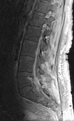
Figure 10 shows the MRI scan of a 56-year-old woman with metastatic breast cancer who now reports progressive paraparesis. Her general health remains good. Treatment should consist of


Explanation

If the patient's medical condition and prognosis remain good in the presence of significant and progressive neurologic deficit from cord compression, then the most reliable means of restoring function is via surgical decompression and fusion. Decompression should be directed toward the compressing structure (eg, anteriorly if the compression is from the anterior side). This procedure can be done via a posterolateral technique, such as costotransversectomy in some cases.

Question 2

When 6 weeks of noninvasive nonsurgical management fails to provide relief for a lumbar disk herniation, a trial of epidural steroid injections is likely to yield which of the following results?


Explanation

Lumbar epidural steroid injections appear to play a role in management of a lumbar disk herniation that has failed to respond to at least 6 weeks of nonsurgical treatment. Approximately 42% to 56% of patients report significant pain relief compared with 92% to 98% of those patients treated with diskectomy. Patients with extruded or sequestered herniations report the greatest and most rapid relief. Similarly, those with well-hydrated disk fragments report rapid relief of symptoms. A smaller percentage of patients report symptom relief compared with those having surgery, but the degree of improvement is similar for both groups and the improvement lasts up to 3 years. Butterman GR: Treatment of lumbar disc herniation: Epidural steroid injection compares with discectomy: A prospective, randomized study. J Bone Joint Surg Am 2004;86:670-679.

Question 3

Which of the following anatomic changes is observed as part of the normal aging process of the adult spine?


Explanation

The primary change that takes place in the aging spine is degeneration of the lumbar disks and loss of the overall lumbar lordosis. This also may be associated with osteopenic-related compression fractures. With these changes, the sagittal vertical line moves anteriorly relative to the sacrum; cervical scoliosis is uncommon and not part of the normal aging process. Overall kyphosis in the thoracic spine gradually increases, but the coronal balance remains essentially the same unless scoliosis develops. Gelb DE, Lenke LG, Bridwell KH, et al: An analysis of sagittal spinal alignment in 100 asymptomatic middle and older aged volunteers. Spine 1995;20:1351-1358.

Question 4

A previously healthy 30-year-old woman has neck pain and bilateral hand and lower extremity tingling with weakness after falling down stairs. She is alert and oriented. Examination reveals incomplete quadriplegia at the C6 level that remains unchanged throughout her evaluation and initial treatment. Radiographs show a bilateral facet dislocation of C6 on C7 without fracture. Attempts at reduction with halo cervical traction up to her body weight are unsuccessful. What is the next most appropriate step?


Explanation

A facet dislocation that cannot be reduced in an alert, awake patient with some preservation of cord function requires MRI to evaluate the disk prior to a reduction under anesthesia. The presence or absence of a disk herniation must be assessed, as this factor may influence the method of reduction. Vaccaro AR, Falatyn SP, Flanders AE, et al: Magnetic resonance evaluation of the intervertebral disc, spinal ligaments, and spinal cord before and after closed traction reduction of cervical spine dislocations. Spine 1999;24:1210-1217. Fardon DF, Garfin SR, Abitbol J (eds): Orthopaedic Knowledge Update: Spine 2. Rosemont, IL, American Academy of Orthopaedic Surgeons, 2002, pp 247-262. Eismont FJ, Arena MJ, Green BA: Extrusion of an intervertebral disc associated with traumatic subluxation or dislocation of cervical facets. J Bone Joint Surg Am 1991;73:1555-1560.

Question 5

Which of the following findings is the best radiographic indicator of segmental instability at L4-L5?


Explanation

Motion segments that demonstrate more than 4 mm of translation or 10 degrees of angulation compared with adjacent motion segments on flexion-extension radiographs have excessive motion and instability. Anterior marginal osteophytes form at the insertion of the annulus from increased forces but do not indicate increased motion. A spondylolisthesis or lateral listhesis is often static without increased motion. More than 3.5 mm of translation or 11 degrees of angulation is considered instability criteria for the cervical spine. Internal disk disruption does not denote instability. Boden SD, Wiesel SW: Lumbosacral segmental motion in normal individuals. Have we been measuring instability properly? Spine 1990;15:571-576.

Question 6

In a patient who has undergone fusion with instrumentation from T4 to the sacrum for adult scoliosis, at which site is a pseudarthrosis most likely to be discovered?


Explanation

Although pseudarthrosis can be found anywhere within the spine that has been fused using long multisegmental fixation to the sacrum, it most commonly occurs at the lumbosacral junction. The thoracolumbar junction is another common site of potential pseudarthrosis. In this location, the anatomy changes from lumbar transverse processes to thoracic through the transition zone, and overlying instrumentation often makes it difficult to obtain enough sound bone on decorticated bone to achieve a successful fusion. Saer EH III, Winter RB, Lonstein JE: Long scoliosis fusion to the sacrum in adults with nonparalytic scoliosis: An improved method. Spine 1990;15;650-653. Kostuik JP, Hall BB: Spinal fusions to the sacrum in adults with scoliosis. Spine 1983;8:489-500.

Question 7

The afferent pain innervation of the L3-L4 facet joint arises from the medial branch nerve of


Explanation

Afferent pain fibers to the lumbar facet joints arise from the medial branch nerves originating from the next two cephalad levels. Therefore, innervation of the L3-L4 facet joint arises from the L2 and L3 medial branch nerves. This effect should be taken into account when considering a medial branch block or facet denervation. The medial branch nerve arises from the dorsal ramus of the exiting nerve root. Nade SL, Bell E, Wyke BD: The innervation of the lumbar spinal joint and its significance. J Bone Joint Surg Br 1980;62:255-261

Question 8

When posterior fusion with instrumentation to the sacrum is used to treat adult scoliosis, what instrumentation technique best increases the chance of a successful lumbosacral fusion?


Explanation

As the chance of success of lumbosacral fusion increases with the stiffness and rigidity of the construct, fixation and stiffness improve with fixation into both the upper sacrum and the ilium. In a review of individuals treated with long constructs to the pelvis for adult scoliosis, Islam and associates reported that the rate of pseudarthrosis was significantly lower with sacral and iliac fixation compared with sacral fixation alone or iliac fixation alone. Iliac screws provide significant fixation anterior to the instantaneous axis of rotation for flexion and extension, as well as provides resistance to lateral bending and rotational forces. Numerous biomechanical studies support the concept of increasing biomechanical stabilization with increased fixation from the sacrum to the ilium. Islam NC, Wood KB, Transfeldt EE, et al: Extension of fusions to the pelvis in idiopathic scoliosis. Spine 2001;26:166-173. O'Brien N, et al: Sacral pelvic fixation and spinal deformity, in DeWald RL (ed): Spinal Deformities: A Comprehensive Text. New York, NY, Thieme, 2003, pp 601-614.

Question 9

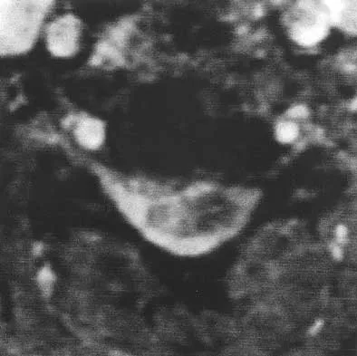
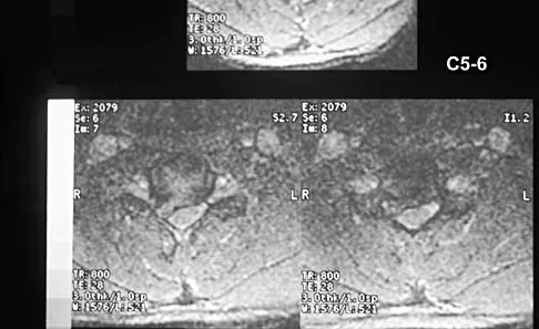
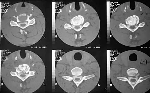
Which of the following structures runs through the site indicated by the arrow in Figure 11?


Explanation

The vertebral artery traverses through the arcuate foramen after exiting the lateral aspect of C1 and before entering the skull. The foramen usually is not fully formed, but a complete foramen such as this one has been reported in up to 18% of patients. Stubbs DM: The arcuate foramen: Variability in distribution related to race and sex. Spine 1992;17:1502-1504.

Question 10

A 42-year-old man has had left lower extremity pain in an L5 radicular pattern for the past 6 weeks. He denies significant axial low back pain. History reveals that he underwent an L4-5 diskectomy with successful relief of similar pain 5 years ago. Which of the following imaging studies would offer the greatest amount of information?


Explanation

MRI with gadolinium will best identify recurrent herniated nucleus pulposus or other root compression and distinguish scar from recurrent disk. CT is unable to distinguish scar from recurrent disk density, and the addition of myelogram dye can reveal compromise of the thecal sac but cannot distinguish the scar from recurrent disk as the source of compression. Although lateral flexion-extension radiographs may be important to rule out any instability, much of that information can be inferred from the associated disk and adjacent bony changes on MRI. Bone scan techniques may identify subtle stress fractures resulting from previous aggressive facet resection, but low back pain also would be expected. Mirowitz SA, Shady KL: Gadopentetate dimeglumine-enhanced MR imaging of the postoperative lumbar spine: Comparison of fat-suppressed and conventional T1-weighted images. Am J Roentgenol 1992;159:385-389.

Question 11

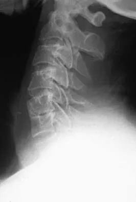
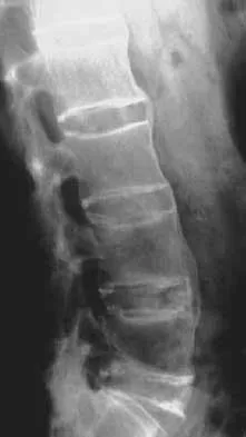
Figure 12 shows the radiograph of an 80-year-old woman who has had an 8-month history of back pain after a fall. What is the most likely diagnosis based on the radiographic findings at the fractured vertebrae?


Explanation

An intravertebral vacuum cleft suggests nonunion of the vertebral fracture with osteonecrosis and is not seen in routine healing fractures. MRI characteristically shows a high T2 signal in the cleft. The cleft is not indicative of an infectious or neoplastic lesion. A vacuum disk phenomenon is associated with end-stage degenerative disk disease, but those findings are not found in the vertebral body. Murakami H, Kawahara N, Gabata T, et al: Vertebral body osteonecrosis without vertebral collapse. Spine 2003;28:E323-E328.

Question 12

Which of the following complications is uniquely associated with an anterior approach to the lumbosacral junction?


Explanation

Retrograde ejaculation is a sequela of injury to the superior hypogastric plexus. The structure needs protection, especially during anterior exposure of the lumbosacral junction. The use of monopolar electrocautery should be avoided in this region. The ideal exposure starts with blunt dissection just to the medial aspect of the left common iliac vein, sweeping the prevertebral tissues toward the patient's right side. Although erectile dysfunction can be seen after spinal surgery, it is not typically related to the surgical exposure because erectile function is regulated by parasympathetic fibers derived from the second, third, and fourth sacral segments that are deep in the pelvis and are not at risk with the anterior approach. The other choices are complications of spinal surgery but are not uniquely associated with an anterior L5-S1 exposure. Flynn JC, Price CT: Sexual complications of anterior fusion of the lumbar spine. Spine 1984;9:489-492. Watkins RG (ed): Surgical Approaches to the Spine, ed 1. New York, NY, Springer-Verlag, 1983, p 107.

Question 13

A 68-year-old woman with a history of rheumatoid arthritis has had neck pain and weakness in all four extremities that has become worse in the past 6 months. She has gone from a community to a household ambulator and uses a wheelchair outside of the home. Examination of the extremities reveals poor coordination, diffuse weakness, hyperactive reflexes, and bilateral sustained clonus. She has a broad-based and unsteady gait. The posterior atlanto-dens interval is 12 mm. Based on these findings and the radiograph and MRI scan shown in Figures 13a and 13b, the treatment of choice is surgical decompression and stabilization. However, the patient inquires about the prognosis with surgery compared to nonsurgical management. Assuming there are no complications from surgery, the patient should be informed that, with surgery, she will most likely


Explanation

13b The patient has a cervical myelopathy with more than 10 mm of space available for the cord; therefore, she has a reasonable chance of improved neurologic function following surgery. If not treated with surgery, however, her neurologic condition likely will worsen and she will die earlier than if she had surgery. Matsunaga S, Sakou T, Onishi T, et al: Prognosis of patients with upper cervical lesions caused by rheumatoid arthritis: Comparison of occipitocervical fusion between C1 laminectomy and nonsurgical management. Spine 2003;28:1581-1587.

Question 14

Five weeks after undergoing a successful L4-L5 diskectomy, with complete relief of his preoperative sciatica, a 36-year-old man has severe, relentless back and buttock pain. Examination and laboratory studies are unremarkable with the exception of an erythrocyte sedimentation rate (ESR) of 90 mm/h. What is the next most appropriate step in management?


Explanation

The patient's history, including the timing and type of symptoms, is typical for postoperative diskitis. The elevated ESR, 5 weeks after surgery, is also consistent with infection; a normal WBC count is not unusual. Management should consist of MRI with gadolinium; if positive, this should be followed by percutaneous biopsy to confirm the organism. Open biopsy may be considered if the percutaneous biopsy is unsuccessful. Anterior debridement and interbody fusion is reserved for the occasional patient that fails to respond to intravenous antibiotics, bed rest, and immobilization. Garfin SR, Vaccaro AR (eds): Orthopaedic Knowledge Update: Spine. Rosemont, IL, American Academy of Orthopaedic Surgeons, 1997, pp 257-271.

Question 15

An 18-year-old man sustained a knife injury to his midback, with the entry wound 2 cm to the left of the midline. He has been diagnosed with a hemicord transection. Neurologic examination will most likely reveal left-sided loss of


Explanation

Brown-Sequard syndrome results from an injury to one half of the spinal cord and is characteristically seen in penetrating injuries. The spinothalamic fibers cross the midline below the level of the lesion, resulting in contralateral loss of pain and temperature sensation. The posterior columns and corticospinal tracts carry vibratory, position, and light touch sensation, as well as motor function from the ipsilateral side of the body. This results in the characteristic neurologic findings seen with Brown-Sequard syndrome. Northrup BE, Evaluation and early treatment of acute injuries to the spine and spinal cord, in Clark CR (ed): The Cervical Spine, ed 3. Philadelphia, PA, Lippincott Raven, 1998, pp 541-549.

Question 16

When using surgery extending to the pelvis to treat long spinal deformity in adults, the addition of anterior interbody structural support at the lumbosacral junction serves what biomechanical function?


Explanation

Shufflebarger and others have reported that the placement of anterior interbody structural support at the lumbosacral junction increases the overall construct stiffness and reduces the strain on posterior instrumentation, thereby reducing the risk of screw pull-out or fracture. The stiffness of the posterior instrumentation actually increases, whereas the actual strength of the instrumentation remains the same. Actual strain measured at an adjacent intervertebral disk to a fusion construct is expected to increase. Shufflebarger HL: Moss-Miami spinal instrumentation system: Methods of fixation of the spondylopelvic junction, in Margulies JI, Floman Y, Farcy JPC, et al (eds): Lumbosacral and Spinal Pelvic Fixation. Philadelphia, PA, Lippincott-Raven, 1996, pp 381-393. Cunningham BW: A biomechanical approach to posterior spinal instrumentation: principles and applications, in DeWald RL (ed): Spinal Deformities: A Comprehensive Text. New York, NY, Thieme, 2003, pp 588-600.

Question 17

A 40-year-old woman has had sciatic pain on the left side for the past 8 weeks. She reports that the pain radiates to her posterior thigh, lateral calf, and into the dorsum of her left foot. Neurologic examination shows weakness of the left extensor hallucis longus. Axial T2-weighted MRI scans through L4-L5 are shown in Figure 14. Management should consist of


Explanation

The MRI scans show hypertrophy of the left L4-L5 facet joint and ligamentum flavum, with a synovial cyst. Appropriate surgical management consists of a hemilaminectomy and direct decompression of the neural elements. Fusion, in addition to the decompression, may be considered, particularly in patients with an associated spondylolisthesis. Epstein NE: Lumbar laminectomy for the resection of synovial cysts and coexisting lumbar spinal stenosis or degenerative spondylolisthesis: An outcome study. Spine 2004;29:1049-1055.

Question 18

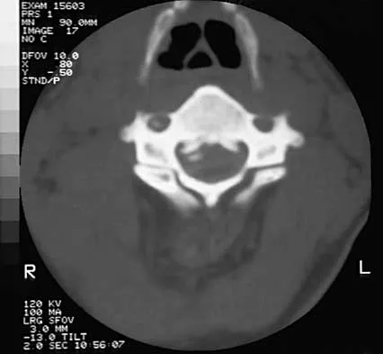
During C1-C2 transarticular screw fixation, screw misplacement is most likely to result in injury to the


Explanation

With C1-C2 transarticular screw fixation, the following structures are potentially at risk: vertebral artery, spinal cord, occiput-C1 joint, and hypoglossal nerve. The vertebral artery is most vulnerable to injury with drill misdirection or anatomic variations in the vertebral foramen. The hypoglossal nerve may be injured if the drill, tap, or screw passes too far anterior to the lateral mass of C1. This complication is extremely rare. The occiput-C1 joint may be injured if the screw trajectory is too cephalad or cranially directed; however,this scenario is very unlikely because the exposure tends to direct the screw into a caudally inclined direction. This caudal orientation has the potential to cause vertebral artery injury, especially in patients who have a large vertebral foramen in the lateral mass of C2 because of erosions (rheumatoid arthritis) or anatomic variation. CT of the vertebral foramen is recommended when C1-C2 transarticular fixation is being considered. Spinal cord injury is extremely unlikely because of the very large size of the spinal canal in the upper cervical spine; the spinal cord lies far away from the lateral masses of C1 and C2. Mueller ME, Allgower M, et al: Manual of Internal Fixation, ed 3. New York, NY, Springer-Verlag, 1991, pp 634-636.

Question 19

A 27-year-old professional soccer player sustained an injury to his cervical spine in a collision with another player. Initially he was diagnosed with a right C6 radiculopathy that resolved with rest, anti-inflammatory medications, and physical therapy. Following a fall in a game, he noted a recurrence of neck pain without radicular signs or symptoms. Additional nonsurgical management over the past few months has failed to provide relief. A cervical MRI scan shows a right-sided C5-6 herniation without any evidence of disk disease at other cervical levels. The patient desires to continue his career as a professional soccer player. What treatment offers the best long-term option for return to play?


Explanation

The patient has chronic neck pain that is affecting his career as a professional soccer player. Although he had signs and symptoms of a right C6 radiculopathy, neck pain is his only current symptom. Therefore, procedures to address the relief of radiculopathy (keyhole foraminotomy and transforaminal epidural steroid injection) are likely to be ineffective. Although Watkins and others have described continuing nonsurgical management for symptomatic herniated disks and return to play only when asymptomatic, the patient has not found relief with these modalities. A single-level cervical fusion (either postoperative or congenital) generally is not considered a contraindication for return to play in collision or contact sports. Therefore, anterior cervical fusion at C5-6 offers the best long-term option for return to play. Watkins RG: Cervical spine injuries in athletes, in Clark CR (ed): The Cervical Spine, ed 3. Philadelphia, PA, Lippincott Raven, 1998, pp 373-386. Watkins RG: Neck injuries in football players. Clin Sports Med 1986;5:215-246. Morganti C, Sweeney CA, Albanese SA, et al: Return to play after cervical spine injury. Spine 2001;26:1131-1136.

Question 20

A collegiate football player who sustained an injury to his neck has significant neck pain and weakness in his extremities. Following immobilization, which of the following steps should be taken prior to transport?


Explanation

Prior to transport, the face mask should be removed so that the airway can be easily accessible. If serious injury is suspected, the helmet and shoulder pads should be left in place until he is assessed at the hospital and radiographs are obtained. Leaving the helmet and shoulder pads in place helps to keep the spine in the most neutral alignment. Removal of the helmet will result in extension of the neck, whereas removal of the shoulder pads will most likely result in flexion of the neck. Clark CR (ed): The Cervical Spine, ed 3. Philadelphia, PA, Lippincott Williams & Wilkins, 1998, p 376. Thomas B, McCullen GM, Yuan HA: Cervical spine injuries in football players. J Am Acad Orthop Surg 1999;7:338-347. Waninger KN, Richards JG, Pan WT, et al: An evaluation of head movement in backboard-immobilized helmeted football, lacrosse, and ice hockey players. Clin J Sport Med 2001;11:82-86. Donaldson WF III, Lauerman WC, Heil B, et al: Helmet and shoulder pad removal from a player with suspected cervical spine injury: A cadaveric model. Spine 1998;23:1729-1732.

Question 21

What is the most common complication following total disk arthroplasty in the lumbar spine?


Explanation

In a midterm (7 to 11 years) follow-up study of lumbar total disk arthroplasty, 5 of 55 patients had transient radicular leg pain without evidence of nerve root compression. Implant migration is rare. Deep venous thrombosis, incisional hernia, and retrograde ejaculation are less common complications of disk arthroplasty.

Question 22

A 42-year-old woman has cervical stenosis and radicular deficits at the C5-6 and C6-7 levels. History reveals that she has smoked one pack of cigarettes a day for 25 years. Because nonsurgical management has failed to provide relief, she is now seeking surgical treatment. After preoperative counseling, it becomes clear that she is not likely to stop smoking. Which of the following surgical procedures should be used?


Explanation

In a review of 190 anterior cervical fusions, Hilibrand and associates reported that only 20 of 40 patients who smoked had solid fusion at all levels, whereas 64 of 91 nonsmokers had solid fusions at all levels when treated with multilevel interbody technique (Smith-Robinson). When fused with strut grafts, 14 of 15 smokers and 41 of 44 nonsmokers had solid fusions with a fusion rate of 93% in the same series. Multilevel allografts have a lower fusion rate than autografts, and diskectomy without fusion has an increased rate of residual neck pain. Hilibrand AS, Fye MA, Emery SE, et al: Impact of smoking on the outcome of anterior cervical arthrodesis with interbody or strut-grafting. J Bone Joint Surg Am 2001;83:668-673.

Question 23

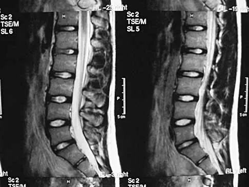
An otherwise healthy 54-year-old man who underwent a successful multilevel lumbar decompression and fusion 4 years ago now reports increasingly severe bilateral thigh claudication with paresthesia and severe back pain for the past 12 months. Physical therapy, bracing, and epidural steroids have failed to provide relief. A radiograph and MRI scans are shown in Figures 15a through 15c. He is afebrile, and laboratory studies show an erythrocyte sedimentation rate of 5 mm/h and a normal WBC count. What is the best course of action?


Explanation

15b 15c The patient has degeneration of an adjacent segment with resultant kyphosis and stenosis. Because he is healthy, has responded well to previous surgery, and has a potentially correctable lesion, he is not a good candidate for an end-stage failed back procedure such as a morphine pump. The stenosis is exacerbated by the deformity; therefore, a simple decompression will contribute to instability. Because of the kyphosis and the patient's relatively young age, the treatment of choice is restoration of sagittal alignment and posterior decompression.

Question 24

Which of the following is considered a risk factor for the development of low back pain?


Explanation

Risk factors associated with low back pain include poor physical fitness, smoking, a history of repetitive bending or stooping on the job, or whole body vibration exposure. Some radiographic factors such as stenosis, spondyloarthropathy, severe deformity, or instability are also associated with low back pain. Gender, weight, transitional anatomy, or facet trophism are not associated with low back pain.

Question 25

A corset-type brace may help reduce symptoms during an episode of acute low back pain as the result of


Explanation

Although there is no significant alteration in motion with a corset, studies have shown a decrease in intradiskal pressure. Nachemson A, Morris JM: In vivo measurements of intradiscal pressure: Discometry, a method for determination of pressure in the low lumbar disc. J Bone Joint Surg Am 1964;46:1077-1092.

Question 26

A 65-year-old female presents with severe neurogenic claudication and L4-L5 Grade 1 degenerative spondylolisthesis. After failing 6 months of conservative management, she is considering surgery. Based on long-term data from the Spine Patient Outcomes Research Trial (SPORT), what is the expected outcome of surgical decompression and fusion compared to non-operative treatment for this condition?


Explanation

The SPORT trial for degenerative spondylolisthesis demonstrated that surgically treated patients maintained significantly greater improvements in pain and function at 4 and 8 years compared to those treated non-operatively. The operative cohort showed durable advantages in patient-reported outcome measures (SF-36 and ODI) despite some crossover between the groups.

Question 27

A 58-year-old man presents with progressive clumsiness in his hands and difficulty with balance. On physical examination, rapidly flicking the nail of his middle finger results in involuntary flexion of the interphalangeal joint of his thumb and the distal interphalangeal joint of his index finger. Which of the following best describes this physical exam finding and its anatomic localizing value?


Explanation

The Hoffmann sign is elicited by flicking the nail of the middle finger, causing reflex flexion of the thumb and index finger. It indicates an upper motor neuron lesion (corticospinal tract) above the C5 or C6 level, and is a classic finding in cervical spondylotic myelopathy. The inverted radial reflex indicates a lesion specifically at the C5-C6 level, while Lhermitte sign is an electric shock-like sensation down the spine upon neck flexion.

Question 28

When evaluating a patient for adult spinal deformity correction, achieving a harmonious sagittal profile is a primary goal to improve health-related quality of life. According to the SRS-Schwab classification, which of the following spinopelvic parameter combinations represents the ideal target for postoperative alignment?


Explanation

The SRS-Schwab classification established threshold values for optimal sagittal alignment in adult spinal deformity: PI - LL < 10 degrees, PT < 20 degrees, and SVA < 50 mm. Failure to achieve these targets strongly correlates with poorer health-related quality of life (HRQOL) scores, persistent pain, and higher rates of revision surgery due to proximal junctional kyphosis or implant failure.

Question 29

A 35-year-old male falls from a height of 15 feet and sustains a L1 fracture.

Imaging shows a burst fracture with 30% canal compromise. He is neurologically intact. MRI demonstrates an intact posterior ligamentous complex (PLC). According to the Thoracolumbar Injury Classification and Severity (TLICS) score, what is his total score and the recommended management?


Explanation

In the Thoracolumbar Injury Classification and Severity (TLICS) system, points are awarded for morphology (Burst = 2 points), neurologic status (Intact = 0 points), and PLC integrity (Intact = 0 points). The total score is 2. A score of 3 or less indicates non-operative management. A score of 4 is indeterminate (surgeon's choice), and 5 or more dictates operative intervention.

Question 30

A 62-year-old male presents with insidious onset of sacral pain and bowel/bladder dysfunction. Imaging reveals a large, destructive midline sacral mass with a 'soap bubble' appearance and an anterior cortical break.

Biopsy demonstrates physaliferous cells. What is the most appropriate definitive management for this lesion?


Explanation

The clinical presentation, radiographic 'soap bubble' appearance, and histologic finding of physaliferous cells are pathognomonic for a chordoma. Chordomas are locally aggressive, chemo-resistant, and relatively radio-resistant tumors. The gold standard treatment for a sacral chordoma is en bloc wide surgical resection with negative margins, which offers the only chance for long-term disease-free survival.

Question 31

A 55-year-old female with a 20-year history of rheumatoid arthritis presents with severe neck pain, suboccipital headaches, and bilateral hand clumsiness. Radiographs show significant basilar invagination.

Which of the following radiographic measurements is the most accurate for diagnosing basilar invagination on a lateral cervical spine radiograph?


Explanation

Basilar invagination (cranial settling) in rheumatoid arthritis is classically assessed using McGregor's line (a line drawn from the posterior edge of the hard palate to the most caudal point of the occipital curve). An odontoid tip extending more than 4.5 mm above this line is diagnostic of basilar invagination. ADI and PADI assess atlantoaxial subluxation, while BDI and Powers ratio assess occipitocervical dissociation in trauma.

Question 32

A 24-year-old male rugby player presents with severe neck pain and bilateral upper extremity weakness (deltoids and biceps 3/5, distal muscles 5/5) following a tackling injury. He is awake, alert, and cooperative. Plain films and CT demonstrate a unilateral jumped facet at C5-C6. What is the most appropriate next step in management?


Explanation

In an awake, alert, and cooperative patient with a cervical facet dislocation and a neurologic deficit, immediate closed reduction with cranial traction (e.g., Gardner-Wells tongs) is indicated to decompress the spinal cord as rapidly as possible. MRI prior to reduction is indicated in patients who are obtunded/unexaminable or those who fail closed reduction, to rule out a herniated disc that could cause secondary cord injury during reduction.

Question 33

A 68-year-old male undergoes a C3-C6 posterior cervical laminectomy and fusion for cervical spondylotic myelopathy. On postoperative day 2, he develops profound weakness in his right deltoid and biceps (1/5), with no other new sensory or motor deficits. What is the most likely etiology of this complication?


Explanation

Postoperative C5 palsy is a well-documented complication of cervical decompression, particularly posterior laminectomy. The most widely accepted mechanism is the posterior drift or shift of the spinal cord following decompression, which puts tension on the relatively short and tethered C5 nerve roots. It presents as isolated deltoid and/or biceps weakness, usually within the first few days post-op, and typically resolves spontaneously over months with supportive care and physical therapy.

Question 34

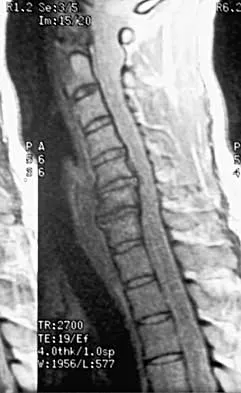
A 50-year-old diabetic male presents with 2 weeks of worsening back pain, low-grade fevers, new-onset urinary retention, and bilateral leg weakness.

MRI reveals a dorsal spinal epidural abscess at T10-T12 with severe cord compression. Which of the following is the most appropriate definitive management?


Explanation

The patient has a spinal epidural abscess complicated by progressive neurologic deficits (weakness, urinary retention), indicating cauda equina/conus medullaris or cord compromise. The standard of care for a symptomatic epidural abscess causing neurologic deficit is emergent surgical decompression (usually via laminectomy for dorsal abscesses) and debridement, combined with organism-specific IV antibiotics. Medical management alone is strictly reserved for patients without neurologic deficits or those entirely unfit for surgery.

Question 35

A 42-year-old male presents with severe right-sided anterior thigh pain, weakness in knee extension, and a diminished patellar reflex. MRI of the lumbar spine reveals a far-lateral (extraforaminal) disc herniation. At which lumbar level is this herniation most likely located to produce these specific neurologic findings?


Explanation

A far-lateral (extraforaminal) disc herniation compresses the exiting nerve root at the same level. Therefore, a far-lateral disc herniation at the L4-L5 level compresses the exiting L4 nerve root (unlike a typical paracentral disc herniation at L4-L5, which compresses the traversing L5 nerve root). L4 radiculopathy is classically characterized by anterior thigh pain, quadriceps weakness (knee extension), and an asymmetric or diminished patellar reflex.

Question 36

An 82-year-old man is evaluated in the emergency department after suffering a ground-level fall. He complains of upper neck pain without radiation. Neurologic examination is completely normal. CT imaging of the cervical spine reveals a Type II odontoid fracture with 2 mm of posterior displacement. Given the patient's age and clinical presentation, what is the most appropriate management strategy?


Explanation

In the elderly population (especially patients >80 years old), the morbidity and mortality associated with surgical intervention and halo vest immobilization are significantly high. Multiple studies have demonstrated that for mildly displaced Type II odontoid fractures in the elderly, immobilization in a rigid cervical collar is the preferred initial treatment. It offers an acceptable rate of stable nonunion (fibrous union) while avoiding the severe respiratory complications and dysphagia associated with halo vests and surgery.

Question 37

During a posterior spinal fusion for adolescent idiopathic scoliosis, the neuromonitoring team reports a sudden, complete loss of Motor Evoked Potentials (MEPs) in both lower extremities. Somatosensory Evoked Potentials (SSEPs) remain at baseline. Which of the following is the most appropriate initial management step?


Explanation

A sudden loss of MEPs with intact SSEPs suggests an isolated insult to the anterior corticospinal tracts, commonly due to hypoperfusion of the anterior spinal artery (anterior cord syndrome). The most critical initial step is to optimize spinal cord perfusion by raising the mean arterial pressure (MAP) to greater than 85 mm Hg. Other immediate steps include correcting anemia, reversing any recent corrective maneuvers, and ensuring that anesthetic agents (like volatile gases) are not depressing the signals.

Question 38

A 45-year-old woman presents with severe neck pain radiating down her right arm. Physical examination reveals a diminished triceps reflex, profound weakness in elbow extension and wrist flexion, and decreased sensation over the dorsal aspect of the middle finger. Which of the following cervical disc herniations is most likely responsible for these findings?


Explanation

The patient's findings of triceps weakness, a diminished triceps reflex, and paresthesias in the middle finger are classic for a C7 radiculopathy. In the cervical spine, exiting nerve roots exit above the correspondingly named pedicle (e.g., the C7 root exits at the C6-C7 neural foramen). Therefore, a C6-C7 disc herniation will typically compress the C7 nerve root.

Question 39

A 62-year-old man with a 30-year history of ankylosing spondylitis presents to the emergency department after a low-speed motor vehicle collision. He complains of localized neck pain but has normal motor and sensory function. Plain radiographs of the cervical spine show extensive syndesmophytes and a 'bamboo spine' appearance but no definitive fracture. What is the most appropriate next step in management?


Explanation

Patients with ankylosing spondylitis have highly rigid, osteopenic spines that act as long lever arms, making them extremely susceptible to fractures even from minor trauma. These fractures are often highly unstable, most common at the cervicothoracic junction, and easily missed on plain radiographs due to distorted anatomy. The standard of care for an ankylosing spondylitis patient presenting with back or neck pain after trauma is a CT scan of the entire spine (cervical, thoracic, and lumbar) to rule out occult fractures. Flexion-extension views are contraindicated due to the high risk of neurologic injury.

Question 40


A 65-year-old woman is evaluated for a debilitating flatback deformity and sagittal imbalance. Figure 39 represents a templated standing lateral radiograph. Measurement of her spino-pelvic parameters reveals a pelvic incidence (PI) of 56 degrees and a sacral slope (SS) of 22 degrees. What is her calculated pelvic tilt (PT), and what is the generally accepted target for her postoperative lumbar lordosis (LL)?


Explanation

The formula relating the key pelvic parameters is Pelvic Incidence (PI) = Pelvic Tilt (PT) + Sacral Slope (SS). Given PI = 56 and SS = 22, the PT is 56 - 22 = 34 degrees. For optimal postoperative sagittal alignment and minimization of adjacent segment disease and hardware failure, the target Lumbar Lordosis (LL) should be restored to within 10 degrees of the patient's PI (i.e., PI - LL ≤ 10 degrees). Therefore, an appropriate target LL for this patient is between 46 and 56 degrees.

Question 41

A 55-year-old diabetic intravenous drug user presents with a 1-week history of worsening severe mid-thoracic back pain, fevers, and new-onset bilateral lower extremity weakness (3/5 strength in iliopsoas and quadriceps) along with urinary retention. MRI reveals a large, dorsal spinal epidural abscess compressing the spinal cord at T8. What is the most appropriate definitive management?


Explanation

The patient is presenting with a spinal epidural abscess complicated by an acute, progressive neurologic deficit (motor weakness and bowel/bladder dysfunction). In the presence of neurologic compromise, emergent surgical decompression (e.g., laminectomy) and debridement is indicated to relieve cord compression and obtain cultures, followed by targeted prolonged antibiotic therapy. Medical management alone is reserved for patients who are neurologically intact, poor surgical candidates, or have extensive pan-spinal disease without focal compression.

Question 42

A 42-year-old man presents with severe low back pain, bilateral sciatica, perineal numbness, and acute urinary retention. Post-void residual volume is 600 mL. MRI confirms a massive L4-L5 central disc herniation causing cauda equina syndrome. He is scheduled for emergent surgical decompression. Which of the following factors is the most significant predictor of full postoperative recovery of bladder and sphincter function?


Explanation

In cauda equina syndrome, the most critical prognostic factor for the recovery of autonomic function (bowel, bladder, and sexual function) is the time elapsed from symptom onset to surgical decompression. Surgery performed within 24 to 48 hours of the onset of symptoms is associated with significantly better neurologic and functional outcomes compared to delayed decompression.

Question 43


A 60-year-old man presents with progressive clumsiness in his hands and a wide-based, unsteady gait. Figure 8 shows his sagittal T2-weighted MRI. Imaging confirms ossification of the posterior longitudinal ligament (OPLL). The K-line is drawn from the mid-canal of C2 to the mid-canal of C7, and the ossified mass crosses the K-line anteriorly (K-line negative). Additionally, the cervical spine demonstrates 15 degrees of kyphosis. Which of the following surgical approaches is most appropriate?


Explanation

The K-line is a critical concept in planning surgery for cervical OPLL. When the ossified mass crosses the K-line (K-line negative) or there is significant cervical kyphosis, posterior decompression alone (like laminoplasty or laminectomy) is contraindicated. This is because the spinal cord will not sufficiently 'drift back' away from the anterior compressive OPLL mass due to the kyphotic tension. Instead, an anterior approach (such as anterior cervical corpectomy and fusion) or a combined anterior-posterior approach is required for direct decompression.

Question 44

The Spine Patient Outcomes Research Trial (SPORT) evaluated outcomes for patients with symptomatic degenerative spondylolisthesis and lumbar spinal stenosis. At the 4-year follow-up, which of the following conclusions was most strongly supported by the data regarding surgical versus nonoperative management?


Explanation

The SPORT trial results for degenerative spondylolisthesis demonstrated a clear, statistically significant advantage for surgical intervention (decompression with or without fusion) over nonoperative treatment in terms of pain relief and functional improvement at 4-year follow-up. While there was significant crossover between groups, the 'as-treated' analysis definitively showed the superiority of surgery for this specific pathology.

Question 45


A 19-year-old man is brought to the trauma bay after a high-speed motor vehicle collision where he was restrained by a lap belt only. He sustains an L2 flexion-distraction injury (classic Chance-type fracture pattern), as demonstrated in Figure 12. Biomechanically, if the axis of rotation is located at the anterior longitudinal ligament, which of the following best describes the mechanism of failure according to the Denis three-column model?


Explanation

In a flexion-distraction injury where the axis of rotation is at the anterior longitudinal ligament or the anterior aspect of the vertebral body, the biomechanical failure involves compression of the anterior column while the middle and posterior columns fail in tension (distraction). If the axis of rotation is displaced further anteriorly (e.g., at the anterior abdominal wall due to a lap belt), a pure tension failure can occur across all three columns (the classic 'bony' Chance fracture). However, the classic Denis description of a flexion-distraction injury involves anterior compression with middle/posterior tension.

Question 46

A 65-year-old man presents with progressive gait instability and fine motor clumsiness in his hands. Examination reveals hyperreflexia in the lower extremities, a positive Hoffmann sign bilaterally, and loss of proprioception in his toes.

Which of the following parameters on MRI is most predictive of poor neurological recovery following surgical decompression?


Explanation

In cervical spondylotic myelopathy (CSM), MRI findings that correlate with a poor prognosis for neurologic recovery after surgical decompression include a decreased T1 signal intensity within the spinal cord. This finding often represents permanent spinal cord damage such as cystic changes, necrosis, or severe myelomalacia. Increased T2 signal intensity alone is frequently observed and can represent reversible edema or gliosis, having a more variable or less definitive negative prognostic value compared to decreased T1 signal changes.

Question 47

A 68-year-old woman presents with severe low back pain, global sagittal imbalance, and difficulty standing upright. Standing full-length lateral radiographs show a pelvic incidence (PI) of 60 degrees, lumbar lordosis (LL) of 30 degrees, and a sagittal vertical axis (SVA) of 12 cm. What is the approximate target lumbar lordosis required to achieve an optimal sagittal balance in this patient if surgical correction is planned?


Explanation

The relationship between pelvic incidence (PI) and lumbar lordosis (LL) is critical in correcting adult spinal deformity. According to the Schwab criteria, normal sagittal balance typically requires the LL to be within 10 degrees of the PI (PI - LL < 10°). In this patient with a PI of 60 degrees, the target LL should be approximately 60 degrees (acceptable range 50-70 degrees). An LL of 30 degrees leaves the patient with a significant PI-LL mismatch of 30 degrees, leading to a positive sagittal vertical axis (SVA) and compensatory mechanisms such as pelvic retroversion and knee flexion.

Question 48

An 82-year-old man falls from a standing height and presents with neck pain.

Imaging reveals a Type II odontoid fracture with 3 mm of posterior displacement. He is neurologically intact. He has a history of severe chronic obstructive pulmonary disease (COPD) and coronary artery disease. What is the most appropriate initial management?


Explanation

In elderly patients with Type II odontoid fractures, rigid cervical collar immobilization is often the preferred initial management, especially in those with significant medical comorbidities (such as COPD and severe coronary artery disease). Halo vest immobilization in the elderly is associated with high morbidity and mortality (including pneumonia, cardiac arrest, and pin site infections) and is generally contraindicated. While surgical intervention (posterior C1-C2 fusion) provides the highest union rate, it carries substantial perioperative risks that must be weighed against the patient's frail health status. Although nonunion is common with a rigid collar, a stable fibrous nonunion frequently results and is well-tolerated by this patient population.

Question 49

A 62-year-old woman presents with a 1-year history of neurogenic claudication and low back pain. Flexion-extension radiographs demonstrate a dynamic L4-L5 degenerative spondylolisthesis with 4 mm of translation. MRI confirms severe central canal stenosis at L4-L5. She has failed 6 months of nonoperative management. Based on classic randomized controlled trials, which of the following surgical interventions has historically demonstrated the most reliable long-term outcomes for this condition?


Explanation

The SPORT trial (Spine Patient Outcomes Research Trial) and other classic studies have traditionally supported laminectomy with instrumented fusion over decompression alone for patients with degenerative spondylolisthesis and spinal stenosis, due to a lower risk of progression of the slip and need for reoperation. For a patient with documented dynamic instability (4 mm of translation on flexion-extension), laminectomy with instrumented posterolateral fusion remains the gold standard to achieve both neural decompression and segmental stability.

Question 50

A 15-year-old male gymnast presents with persistent low back pain that is worsened by spinal extension. Oblique radiographs demonstrate a "Scottie dog with a collar" sign at L5. MRI shows increased signal in the pars interarticularis on STIR sequences but no obvious gap on T1. What is the most appropriate initial management?


Explanation

The patient has an acute or stress-reactive spondylolysis, indicated by the increased STIR signal on MRI without a definite fracture gap (which would be more typical of a chronic nonunion). The most appropriate initial management for an acute symptomatic spondylolysis is activity modification, core strengthening, and often the use of an anti-lordotic (Boston-style) brace to limit extension. Surgery is reserved for patients who fail prolonged nonoperative management (usually at least 6 months) or those with progressive spondylolisthesis and neurological symptoms.

Question 51

A 45-year-old man with a long-standing history of ankylosing spondylitis presents to the emergency department after a low-speed motor vehicle collision. He complains of severe lower neck pain. Neurological examination is normal. Standard anteroposterior and lateral cervical spine radiographs are interpreted as negative. What is the most appropriate next step in management?


Explanation

Patients with ankylosing spondylitis have a highly rigid, osteopenic spine that acts as long bone. It is extremely susceptible to fractures even from minor trauma. These fractures are often highly unstable, involve all three columns, and can be easily missed on standard radiographs due to altered anatomy, osteopenia, and superimposition of the shoulders. Therefore, any patient with ankylosing spondylitis presenting with neck or back pain after trauma must undergo a CT scan of the entire involved spinal region to rule out a fracture. Flexion-extension radiographs are strictly contraindicated due to the high risk of catastrophic neurological injury in the presence of an occult unstable fracture.

Question 52

A 54-year-old diabetic man presents with a 1-week history of severe mid-back pain, low-grade fevers, and new-onset urinary retention. Examination reveals bilateral lower extremity weakness (motor strength 3/5) and decreased sensation below the T8 dermatome. MRI with gadolinium demonstrates a dorsal epidural collection spanning T6 to T9 with peripheral rim enhancement, severely compressing the spinal cord. What is the most appropriate definitive management?


Explanation

The patient is presenting with a spinal epidural abscess complicated by an acute, progressive neurological deficit (myelopathy and neurogenic bladder). In the setting of a spinal epidural abscess with progressive or acute neurological compromise, emergent surgical decompression (via posterior laminectomy for a dorsal abscess) and debridement is the standard of care. Nonoperative management with IV antibiotics is generally reserved for patients who are neurologically intact, extremely poor surgical candidates, or have pan-spinal epidural abscesses without focal cord compression.

Question 53

A 42-year-old woman presents with acute onset of severe low back pain, bilateral sciatica, and perineal numbness. She reports one episode of urinary incontinence earlier in the day. Post-void residual (PVR) volume is 400 mL. MRI reveals a massive L4-L5 central disc herniation filling the spinal canal. Which of the following is the most critical prognostic factor for full recovery of her bladder function?


Explanation

In cauda equina syndrome (CES), the most important prognostic factor for the recovery of neurological function, particularly bladder and bowel function, is the timing of surgical decompression. Decompression within 24 to 48 hours of the onset of autonomic (bladder/bowel) dysfunction or perineal numbness is generally recommended to maximize the chance of full recovery. The presence of urinary incontinence with a high post-void residual indicates CES with retention (CES-R), which typically has a poorer prognosis than incomplete CES (CES-I), making urgent surgical decompression the absolute priority.

Question 54

A 60-year-old man with a history of prostate cancer presents with progressive mechanical back pain.

Imaging shows a metastatic lesion at L2 involving the vertebral body and the left pedicle. The Spine Instability Neoplastic Score (SINS) is calculated to be 14. He has no neurological deficits. Based on this score, what is the most appropriate recommendation regarding his spinal stability?


Explanation

The Spine Instability Neoplastic Score (SINS) evaluates six components: location, pain, bone lesion type, radiographic alignment, vertebral body collapse, and posterolateral involvement. A score of 0-6 indicates stability, 7-12 indicates potential instability, and 13-18 indicates instability. A SINS of 14 falls into the unstable category. Therefore, the patient should be referred for surgical consultation for stabilization before undergoing radiation therapy. Radiation alone on an unstable spine may lead to progressive collapse and secondary neurological compromise.

Question 55

A 55-year-old woman with a 20-year history of severe rheumatoid arthritis presents with neck pain and paresthesias in her hands. Flexion-extension radiographs of the cervical spine demonstrate an anterior atlanto-dens interval (ADI) of 11 mm. What is the most appropriate management?


Explanation

In rheumatoid arthritis, atlantoaxial subluxation is a serious complication. An anterior atlanto-dens interval (ADI) greater than 3 mm is abnormal in adults. An ADI > 9-10 mm indicates disruption of all supporting ligamentous structures (transverse, alar, and apical ligaments) and places the patient at a high risk for neurologic injury, as the space available for the cord is significantly compromised. In the presence of an ADI of 11 mm and neurologic symptoms (paresthesias), surgical stabilization—typically a posterior C1-C2 fusion—is firmly indicated to prevent progressive myelopathy or sudden death.

Question 56

A 68-year-old woman presents with severe mechanical back pain and difficulty standing upright. Radiographs reveal a pelvic incidence (PI) of 65°, lumbar lordosis (LL) of 30°, pelvic tilt (PT) of 35°, and a sagittal vertical axis (SVA) of +12 cm. She has failed extensive nonoperative management. If surgical correction is planned, what is the primary sagittal alignment goal to optimize her clinical outcome?


Explanation

In adult spinal deformity, restoring sagittal balance is highly correlated with improved clinical outcomes (e.g., ODI scores). The key parameters include a sagittal vertical axis (SVA) < 5 cm, a pelvic tilt (PT) < 20°, and a mismatch between pelvic incidence (PI) and lumbar lordosis (LL) of < 10° (PI - LL < 10°). Because PI is a fixed anatomic parameter, the surgical goal is to increase LL to closely match the PI.

Question 57

A 45-year-old man presents with severe right-sided neck and arm pain. Physical examination reveals weakness in right elbow extension, wrist flexion, and finger extension. His triceps reflex is diminished on the right. Sensation is decreased over the middle finger. Which of the following nerve roots is most likely compressed?


Explanation

The clinical presentation is classic for a C7 radiculopathy. C7 nerve root compression typically causes weakness in elbow extension (triceps), wrist flexion (flexor carpi radialis), and finger extension (extensor digitorum communis). The triceps reflex is mediated by C7. Sensory changes are typically noted in the middle finger. For differentiation, C5 affects the deltoid/biceps; C6 affects wrist extension/biceps reflex/thumb sensation; C8 affects finger flexion/hand intrinsics.

Question 58

A 72-year-old man with long-standing ankylosing spondylitis presents to the emergency department after a low-energy ground-level fall. He complains of severe neck pain but has no neurologic deficits. Initial plain radiographs of the cervical spine are obscured by his severe cervicothoracic kyphosis. What is the most appropriate next step in management?


Explanation

Patients with ankylosing spondylitis are at a high risk for highly unstable, often unrecognized, spinal fractures even after low-energy trauma. The fused spine acts as a long, brittle bone. Because plain radiographs are frequently difficult to interpret due to deformity and osteopenia, a CT scan of the spine is the diagnostic modality of choice to evaluate for fracture. These fractures are often 3-column injuries and carry a high risk of neurologic injury or epidural hematoma.

Question 59

A 55-year-old woman presents with progressive leg weakness, numbness in her perineal region, and recent onset of urinary incontinence. She reports an acute exacerbation of lower back pain after lifting a heavy box. Post-void residual (PVR) bladder volume is 400 mL. MRI reveals a massive L4-L5 central disc herniation. Which of the following is the most significant prognostic factor for recovery of normal bladder function following emergency surgical decompression?


Explanation

The clinical presentation is consistent with Cauda Equina Syndrome (CES), a surgical emergency. The most significant prognostic factor for the recovery of neurologic and bladder/bowel function is the duration of symptoms (specifically autonomic/sphincter dysfunction) prior to surgical decompression. Decompression within 24 to 48 hours of symptom onset is generally associated with the most favorable outcomes.

Question 60

In the management of pyogenic vertebral osteomyelitis, which of the following scenarios is an absolute indication for surgical intervention rather than treatment with prolonged intravenous antibiotics alone?


Explanation

The majority of pyogenic vertebral osteomyelitis cases can be managed successfully with image-guided biopsy followed by a prolonged course of culture-directed intravenous antibiotics. Absolute indications for surgical intervention include progressive neurologic deficit, spinal instability or significant deformity, an epidural abscess causing neurologic compromise, and failure of medical management despite appropriate targeted antibiotic therapy.

Question 61

A 32-year-old construction worker falls from a height of 10 feet and sustains an isolated L1 burst fracture. He is neurologically intact. Upright radiographs demonstrate 15° of regional kyphosis and 30% loss of anterior vertebral body height. CT scan shows retropulsion of the posterosuperior vertebral body fragment occluding 25% of the spinal canal. The posterior ligamentous complex (PLC) is intact on MRI. According to the Thoracolumbar Injury Classification and Severity (TLICS) system, what is his total score and the recommended treatment pathway?


Explanation

The Thoracolumbar Injury Classification and Severity (TLICS) score evaluates injury morphology, integrity of the posterior ligamentous complex (PLC), and neurologic status. For this patient: Morphology is a burst fracture (2 points); PLC is intact (0 points); Neurologic status is intact (0 points). The total TLICS score is 2. A score of 3 or less indicates nonoperative management. A score of 4 can be treated operatively or nonoperatively (surgeon's choice), and a score of 5 or more dictates operative stabilization.

Question 62


A 60-year-old man presents with neurogenic claudication. Figure 32 shows an imaging study demonstrating degenerative spondylolisthesis at L4-L5. Based on the Spine Patient Outcomes Research Trial (SPORT) for degenerative spondylolisthesis, patients treated surgically with decompression and fusion compared to those treated nonoperatively demonstrated:


Explanation

The SPORT trial for degenerative spondylolisthesis found that patients treated surgically had significantly improved pain and function. However, due to very high rates of crossover between the two randomized groups, the pure intention-to-treat analysis failed to show a statistically significant difference. When evaluated by the 'as-treated' method (analyzing patients based on the treatment they actually received), surgical intervention showed a highly significant and sustained advantage in pain relief, functional recovery, and patient satisfaction over nonoperative treatment.

Question 63

A 40-year-old woman undergoes a posterior cervical foraminotomy for a C5-C6 soft disc herniation causing C6 radiculopathy. Postoperatively, she develops new-onset weakness in her ipsilateral deltoid and biceps (MRC grade 2/5) without any sensory changes. MRI confirms adequate decompression of the C5 and C6 nerve roots with no evidence of an epidural hematoma. What is the most likely diagnosis?


Explanation

Postoperative C5 palsy is a well-recognized complication following cervical spine surgery, occurring after both anterior and posterior approaches. It typically presents as a new-onset, isolated motor deficit of the deltoid and/or biceps muscles, usually without sensory changes or long tract signs. Its exact etiology is debated but is thought to be related to nerve root tethering, shifting of the spinal cord after decompression, or local reperfusion injury. The majority of cases resolve spontaneously with conservative management and physical therapy.

Question 64


Figure 12 displays the MRI of a 50-year-old man who presents with right leg pain radiating down the anterior aspect of his thigh to the medial malleolus, along with weakness in knee extension and a diminished patellar reflex. MRI reveals a far-lateral (extraforaminal) disc herniation at the L4-L5 level. Which nerve root is primarily compressed by this specific herniation?


Explanation

In the lumbar spine, standard paracentral disc herniations compress the traversing nerve root (e.g., an L4-L5 paracentral disc affects the L5 root). In contrast, a far-lateral (extraforaminal) disc herniation compresses the exiting nerve root at the same level in the neural foramen or further laterally. Thus, a far-lateral herniation at L4-L5 compresses the L4 nerve root. This results in L4 radiculopathy, characterized by anterior thigh pain, weakness in knee extension (quadriceps), and a diminished or absent patellar reflex.

Question 65

A 65-year-old man with metastatic prostate cancer presents with progressively worsening midthoracic back pain. Neurologic examination reveals 4/5 strength in bilateral hip flexors and a positive Babinski sign. MRI demonstrates a metastatic lesion at T8 with significant epidural spinal cord compression. His estimated life expectancy is 18 months, and the tumor is considered radioresistant. According to the landmark Patchell criteria and current literature, what is the most appropriate management?


Explanation

The landmark study by Patchell et al. demonstrated that for patients with metastatic epidural spinal cord compression (MESCC) caused by solid tumors, surgical decompression followed by radiation therapy results in significantly better outcomes (preservation of ambulation, regained ambulation, and prolonged survival) compared to radiation therapy alone. Surgery is highly indicated for patients with radioresistant tumors (like prostate, thyroid, melanoma, GI), mechanical instability, or neurologic progression, provided they are surgical candidates with a life expectancy of at least 3 months.

Question 66

A 60-year-old male presents with a 6-month history of progressive clumsiness in bilateral hands and frequent tripping. Physical examination reveals a positive Hoffmann's sign and an inverted brachioradialis reflex bilaterally. MRI demonstrates severe central canal stenosis from C3 to C6. A standing lateral cervical radiograph shows a fixed cervical kyphosis of 18 degrees. Which of the following surgical approaches is most appropriate?


Explanation

Cervical kyphosis greater than 13 to 15 degrees is a well-established contraindication for posterior indirect decompression techniques (such as laminectomy alone or laminoplasty). In a kyphotic spine, the spinal cord is tethered over the anterior compressive lesions and will not drift posteriorly (the 'bowstring effect') following a posterior-only decompression, leading to persistent anterior cord compression. An anterior approach (like ACDF or corpectomy) or a combined anterior-posterior approach is necessary to restore lordosis and directly decompress the cord.

Question 67

A 40-year-old male is brought to the trauma bay after falling from a 15-foot ladder. He is neurologically intact with full motor strength and normal sensation in the lower extremities.

CT imaging shows an L1 burst fracture with a 30% loss of anterior vertebral body height and 15% canal compromise. An MRI reveals an intact posterior ligamentous complex (PLC). According to the Thoracolumbar Injury Classification and Severity (TLICS) score, what is his score and the recommended management?


Explanation

The TLICS system scores based on injury morphology, neurologic status, and the integrity of the posterior ligamentous complex (PLC). Burst fracture morphology = 2 points. Neurologically intact = 0 points. Intact PLC = 0 points. The total score is 2. A TLICS score of 3 or less suggests nonoperative management (e.g., TLSO bracing). A score of 4 is considered a gray area (surgeon's choice), and a score of 5 or more indicates operative intervention.

Question 68

A 55-year-old male with a history of renal cell carcinoma presents with intractable mechanical back pain that worsens significantly with standing.

MRI of the thoracic spine reveals a metastatic lesion at T8 causing epidural spinal cord compression (ESCC grade 2) with deformation of the thecal sac but no cord signal change. The patient is neurologically intact. According to the Neurologic, Oncologic, Mechanical, and Systemic (NOMS) framework, what is the most appropriate management?


Explanation

The NOMS framework guides decision-making in metastatic spine disease. Renal cell carcinoma is considered a radioresistant tumor, making cEBRT ineffective. While SBRT can overcome radioresistance, it is contraindicated as a standalone treatment when there is high-grade epidural spinal cord compression (ESCC grade 2 or 3) because the target volume is too close to the spinal cord. Furthermore, the patient has mechanical instability (intractable pain with loading). Therefore, the correct approach is posterior separation surgery (to decompress the cord and create a safe margin for radiation) combined with mechanical stabilization, followed postoperatively by SBRT.

Question 69

A 14-year-old competitive gymnast presents with a 9-month history of severe, unrelenting low back pain. She denies any leg pain, numbness, or weakness. Radiographs demonstrate a Grade II L5-S1 isthmic spondylolisthesis. She has failed to improve despite 6 months of rest, NSAIDs, and a targeted physical therapy program. What is the most appropriate surgical intervention?


Explanation

In an adolescent with a symptomatic Grade II isthmic spondylolisthesis who has failed exhaustive conservative management, the gold standard surgical treatment is an L5-S1 posterolateral fusion (with or without interbody fusion). Direct pars repair is generally reserved for patients with Grade I or less slips, typically at L4 or above, without significant disc degeneration. Laminectomy alone is contraindicated in this age group due to the risk of progressive instability and slip progression.

Question 70

Which of the following best summarizes the 4-year outcome data from the Spine Patient Outcomes Research Trial (SPORT) comparing surgical discectomy versus nonoperative treatment for lumbar disc herniation?


Explanation

The SPORT trial for lumbar disc herniation experienced high crossover rates between the operative and nonoperative cohorts (many nonoperative patients eventually chose surgery, and some surgical patients declined surgery). Due to this high crossover, the primary intention-to-treat analysis failed to show a statistically significant difference. However, the 'as-treated' analysis, which evaluated patients based on the actual treatment they received, demonstrated that surgical discectomy provided significantly greater improvements in pain, function, and satisfaction at 4 years.

Question 71

A 58-year-old male with poorly controlled type 2 diabetes presents to the emergency department with a 3-day history of worsening back pain, fevers, and new-onset inability to void. Examination reveals 3/5 strength in bilateral ankle dorsiflexion and decreased perianal sensation.

MRI of the lumbar spine reveals a substantial ventral epidural abscess spanning L2 to L4. What is the most appropriate immediate step in management?


Explanation

Spinal epidural abscesses can be managed medically (IV antibiotics) in select patients who are neurologically intact and clinically stable. However, the presence of a progressive neurologic deficit or cauda equina syndrome (indicated by bilateral weakness, perianal numbness, and urinary retention) is an absolute indication for emergent surgical decompression and debridement to prevent permanent neurologic injury.

Question 72

A 68-year-old female presents with progressive difficulty standing upright and severe mechanical low back pain. Full-length standing radiographs demonstrate significant adult spinal deformity. Her measured spino-pelvic parameters are: Pelvic Incidence (PI) = 58 degrees, Pelvic Tilt (PT) = 32 degrees, and Lumbar Lordosis (LL) = 20 degrees. To restore optimal sagittal alignment and minimize the risk of mechanical failure or adjacent segment disease postoperatively, what should the target Lumbar Lordosis be?


Explanation

According to the Schwab criteria for adult spinal deformity correction, optimal sagittal balance is achieved when the mismatch between Pelvic Incidence (PI) and Lumbar Lordosis (LL) is within 10 degrees (PI - LL ≤ 10°). Given a PI of 58 degrees, the ideal postoperative LL should be at least 48 degrees, making 48 to 58 degrees the correct target range.

Question 73

An 84-year-old frail female is evaluated after a mechanical fall at her nursing home. She complains of high neck pain but is neurologically intact.

A CT scan of the cervical spine reveals a Type II odontoid fracture with 3 mm of posterior displacement. She has severe COPD, congestive heart failure, and osteoporosis, making her a prohibitive surgical risk. What is the most appropriate management?


Explanation

In the elderly, frail population, the use of a halo vest is associated with unacceptably high morbidity and mortality rates (from respiratory complications, pin site infections, and falls). While surgery (posterior C1-C2 fusion) is indicated for fit patients with Type II odontoid fractures, those with prohibitive surgical risk are best managed with a hard cervical collar. Although the nonunion rate is high with a collar, the resulting fibrous nonunion is typically stable and asymptomatic in this low-demand population.

Question 74

A 35-year-old male presents to the emergency department with an acute massive L4-L5 disc herniation. He reports saddle anesthesia and an inability to urinate for the past 12 hours. Which of the following urodynamic findings is most characteristic of early cauda equina syndrome in this patient?


Explanation

Cauda equina syndrome (CES) is a lower motor neuron lesion that disrupts the parasympathetic innervation to the detrusor muscle, resulting in detrusor areflexia (a flaccid bladder). Clinically, this manifests as urinary retention and overflow incontinence. Urodynamically, the hallmark of early CES is a significantly increased post-void residual volume.

Question 75

When performing a multi-level posterior lumbar instrumented fusion for degenerative scoliosis, which of the following is the most significant modifiable radiographic risk factor for the subsequent development of adjacent segment disease (ASD)?


Explanation

Sagittal malalignment is one of the strongest predictors of adjacent segment disease (ASD) following lumbar fusion. Specifically, failure to adequately restore lumbar lordosis—resulting in a PI-LL mismatch greater than 10 degrees—places excessive biomechanical stress on the adjacent unfused segments, significantly accelerating their degeneration. An SVA < 5 cm is considered normal sagittal balance, not a risk factor.

Question 76

A 78-year-old man presents with neck pain after a low-speed motor vehicle collision. CT scan shows a displaced Type II odontoid fracture. He has a history of severe COPD and ischemic heart disease. What is the most appropriate management?


Explanation

In elderly patients with significant comorbidities, rigid cervical collar immobilization is often preferred for Type II odontoid fractures. This is due to the high morbidity and mortality associated with surgical intervention and halo vest immobilization in this age group. Although nonoperative management with a collar carries a higher rate of nonunion, it is often a stable fibrous nonunion and is well-tolerated by the patient.

Question 77

A 65-year-old man presents with severe weakness in his upper extremities and mild weakness in his lower extremities following a hyperextension injury to his neck. MRI of the cervical spine reveals severe spondylosis without fracture, but with increased T2 signal intensity in the central portion of the spinal cord at C4-C5. Which of the following is the most likely prognosis regarding his recovery?


Explanation

Central cord syndrome typically occurs after hyperextension injuries in patients with pre-existing cervical spondylosis. The upper extremities (particularly distal hand function) are more severely affected than the lower extremities due to the somatotopic organization of the corticospinal tracts. The typical pattern of recovery is lower extremities first (allowing for ambulation), followed by bowel/bladder function, then proximal upper extremities, and finally distal upper extremities. Complete recovery of fine motor hand function is often poor.

Question 78

In evaluating a 60-year-old woman for adult spinal deformity, her standing full-length lateral radiograph reveals a pelvic incidence (PI) of 65 degrees, pelvic tilt (PT) of 30 degrees, and lumbar lordosis (LL) of 35 degrees. Which of the following best describes her spinopelvic alignment?


Explanation

The PI-LL mismatch is calculated as Pelvic Incidence minus Lumbar Lordosis. In this patient, 65 - 35 = 30 degrees. A normal PI-LL mismatch should be within 10 degrees (ideally PI = LL +/- 9 degrees). A mismatch of 30 degrees indicates a significant flatback deformity. Her PT is also elevated (normal < 20 degrees), indicating pelvic retroversion as a compensatory mechanism to maintain upright posture.

Question 79

A 72-year-old woman with a history of osteoporosis presents with severe, progressive back pain three months after a minor fall. Initial radiographs at the time of injury were read as normal. Current radiographs demonstrate a T12 compression fracture with an intravertebral vacuum cleft on extension views. What is the most likely diagnosis?


Explanation

Kummell disease is delayed post-traumatic avascular necrosis of a vertebral body, typically occurring in patients with osteoporosis. It presents with progressive pain following seemingly minor trauma with initially normal radiographs. The hallmark radiographic finding is the intravertebral vacuum cleft sign, which represents gas (mainly nitrogen) filling the pseudoarthrosis or necrotic cavity. This cleft often becomes more prominent on extension radiographs.

Question 80

A 45-year-old man presents with right-sided neck pain radiating down his arm into his thumb and index finger. Physical examination reveals weakness in wrist extension and decreased sensation over the dorsal aspect of the thumb. The biceps reflex is diminished. Which cervical nerve root is most likely compressed?


Explanation

Compression of the C6 nerve root (usually from a C5-C6 disc herniation) typically causes pain and numbness radiating to the lateral forearm, thumb, and index finger. Motor weakness is often seen in wrist extension (extensor carpi radialis longus and brevis) and elbow flexion (biceps and brachioradialis). Both the brachioradialis reflex and biceps reflex may be diminished.

Question 81

A 55-year-old man of Japanese descent presents with progressive clumsiness in his hands and difficulty walking. A lateral cervical radiograph demonstrates a dense, continuous band of ossification posterior to the vertebral bodies from C3 to C6. During surgical planning for decompression, which of the following represents the most significant specific intraoperative risk associated with the anterior approach for this condition?


Explanation

Ossification of the posterior longitudinal ligament (OPLL) is commonly seen in patients of East Asian descent. When performing an anterior decompression (e.g., corpectomy or discectomy) for OPLL, it is well documented that the ossified ligament is frequently adherent to, or directly incorporates, the underlying dura mater. Therefore, there is a significantly higher risk of dural tears and CSF leaks compared to decompression for typical cervical spondylotic myelopathy.

Question 82

A 42-year-old man with a long-standing history of ankylosing spondylitis is brought to the emergency department after a low-energy fall from a standing height. He complains of severe neck pain but has no neurologic deficits. Initial plain radiographs of the cervical spine are difficult to interpret due to marked cervicothoracic kyphosis but show no obvious fracture. What is the most appropriate next step in management?


Explanation

Patients with ankylosing spondylitis have highly rigid, osteoporotic spines that act like long bones and are extremely susceptible to fractures, even from trivial trauma. These fractures are often highly unstable and can be easily missed on plain radiographs due to altered anatomy, osteopenia, and superimposition of shoulders. A CT scan of the entire cervical and upper thoracic spine is mandatory in any patient with AS who sustains trauma and has new neck or back pain to rule out an occult, unstable fracture.

Question 83

A 60-year-old man presents with severe, acute right-sided leg pain radiating down the anterior aspect of his thigh to the knee. Physical examination shows weakness in knee extension and an absent right patellar reflex. An MRI of the lumbar spine reveals a far-lateral (extraforaminal) disc herniation at the L4-L5 level. Which nerve root is most likely compressed?


Explanation

In the lumbar spine, a typical paracentral disc herniation affects the traversing nerve root (e.g., an L4-L5 paracentral disc affects the L5 root). However, a far-lateral (extraforaminal) disc herniation compresses the exiting nerve root at the same level. Therefore, an L4-L5 far-lateral disc herniation compresses the exiting L4 nerve root, leading to L4 radiculopathy symptoms (anterior thigh pain, weak quadriceps/knee extension, and an absent or diminished patellar reflex).

Question 84

A 54-year-old man with a history of intravenous drug use presents with a 2-week history of worsening back pain, low-grade fevers, and new-onset bilateral lower extremity weakness and urinary retention over the past 24 hours. Laboratory studies show an elevated ESR and CRP. MRI with contrast reveals an extensive posterior epidural abscess from T8 to T11 causing severe spinal cord compression. What is the most appropriate management?


Explanation

This patient presents with a spinal epidural abscess causing progressive neurologic deficits (myelopathy and early cauda equina-like symptoms). The presence of acute or progressive neurologic deficits, such as profound weakness and urinary retention, is an absolute indication for emergent surgical decompression (via laminectomy) and evacuation of the abscess. Intravenous antibiotics alone are reserved for patients strictly without neurologic deficits or those entirely medically unfit for surgery.

Question 85

A 70-year-old woman presents with bilateral buttock and posterior thigh pain that worsens with walking and standing, but is relieved when she sits or leans forward over a shopping cart. She has a normal neurologic examination at rest. MRI confirms severe lumbar spinal stenosis at L3-L4 and L4-L5. She has failed 6 months of conservative management including physical therapy and epidural steroid injections. Which of the following surgical interventions is most commonly indicated to provide long-term symptomatic relief of her leg pain?


Explanation

The patient's symptoms are classic for neurogenic claudication secondary to lumbar spinal stenosis. Relief with sitting or leaning forward (flexion) temporarily increases the central canal diameter. After exhausting conservative measures, an open decompressive lumbar laminectomy is the gold standard surgical treatment. It directly relieves pressure on the neural elements and reliably provides significant, long-term improvement in leg symptoms and walking tolerance in patients without underlying instability.

Question 86

Which of the following MRI findings in a patient with cervical spondylotic myelopathy is the strongest predictor of poor neurological recovery following decompression surgery?


Explanation

T1 hypointensity in the spinal cord indicates myelomalacia or cystic necrosis and is a strong predictor of poor neurologic recovery compared to T2 hyperintensity alone, which often reflects reversible edema. The presence of a T1 black hole correlates with irreversible cord damage and worse postoperative mJOA scores.

Question 87

An 84-year-old man sustains a Type II odontoid fracture after a ground-level fall. He has a history of COPD and mild heart failure. Which of the following management strategies is associated with the highest rate of major complications and mortality in this specific patient population?


Explanation

Halo vest immobilization in the elderly (especially >65-80 years) is associated with significant morbidity (e.g., pin site infections, respiratory distress, pneumonia) and increased mortality compared to a rigid cervical collar or surgical fixation. Therefore, it is generally contraindicated in elderly patients.

Question 88

Based on the Spine Patient Outcomes Research Trial (SPORT) for degenerative spondylolisthesis, which of the following statements regarding surgical versus nonoperative treatment is true at 4-year follow-up?


Explanation

The SPORT trial for degenerative spondylolisthesis demonstrated that patients treated surgically maintained significantly greater improvements in pain and function at 4 years compared to those treated nonoperatively. The as-treated analysis highlighted a clear benefit of decompression and fusion over conservative management.

Question 89

In the evaluation of Adolescent Idiopathic Scoliosis (AIS) using the Lenke classification system, a proximal thoracic curve is considered "structural" and must be included in the fusion construct if the Cobb angle on the side-bending radiograph is at least:


Explanation

In the Lenke classification system for AIS, a minor curve is considered structural if it does not bend out to less than 25 degrees on side-bending radiographs, or if there is local kyphosis of +20 degrees or more across that region. Identifying structural minor curves is critical for determining the proper levels for spinal fusion.

Question 90

A 24-year-old man is involved in a high-speed motor vehicle collision while wearing a lap belt. Radiographs and CT show a fracture line extending horizontally through the spinous process, pedicles, and vertebral body of L2.

Which of the following associated injuries has the highest incidence in this patient?


Explanation

The description and mechanism represent a Chance fracture (flexion-distraction injury), typically caused by a lap belt. There is a high association (up to 40-50%) with intra-abdominal injuries, most commonly involving the hollow viscus (bowel) or mesentery, secondary to the fulcrum effect of the seatbelt during rapid deceleration.

Question 91

In the assessment of sagittal balance for adult spinal deformity, Pelvic Incidence (PI) is a constant morphological parameter unaffected by posture. Which of the following equations correctly describes the relationship between Pelvic Incidence (PI), Pelvic Tilt (PT), and Sacral Slope (SS)?


Explanation

Pelvic incidence (PI) is a fixed anatomical parameter unique to each individual and is defined geometrically as the sum of Pelvic Tilt (PT) and Sacral Slope (SS). Therefore, PI = PT + SS. As a patient loses lumbar lordosis, they often retrovert their pelvis to compensate, which increases PT and decreases SS, while PI remains constant.

Question 92

A 68-year-old man with a long-standing history of ankylosing spondylitis presents to the emergency department after a minor ground-level fall. He complains of severe neck pain but has a normal neurological examination. A CT scan reveals a transverse fracture through the C5-C6 intervertebral disc space extending into the posterior elements.

What is the most critical next step in management?


Explanation

Patients with ankylosing spondylitis who sustain spinal fractures are at extremely high risk for epidural hematomas (up to 20%), which can lead to delayed, catastrophic neurologic deficits. MRI is mandatory to evaluate for an epidural hematoma. Immediate surgical stabilization is usually required due to the highly unstable (often three-column) nature of these fractures. Halo vests are poorly tolerated and closed reduction/flexion-extension views are dangerous.

Question 93

Following a posterior C4-C7 laminectomy and instrumented fusion for severe cervical spondylotic myelopathy, a 55-year-old patient develops new-onset weakness in the right deltoid and biceps (Medical Research Council grade 2/5) on postoperative day 2. Sensation is decreased over the lateral shoulder. His long-tract signs have otherwise improved. What is the most likely etiology of this new deficit?


Explanation

Postoperative C5 palsy is a known complication following cervical decompression procedures, particularly laminectomy and fusion or laminoplasty. It is generally hypothesized to be caused by traction/tethering of the short C5 nerve root due to the posterior drift of the spinal cord after decompression. Treatment is usually supportive with physical therapy, and the majority of patients recover spontaneously over several months.

Question 94

A 62-year-old man presents with chronic lower back pain and new-onset bowel/bladder dysfunction. Imaging reveals a large, destructive, midline sacral mass.

Biopsy confirms the diagnosis of chordoma. Which of the following statements regarding the treatment and prognosis of this lesion is most accurate?


Explanation

Chordomas are malignant, locally aggressive tumors arising from remnants of the notochord. They are notoriously radioresistant and chemoresistant. The gold standard of treatment for sacral chordomas is wide en bloc surgical resection with negative margins, which provides the best chance for long-term local control and survival. This often requires sacrificing sacral nerve roots, leading to predictable bowel, bladder, and sexual dysfunction.

Question 95

A 45-year-old immunocompromised patient presents with progressive back pain, night sweats, and a low-grade fever. MRI of the thoracic spine demonstrates relative preservation of the intervertebral disc spaces, large paraspinal fluid collections with calcification, and destruction of the anterior vertebral body elements over three consecutive levels leading to focal kyphosis.

What is the most likely causative organism?


Explanation

Spinal tuberculosis (Pott's disease) classically presents with relative preservation of the intervertebral disc spaces until late in the disease process, extensive paraspinal abscesses (often containing calcifications), and anterior vertebral body destruction leading to kyphosis (gibbus deformity). In contrast, pyogenic osteomyelitis (e.g., S. aureus) typically involves early and rapid destruction of the intervertebral disc.

Question 96

A 65-year-old man presents with bilateral leg pain that worsens with walking and improves with leaning forward on a shopping cart. Physical exam shows normal lower extremity pulses and intact deep tendon reflexes. An MRI is obtained, demonstrating severe central canal stenosis at L4-L5. Which of the following is the most appropriate initial management?


Explanation

Intermittent neurogenic claudication is the hallmark of lumbar spinal stenosis. The initial management for mild to moderate symptoms is typically nonsurgical, comprising NSAIDs, physical therapy (focused on flexion exercises), and activity modification. Epidural steroid injections may be considered if initial conservative measures fail or if symptoms are severe and acute, before opting for surgery.

Question 97

A 54-year-old man presents with progressive clumsiness in his hands and a feeling of unsteadiness while walking over the past 6 months. Examination reveals hyperreflexia in the lower extremities, a positive Hoffmann sign bilaterally, and an inverted brachioradialis reflex. MRI of the cervical spine shows severe stenosis with cord signal change at C4-C5.

Without surgical intervention, what is the most likely natural history of his condition?


Explanation

The clinical presentation is consistent with cervical spondylotic myelopathy (CSM). The natural history of CSM typically involves a stepwise progression of neurologic deterioration, with periods of stable symptoms followed by acute declines. Spontaneous improvement is rare. Surgical decompression is generally recommended for moderate to severe or progressive myelopathy to halt disease progression.

Question 98

A 50-year-old man with a history of intravenous drug use presents with severe lower back pain, fever, and progressive weakness in both legs over the last 48 hours. Laboratory studies show an elevated erythrocyte sedimentation rate (ESR) and C-reactive protein (CRP). MRI reveals a fluid collection in the epidural space at L3-L4 compressing the thecal sac.

Which of the following is the most likely causative organism?


Explanation

Spinal epidural abscesses present with a classic triad of back pain, fever, and neurologic deficits. Intravenous drug use, diabetes mellitus, and immunocompromised states are significant risk factors. The most common causative organism overall, including in intravenous drug users, is Staphylococcus aureus. Pseudomonas is also seen in IV drug users but remains less common than S. aureus.

Question 99

A 12-year-old girl is evaluated for a spinal deformity. Radiographs demonstrate a right thoracic curve of 25 degrees. She has not yet reached menarche. Her Risser stage is 0, and her Sanders bone age stage is 2. Which of the following factors is most predictive of curve progression in this patient?


Explanation

The risk of curve progression in adolescent idiopathic scoliosis is primarily determined by two main factors: the magnitude of the curve at presentation and the amount of remaining skeletal growth. Remaining growth is assessed using indicators such as Risser stage, menarcheal status, and Sanders bone age. A 25-degree curve in a pre-menarcheal girl with a Risser stage of 0 has a high risk of progression, and bracing is typically indicated.

Question 100

A 35-year-old man falls from a roof and sustains a T12 burst fracture. He is neurologically intact on examination. CT of the spine shows 40% loss of anterior vertebral body height, 15 degrees of local kyphosis, and no evidence of posterior ligamentous complex (PLC) injury. MRI confirms the PLC is intact. According to the Thoracolumbar Injury Classification and Severity (TLICS) score, what is the recommended treatment?


Explanation

The Thoracolumbar Injury Classification and Severity (TLICS) system assigns points based on injury morphology, neurologic status, and posterior ligamentous complex (PLC) integrity. A burst fracture (2 points), neurologically intact status (0 points), and intact PLC (0 points) gives a total TLICS score of 2. A score of 3 or less is typically treated nonoperatively with an orthosis (e.g., TLSO) and early mobilization. Scores of 4 represent a gray area, while scores of 5 or more are considered surgical indications.

None

Clinic OS
Medically Verified Content by
Prof. Clinic OS
Consultant Orthopedic & Spine Surgeon
Chapter Index