العربية
Part of the Master Guide

AAOS Orthopedic MCQs (Set 1): Spine Disorders, Sports Medicine & Arthroplasty | 2026 Board Review

AAOS Spine Surgery MCQs (Set 3): Degenerative, Trauma & Deformity | ABOS Board Review

23 Apr 2026 61 min read 103 Views
Spine 2006 MCQs - Part 3

Key Takeaway

This high-yield question set (Set 3) for AAOS, ABOS, and OITE exams covers essential spine surgery topics. Focus on diagnosis and management of degenerative spine disease, acute spinal trauma, and complex spinal deformities, crucial for board preparation.

AAOS Spine Surgery MCQs (Set 3): Degenerative, Trauma & Deformity | ABOS Board Review

Comprehensive 100-Question Exam


00:00

Start Quiz

Question 1

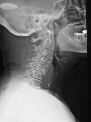
Figure 16 shows the radiograph of a 56-year-old man who has neck pain after a rollover accident on his lawnmower. The injury appears to be isolated, and he is neurologically intact. Management of the fracture should consist of


Explanation

The radiograph shows a type IIa Hangman's fracture, and the classic treatment is halo vest immobilization. Traction should be avoided in type IIa injuries because of the risk of overdistraction. A lesser form of immobilization such as a hard collar or a Minerva jacket can be used for nondisplaced (type I) fractures. Surgery generally is reserved for type III fractures (includes C2-3 facet dislocation), or extenuating circumstances such as multiple trauma or other fractures of the cervical spine that require surgical stabilization. Levine AM, Edwards CC: The management of traumatic spondylolisthesis of the axis. J Bone Joint Surg Am 1985;67:217-226.

Question 2

Degenerative spondylolisthesis of the cervical spine is most commonly seen at which of the following levels?


Explanation

Degenerative spondylolisthesis of the cervical spine is seen almost exclusively at C3-4 and C4-5; this is in contrast to degenerative changes, which are most commonly seen at C5-6 and C6-7. Tani T, Kawasaki M, Taniguchi S, et al: Functional importance of degenerative spondylolisthesis in cervical spondylotic myelopathy in the elderly. Spine 2003;28:1128-1134.

Question 3

Thoracic disk herniations are most frequently found in what area of the spine?


Explanation

Although thoracic disk herniations have been reported at all levels of the thoracic spine, more than two thirds are found at T9-T12, which is the more mobile lower third of the thoracic region. Belanger TA, Emery SE: Thoracic disc disease and myelopathy, in Frymoyer JW, Wiesel SW (eds): The Adult and Pediatric Spine. Philadelphia, PA, Lippincott Williams and Wilkins, 2004, pp 855-864.

Question 4

In a patient who has had low back pain for less than 2 weeks, which of the following findings is an indication for continued observation and symptomatic treatment rather than more aggressive evaluation and/or treatment?


Explanation

An inability to participate in athletics generally is considered an indication for continued symptomatic treatment only. All of the other answers suggest the possibility of more significant pathology that may require more urgent treatment. Frymoyer JW: Back pain and sciatica. N Engl J Med 1988;318:291-300.

Question 5

Radiographs of an 80-year-old woman with back pain reveal a compression fracture. Which of the following imaging studies best evaluates the acuity of the fracture?


Explanation

The best method of evaluating the acuity of osteoporotic compression fractures is to look for edema in the vertebral body. This is best accomplished with a STIR-weighted MRI scan. Bone scans can show increased uptake at the site of fracture for many months after the fracture. T1-weighted MRI scans show loss of normal marrow fat that may not necessarily correspond with acuity of the fracture. CT scans and radiographs show fracture deformity but cannot be used to judge acuity. Phillips FM: Minimally invasive treatments of osteoporotic vertebral compression fractures. Spine 2003;28:S45-S53.

Question 6

A 24-year-old professional football player underwent surgery for a symptomatic cervical disk herniation with radiculopathy 9 months ago. A current radiograph is shown in Figure 17. He has normal neurologic findings, no pain, and full range of motion. A CT scan shows a solid fusion. When can he expect to return to play?


Explanation

The radiograph shows that the two-level anterior cervical diskectomy and fusion has healed. In addition, the patient has good range of motion and the neurologic examination is normal. Based on these findings, the patient can return to play immediately. Patients with one- or two-level anterior cervical diskectomies and fusions that have healed fully can return to play. Any loss of motion, persistent neurologic deficit, or significant adjacent segment degeneration may preclude a player from returning. Thomas B, McCullen GM, Yuan HA: Cervical spine injuries in football players. J Am Acad Orthop Surg 1999;7:338-347.


Question 7

When treating thoracic disk herniations, which of the following surgical approaches has the highest reported rate of neurologic complications?


Explanation

Numerous surgical approaches have been used for thoracic diskectomy, including the most recent VATS. One of the first approaches described, posterior laminectomy, involves manipulation of the spinal cord, which the other approaches avoid. The posterior approach had dismal results, including further neurologic deterioration and even paralysis. Belanger TA, Emery SE: Thoracic disc disease and myelopathy, in Frymoyer JW, Wiesel SW (eds): The Adult and Pediatric Spine. Philadelphia, PA, Lippincott Williams and Wilkins, 2004, pp 855-864. Benjamin V: Diagnosis and management of thoracic disc disease. Clin Neurosurg 1983;30:577-605. Russell T: Thoracic intervertebral disc protrusion: Experience of 67 cases and review of the literature. Br J Neurosurg 1989;3:153-160.

Question 8

When harvesting iliac crest bone graft during a posterior spinal decompression and fusion, injury to what structure can result in painful neuromas or numbness over the skin of the buttocks?


Explanation

The superior cluneal nerves (L1, L2, and L3) are most at risk when harvesting iliac crest bone graft during a posterior decompression and fusion. These nerves pierce the lumbodorsal fascia and cross the posterior iliac crest, beginning 8 cm lateral to the posterior superior iliac spine. The ilioinguinal nerve is more at risk during exposure of the anterior ilium during retraction of the iliacus and abdominal wall muscles. Iliohypogastric nerve injury may arise in a similar fashion to ilioinguinal neuralgia. The lateral femoral cutaneous nerve lies in close proximity to the anterior superior iliac spine and is also at risk with anterior iliac crest bone graft harvesting. The superior gluteal nerve courses through the sciatic notch and supplies motor branches to the gluteus medius, minimus, and tensor fascia lata muscles. Injury results in hip abduction weakness. An HS: Principles and Techniques of Spine Surgery. Baltimore, MD, Williams and Wilkins 1998, pp 770-773. Kurz LT, Garfin SR, Booth RE Jr: Harvesting autogenous iliac bone grafts: A review of complications and techniques. Spine 1989;14:1324-1331.

Question 9

A 42 year-old-woman who underwent surgery for lumbar scoliosis 2 years ago now has fixed sagittal plane imbalance and severe back pain. Which of the following is considered a contraindication to isolated pedicle subtraction osteotomy for the treatment of iatrogenic flatback syndrome in this patient?


Explanation

Pedicle subtraction osteotomy is the preferred osteotomy technique for the treatment of many patients with iatrogenic flatback syndrome. In the presence of an anterior pseudarthrosis, however, it must be done in conjunction with an anterior procedure. Prior laminectomy is not a contraindication. Significant correction, usually averaging about 30 degrees, can be obtained through each osteotomy. Osteotomies should be performed at L2 or below in the presence of kyphosis at the thoracolumbar junction. The pedicle subtraction technique is preferred with vascular calcifications because it does not lengthen the anterior column, which could risk vascular injury. Potter BK, Lenke LG, Kuklo TR: Prevention and management of iatrogenic flatback deformity. J Bone Joint Surg Am 2004;86:1793-1808.

Question 10

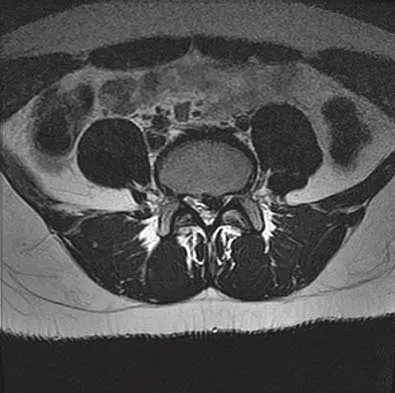
A 42-year-old man sustained a burst fracture at L2 in a motor vehicle accident. Examination reveals that he is neurologically intact. Figure 18 shows a cross-sectional CT scan through the fracture. If the fracture is managed nonsurgically for the next 2 years, the retained fragments can be expected to


Explanation

Numerous articles have reported that both surgical and nonsurgical management of burst fractures are associated with resolution of impingement at long-term follow-up. If the patient is neurologically intact and appropriately treated at the time of injury, neurologic deterioration is not expected nor is there a risk of injury to the dural sac. The retained fragments can be expected to gradually resorb and widen the spinal canal. Mumford J, Weinstein JN, Spratt KF, et al: Thoracolumbar burst fractures: The clinical efficacy and outcome of nonoperative management. Spine 1993;18:955-970.


Question 11

A 50-year-old man reports the onset of back pain and incapacitating pain radiating down his left leg posterolaterally and into the first dorsal web space of his foot 1 day after doing some yard work. He denies any history of trauma. Examination reveals ipsilateral extensor hallucis longus weakness. MRI scans are shown in Figures 19a through 19c. What nerve root is affected?


Explanation

The MRI scans clearly show an extruded L4-5 disk that is affecting the L5 root on the left side. In addition, the L5 root has a cutaneous distribution in the first dorsal web space. S1 affects the lateral foot, and L4 affects the medial calf. An HS: Principles and Techniques of Spine Surgery. Baltimore, MD, Williams and Wilkins, 1998, pp 98-100.


Question 12

Which of the following pharmacologic agents is most likely to adversely affect the success rate of bony union after lumbar arthrodesis?


Explanation

Glassman and associates reported a significantly higher pseudarthrosis rate when ketorolac was used postoperatively compared to a similar group of patients who were not given ketorolac. Animal studies from the same institution support these clinical findings. To reduce narcotic dosage, nonsteroidal anti-inflammatory drugs (NSAIDs) have been promoted as an adjunct for postoperative analgesia in patients undergoing spinal fusion. However, a high failure rate of arthrodesis has been associated with postoperative use of NSAIDs. The analgesics oxycodone hydrochloride, hydrocodone/acetaminophen, and tramadol, as well as the tricyclic antidepressant imipramine, have not been shown to inhibit fusion. Glassman SD, Rose SM, Dimar JR, et al: The effect of postoperative nonsteroidal anti-inflammatory drug administration on spinal fusion. Spine 1998;23:834-838.

Question 13

A 69-year-old woman is seen in the emergency department with a bilateral C5-6 facet dislocation and complete quadriplegia after falling down a flight of stairs. After initial evaluation and treatment by the trauma service, she is moved to the intensive care unit. Examination reveals a blood pressure of 90/50 mm/Hg, a pulse rate of 50/min, a respiration rate of 12/min, and urine output of 1 mL/kg/h. Her hemodynamic status should be addressed by


Explanation

The patient's heart rate is not responding to hypotension with tachycardia, as would be expected in the event of hypovolemic shock. Additionally, the adequate urine output suggests proper fluid resuscitation. Instead, she is bradycardic, possibly indicating neurogenic shock and loss of sympathetic tone to the heart. A Swan-Ganz catheter should be used to help differentiate these problems and guide appropriate fluid resuscitation and use of vasopressor agents. Hadley MN: Management of acute spinal cord injuries in an intensive care unit or other monitored setting. Neurosurgery 2002;50:S51-S57.

Question 14

What region of the spine is most susceptible to changes in the vascular supply to the spinal cord during an anterior approach?


Explanation

The thoracic spinal cord is characterized by a variable and, at times, complicated blood supply. The artery of Adamkiewicz, also known as the great anterior medullary artery, most typically arises off the left side of the aorta between T8 and T12. It represents the sole medullary blood supply to the thoracic spine. When this artery is divided or injured, the blood supply to the thoracic cord may be interrupted. It is important to avoid electocautery of blood vessels within or near the thoracic foramen because this is a site of important, albeit limited, collateral circulation. Sharma M, Anderson FC: Spinal vascular lesions, in Frymoyer JW, Wiesel SW (eds): The Adult and Pediatric Spine. Philadelphia, PA, Lippincott Williams and Wilkins, 2004, pp 301-306.

Question 15

What is the most common presenting sign or symptom in an adult with lumbar pyogenic infection?


Explanation

Pain is very common but is often nonspecific; therefore, the diagnosis of spinal infection is often delayed. Fever and sepsis can occur but are not common. Neurologic manifestations also can occur but are absent in most patients. In findings reported by Carragee, the urinary tract is a common source for hematogenous spinal infection, but the source was found in only 27% of 111 patients. Direct inoculation during spinal surgery is uncommon. Carragee EJ: Pyogenic vertebral osteomyelitis. J Bone Joint Surg Am 1997;79:874-880. Frazier DD, Campbell DR, Garvey TA, et al: Fungal infections of the spine: Report of eleven patients with long-term follow-up. J Bone Joint Surg Am 2001;83:560-565.

Question 16

The natural history of cervical spondylolytic myelopathy is best described as


Explanation

The natural history of cervical myelopathy has been described by Lees and Turner as exacerbations of symptoms followed by often long periods of static or deteriorating function (or very rarely improvement). This stepwise pattern of decreasing function has been corroborated by Clarke and Robinson. These authors described long periods of stable neurologic function, sometimes lasting for years, in about 75% of their patients. In the majority of the patients, however, the condition deteriorated between quiescent streaks. About 20% of their patients showed a slow, steady progression of symptoms and signs without a stable period, and 5% had rapid deterioration of neurologic function. Emery SF: Cervical spondylotic myelopathy: Diagnosis and treatment. J Am Acad Orthop Surg 2001;9:376-388. Lees F, Turner JA: The natural history and prognosis of cervical spondylosis. Brit Med J 1963;2:1607-1610.

Question 17

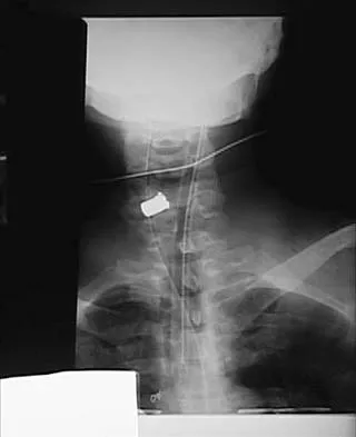
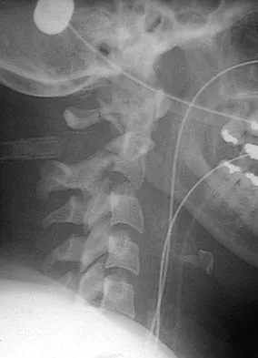
Figures 20a and 20b show lateral and AP radiographs of a 49-year-old man who sustained a gunshot wound through the left shoulder. He reports neck pain and examination reveals weakness in all four extremities. What is the priority of evaluation?


Explanation

The projectile entered the left shoulder and traveled to the right neck; therefore, a high incidence of suspicion must be directed to the airway, great vessels of the neck, and contents of the mediastinum. Immediate assessment of airway, breathing, and circulation takes priority, followed by examination of the neurologic status and other systems, as determined by the examination findings. Subcommittee on ATLS of the American College of Surgeons Committee on Trauma 1993-1997, Spine and Spinal Cord Trauma; Advanced Trauma Life Support Student Manual, ed 6, 1997. International Standards for Neurological and Functional Classification of Spinal Cord Injury. American Spinal Injury Association and International Medical Society of Paraplegia (ASIA/IMSOP).


Question 18

A 35-year-old woman undergoes an L4-5 anterior fusion via a left retroperitoneal approach. Postoperative examination reveals that her right foot is cool and pale. Her neurologic examination is normal, and her pedal pulses are asymmetric. What is the most likely reason for the right foot finding?


Explanation

The lower extremity symptoms are consistent with a sympathectomy that is the result of an injury to the sympathetic chain, ipsilateral to the approach along the anterior border of the lumbar spine. This results in a warm, red foot, which creates the appearance that the normal cooler foot may have compromised circulation. The latter generally attracts greater attention because of the risks associated with limb ischemia. The condition usually is self-limited and does not require any specific treatment. Rothman RH, Simeone FA (eds): The Spine, ed 4. Philadelphia PA, WB Saunders, 1999, p1550.

Question 19

What type of thoracolumbar spinal injury is associated with an increased risk of neurologic deterioration following admission to the hospital?


Explanation

Gertzbein's Scoliosis Research Society Morbidity and Mortality report noted that neurologic deterioration developed in approximately 16% of patients who were hospitalized with fracture-dislocations of the thoracolumbar spine, a particular concern with rotational burst fractures (AO type C). Patients with standard burst fractures and Chance fractures had a markedly lower incidence of neurologic involvement and tended to remain neurologically stable. Gertzbein SD: Neurologic deterioration in patients with thoracic and lumbar fractures after admission to the hospital. Spine 1994;19:1723-1725.

Question 20

A 30-year-old man has had a 3-day history of severe, incapacitating lower back pain without radiation. He reports improvement with rest. He denies any history of trauma, has no constitutional symptoms, and his neurologic examination is normal. What is the best course of action?


Explanation

There are no red flags in the history or examination to warrant MRI. Limited bed rest (less than 3 days) has been shown to be more beneficial to early recovery compared with prolonged bed rest (more than 7 days). No data support the use of epidural or facet steroid injections for acute low back pain.

Question 21

Which of the following patient factors is associated with recurrent radicular pain following lumbar diskectomy for sciatica?


Explanation

A large annular defect at the site of a lumbar disk herniation is associated with persistent radicular pain postoperatively. Large sequestered herniations and a positive SLR preoperatively correlate with good outcomes after diskectomy. Neither symptoms of more than 3 months' duration nor preoperative epidural steroid injections correlate with postoperative results after diskectomy. Carragee EJ, Han MY, Suen PW, et al: Clinical outcomes after lumbar discectomy for sciatica: The effects of fragment type and anular competence. J Bone Joint Surg Am 2003;85:102-108.

Question 22

Figure 21 shows the tomogram of a 26-year-old woman who sustained an axial load injury to her neck in a fall off a horse. What ligament is injured?


Explanation

Levine and Edwards, in their description of the classic C1 burst (Jefferson) fracture, noted that spread of the lateral masses of more than 7 mm is indicative of a transverse ligament rupture. Long-term C1-C2 instability, however, has not been described with this fracture pattern. Although long-term traction followed by halo vest immobilization has been described as the best technique for achieving an ideal result, treatment of this injury remains somewhat controversial. Levine AM, Edwards CC: Fractures of the atlas. J Bone Joint Surg Am 1991;73:680-691.


Question 23

Based on the findings shown in Figures 22a and 22b, corrective surgery to obtain maximal safe correction and optimal instrumentation fixation should be performed at which of the following locations?


Explanation

The clinical photograph and radiograph show an iatrogenic flatback deformity with loss of the normal lumbar lordosis. The safest correction for this malalignment typically is performed away from the spinal cord in the midlumbar spine, most commonly at L2 or L3. The more distal the correction is performed, the more sagittal plane translation of the C7 plumb line with respect to the posterior sacrum. Performing the osteotomy too distally, however, makes it difficult to obtain adequate distal fixation. Shufflebarger HL, Clark CE: Thoracolumbar osteotomy for postsurgical sagittal imbalance. Spine 1992;17:S287-S290.


Question 24

A 65-year-old woman has significant neck pain after falling and striking her head. A radiograph and sagittal CT scan are shown in Figures 23a and 23b. What is the most likely diagnosis?


Explanation

The radiograph shows a displacement of C5 on C6 of approximately 25%. The CT scan shows a perched facet at C5-6. There is no evidence of a facet fracture. A bilateral facet dislocation would show a displacement of more than 50%. Rothman RH, Simeone FA (eds): The Spine, ed 4. Philadelphia PA, WB Saunders, 1999, pp 927-937.


Question 25

Immediately after undergoing lumbar instrumentation, a patient reports severe right leg pain and has 4+/5 weakness. Figure 24 shows an axial CT scan of L5. Exploratory surgery will most likely reveal


Explanation

The most common finding at exploration of an inappropriately placed pedicle screw is displacement of the nerve. Pedicle breach is common, ranging from 2% to 20%, but most are asymptomatic. All of the choices are possible, but in a large series conducted by Lonstein and associates, the authors reported that displacement of the root, most often medial, was the most common finding. Laceration, contusion, or transfixion usually was not seen. Spinal fluid leakage occurs less frequently and is not expected in the minimal broach illustrated. Esses SI, Sachs BL, Dreyzin V: Complications associated with the technique of pedicle screw fixation: A selected survey of ABS members. Spine 1993;18:2231-2238. Laine T, Lund T, Ylikoski M, et al: Accuracy of pedicle screw insertion with and without computer assistance: A randomised controlled clinical study in 100 consecutive patients. Eur Spine J 2000;9:235-240.


Question 26

A 78-year-old man presents with severe neck pain following a ground-level fall. Imaging reveals a Type II odontoid fracture with 3 mm of posterior displacement. He is neurologically intact. What is the most appropriate definitive management?


Explanation

In elderly patients, Type II odontoid fractures have a high nonunion rate, and halo vest immobilization carries significant morbidity and mortality. Posterior C1-C2 fusion is the most reliable treatment to ensure stability and union in this demographic.

Question 27

A 65-year-old woman with adult spinal deformity is undergoing surgical planning. Her pelvic incidence (PI) is 60 degrees. To optimize her postoperative sagittal alignment, what should be the target lumbar lordosis (LL)?


Explanation

The primary goal for sagittal balance in adult spinal deformity is a PI-LL mismatch of less than 10 degrees (ideally PI = LL). Therefore, a lumbar lordosis of approximately 60 degrees would be optimal for a PI of 60 degrees.

Question 28

A 35-year-old construction worker falls from a height and presents with paraplegia at the T10 level.

Imaging demonstrates a T10 flexion-distraction injury with posterior ligamentous complex disruption. What associated injury must be urgently ruled out?


Explanation

Flexion-distraction injuries (Chance fractures) are highly associated with intra-abdominal pathology, particularly hollow viscus injuries, in up to 50% of cases. A high index of suspicion and appropriate abdominal imaging or surgical consultation are critical.

Question 29

In a patient with cervical spondylotic myelopathy (CSM), which of the following physical examination findings is considered the earliest indicator of myelopathy?


Explanation

Gait disturbance, often presenting as difficulty with tandem gait or a wide-based stance, along with fine motor clumsiness of the hands, are typically the earliest clinical signs of cervical myelopathy. Bowel/bladder dysfunction and severe atrophy present much later.

Question 30

A 14-year-old girl with adolescent idiopathic scoliosis (AIS) has a right main thoracic curve of 55 degrees and a left lumbar curve of 35 degrees. On lateral bending radiographs, the lumbar curve reduces to 15 degrees. According to the Lenke classification, what type of curve pattern is this?


Explanation

A structural main thoracic curve with a non-structural lumbar curve (which bends out to less than 25 degrees) is classified as a Lenke 1 curve. This typically requires a selective thoracic fusion.

Question 31

A 45-year-old man presents with right leg pain, numbness over the dorsum of his foot, and weakness in great toe extension. MRI shows a paracentral disc herniation. Which nerve root is most likely compressed?


Explanation

Weakness in the extensor hallucis longus (EHL) and numbness over the dorsum of the foot are classic signs of an L5 radiculopathy. In the lumbar spine, a paracentral disc herniation at L4-L5 typically compresses the traversing L5 nerve root.

Question 32

A 22-year-old man arrives in the trauma bay after a diving accident. He is awake, alert, and cooperative. He has no movement or sensation below the C6 level. Plain films show a unilateral facet dislocation at C5-C6. What is the most appropriate next step in management?


Explanation

In an awake, alert, and testable patient with a cervical spine dislocation and a neurologic deficit, immediate closed reduction with cranial traction is indicated to decompress the spinal cord. An MRI is not required prior to reduction in an awake, reliable patient.

Question 33

A 68-year-old man with ankylosing spondylitis presents to the ED with neck pain after a minor low-speed motor vehicle collision.

Neurologic examination is normal. Initial AP and lateral cervical spine radiographs show no obvious fracture. What is the next most appropriate step?


Explanation

Patients with ankylosing spondylitis are at a high risk for highly unstable extension fractures, which are often occult on plain radiographs, even after minor trauma. A CT scan of the spine is mandatory to rule out fractures and epidural hematomas.

Question 34

A 60-year-old woman underwent a primary L4-L5 microdiscectomy 6 months ago. She was symptom-free for 5 months but now presents with recurrent, severe right-sided L5 radiculopathy failing 6 weeks of conservative treatment. MRI shows a recurrent focal disc extrusion at L4-L5. What is the recommended surgical intervention?


Explanation

For a first-time recurrent disc herniation presenting primarily with radicular pain and no mechanical back pain or gross instability, revision microdiscectomy is the procedure of choice. Fusion is reserved for instability, mechanical back pain, or multiple recurrences.

Question 35

Which of the following congenital spinal anomalies has the highest risk of rapid deformity progression, nearly always requiring early surgical intervention?


Explanation

A unilateral unsegmented bar combined with a contralateral hemivertebra creates a massive growth mismatch, leading to rapid and severe deformity progression. Early surgical intervention (e.g., fusion or hemivertebra excision) is typically required to prevent severe curvature.

Question 36

A 75-year-old man with known cervical spinal stenosis falls forward, striking his forehead. He presents with severe weakness in his bilateral hands and arms, but is able to move his legs against gravity. Proprioception and pain sensation are diminished distally. What is the most likely diagnosis?


Explanation

Central cord syndrome typically occurs after a hyperextension injury in a patient with pre-existing cervical stenosis. It is characterized by disproportionately greater motor impairment in the upper extremities compared to the lower extremities.

Question 37

A 65-year-old man presents with progressive clumsiness in his hands and a wide-based gait. Physical exam shows a positive Hoffman's sign and hyperreflexia. MRI of the cervical spine is ordered. Which of the following MRI findings is the strongest independent predictor of a poor surgical outcome after decompression for degenerative cervical myelopathy?


Explanation

T1 hypointensity in the spinal cord indicates myelomalacia and cystic necrosis, representing irreversible cord damage. While T2 hyperintensity represents edema or gliosis, T1 changes correlate strongly with poor postoperative neurological recovery.

Question 38


A 55-year-old man with long-standing ankylosing spondylitis falls and sustains an undisplaced C6-C7 fracture seen on initial CT. He is neurologically intact on presentation. Twelve hours later, he develops progressive bilateral lower extremity weakness. What is the most likely cause of his delayed neurological deterioration?


Explanation

Patients with an ankylosed spine are at extremely high risk for epidural hematomas following even minor trauma due to altered biomechanics and tearing of the epidural venous plexus. Prompt MRI is indicated for any delayed neurological deficit to rule out this reversible cause.

Question 39

Degenerative spondylolisthesis in the lumbar spine most commonly occurs at the L4-L5 level. Which of the following anatomic variations is most strongly associated with the development of this specific condition?


Explanation

A more sagittal orientation of the L4-L5 facet joints provides less bony resistance to anterior shear forces compared to coronally oriented facets. This predisposes the segment to excessive anterior translation as the disc degenerates.

Question 40

A 72-year-old man presents with an isolated Type II odontoid fracture after a ground-level fall. Anterior odontoid screw fixation is being considered. Which of the following is an absolute contraindication to this specific procedure?


Explanation

Anterior screw fixation depends on an intact transverse ligament to provide stability to the C1-C2 articulation; its rupture is an absolute contraindication. A reverse obliquity fracture pattern is also a classic contraindication as the screw trajectory causes fracture distraction.

Question 41

In adult spinal deformity surgery, achieving optimal sagittal balance is critical to prevent hardware failure and adjacent segment disease. If a patient has a measured Pelvic Incidence (PI) of 58 degrees, what is the ideal postoperative target for Lumbar Lordosis (LL)?


Explanation

The generally accepted goal for sagittal balance in adult spinal deformity is achieving a Pelvic Incidence minus Lumbar Lordosis (PI-LL) mismatch of less than 10 degrees. Therefore, a target LL of approximately 58-60 degrees is ideal for a PI of 58.

Question 42

A 68-year-old man with underlying cervical spondylosis presents after a hyperextension injury. Examination reveals 2/5 motor strength in his upper extremities and 4/5 strength in his lower extremities. The disproportionate upper extremity weakness is primarily due to the anatomic arrangement of which spinal tract?


Explanation

Central cord syndrome preferentially affects the upper extremities because the cervical motor fibers within the lateral corticospinal tract are located medially, closer to the central canal. Lumbar and sacral fibers are situated more laterally and are thus spared.

Question 43

A 65-year-old woman presents with severe back pain, forward-leaning posture, and difficulty standing upright. Radiographs reveal adult spinal deformity. Her pelvic incidence (PI) is 60 degrees. To achieve a harmonious sagittal alignment postoperatively, what is the ideal target for her lumbar lordosis (LL)?


Explanation

For optimal sagittal balance in adult spinal deformity, the lumbar lordosis (LL) should be within 10 degrees of the pelvic incidence (PI). A PI-LL mismatch > 10 degrees is associated with poor health-related quality of life outcomes and adjacent segment disease.

Question 44

An 82-year-old man presents with neck pain after a low-speed motor vehicle collision. CT scan of the cervical spine demonstrates a displaced Type II odontoid fracture. He is neurologically intact but has significant medical comorbidities. Which of the following is the most appropriate initial management?


Explanation

In elderly patients (typically >80 years) with significant comorbidities, a rigid cervical collar is often preferred as initial treatment for Type II odontoid fractures. Surgery carries high morbidity, and halo vest immobilization has an unacceptably high complication and mortality rate in this age group.

Question 45

A 60-year-old man undergoes a posterior C3-C6 laminectomy and fusion for cervical spondylotic myelopathy. On postoperative day 2, he develops isolated right deltoid and biceps weakness (grade 2/5) without sensory deficits or lower extremity changes. What is the most likely etiology?


Explanation

C5 palsy is a well-known complication after cervical decompression (especially posterior laminectomy and fusion), occurring in up to 5-10% of cases. It is thought to result from spinal cord drift and subsequent tethering of the short C5 nerve roots.

Question 46

A 68-year-old man with known cervical spondylosis presents after a hyperextension injury. He has 2/5 motor strength in his bilateral upper extremities, particularly the hands, and 4/5 strength in his lower extremities. He has variable sensory loss but retains bladder function. What is the classic mechanism and diagnosis?


Explanation

Central cord syndrome typically occurs after a hyperextension injury in a patient with pre-existing cervical spondylosis. It affects the medially located cervical motor tracts more than the laterally located sacral/lumbar tracts, causing upper extremity weakness out of proportion to the lower extremities.

Question 47

A 14-year-old girl with adolescent idiopathic scoliosis has a primary right thoracic curve of 55 degrees and a left lumbar curve of 35 degrees. On lateral bending radiographs, the lumbar curve corrects to 15 degrees. According to the Lenke classification, what type of curve does she have?


Explanation

This is a Lenke Type 1 (Main Thoracic) curve. The lumbar curve is nonstructural because it corrects to less than 25 degrees on lateral bending radiographs.

Question 48

A 45-year-old man presents with progressive lower extremity weakness and myelopathy. MRI reveals a large, calcified central disc herniation at T8-T9 causing severe cord compression. Which of the following surgical approaches is CONTRAINDICATED?


Explanation

Posterior laminectomy for a central thoracic disc herniation is strictly contraindicated due to the high risk of catastrophic spinal cord injury. Accessing a central thoracic disc requires an anterior, lateral, or posterolateral approach to avoid manipulating the thoracic cord.

Question 49

A 55-year-old man with a long-standing history of ankylosing spondylitis presents with severe neck pain after a low-energy fall. Radiographs are inconclusive, but CT reveals a transverse fracture through the C5-C6 intervertebral space extending through the posterior elements. What is the most appropriate management?


Explanation

Fractures in ankylosing spondylitis are highly unstable and often behave like long-bone fractures. Due to the high risk of displacement, epidural hematoma, and nonunion, robust long-segment fixation, often combined anteriorly and posteriorly, is recommended.

Question 50

A 16-year-old boy presents with back pain and a prominent thoracic kyphosis. Radiographs reveal a thoracic kyphosis of 65 degrees. Which of the following radiographic criteria is required to confirm the diagnosis of Scheuermann's disease?


Explanation

Sorensen's criteria for classic Scheuermann's kyphosis require the presence of at least 3 adjacent vertebrae, each with a minimum of 5 degrees of anterior wedging. Additional findings often include Schmorl's nodes and irregular endplates.

Question 51

A 68-year-old woman presents with neurogenic claudication and L4 radiculopathy. Imaging demonstrates a L4-L5 degenerative spondylolisthesis with severe central canal and lateral recess stenosis. She has failed 6 months of conservative management. According to the SPORT trial, which of the following statements regarding surgical intervention is true?


Explanation

The Spine Patient Outcomes Research Trial (SPORT) demonstrated that patients with degenerative spondylolisthesis and spinal stenosis treated surgically had significantly greater improvement in pain and function compared to those treated nonoperatively.

Question 52

A 22-year-old woman involved in a high-speed motor vehicle collision as a rear-seat passenger wearing a lap belt presents with severe back pain. Imaging shows an L2 fracture with a horizontal split through the spinous process, pedicles, and vertebral body. What associated injury must be most highly suspected?


Explanation

This describes a bony Chance fracture (flexion-distraction injury) classically associated with lap seatbelts. These injuries have a very high association (up to 50%) with intra-abdominal hollow viscus injuries, particularly bowel perforations.

Question 53

A 35-year-old man fell 30 feet from a roof. Pelvic radiographs and CT demonstrate a transverse sacral fracture through the S1/S2 neural foramina connecting bilateral vertical sacral fractures. He has saddle anesthesia and sphincter dysfunction. This injury pattern represents:


Explanation

A transverse fracture line connecting bilateral vertical sacral fractures forms a U-type or H-type pattern, characteristic of spinopelvic dissociation. This highly unstable injury disconnects the spine from the pelvis and frequently causes profound neurologic deficits.

Question 54

A 48-year-old man presents with right arm pain, numbness in his index and middle fingers, and weakness with triceps extension and wrist flexion. His triceps reflex is diminished. Which cervical nerve root is most likely compressed?


Explanation

Compression of the C7 nerve root classically presents with weakness in elbow extension (triceps) and wrist flexion. Sensory changes are typically noted in the middle finger, along with an absent or diminished triceps reflex.

Question 55

A 14-year-old gymnast presents with chronic lower back pain. Radiographs reveal a Grade II isthmic spondylolisthesis at L5-S1. She has failed 6 months of bracing and physical therapy. If surgery is performed, what is the most appropriate procedure?


Explanation

For a symptomatic Grade II isthmic spondylolisthesis in an adolescent failing conservative care, an in-situ L5-S1 fusion is the standard surgical treatment. Direct pars repair is generally reserved for higher level defects (L1-L4) or L5 defects without significant slip (Grade 0-I).

Question 56

A 65-year-old woman presents with severe low back pain and an inability to stand up straight. Preoperative standing radiographs demonstrate a pelvic incidence (PI) of 60 degrees. To achieve optimal sagittal balance postoperatively and minimize the risk of adjacent segment disease, her lumbar lordosis (LL) should be restored to within what range?


Explanation

In adult spinal deformity, restoring sagittal balance is critical for favorable clinical outcomes. The lumbar lordosis (LL) should match the pelvic incidence (PI) within +/- 10 degrees (PI-LL < 10 degrees).

Question 57

A 60-year-old man presents with progressive clumsiness in his hands and difficulty walking. Examination reveals a positive Hoffmann sign and hyperreflexia in the lower extremities. MRI shows severe cervical stenosis at C4-C5 and C5-C6. Which of the following MRI findings is the most significant predictor of a poor neurological recovery following surgical decompression?


Explanation

A focal T1-weighted hypointensity in the spinal cord indicates myelomalacia or cystic necrosis. It is the strongest MRI predictor of poor neurological recovery and irreversible cord damage after surgical decompression.

Question 58

A 45-year-old man falls from a height and sustains an L1 burst fracture. He is neurologically intact. MRI demonstrates that the integrity of the posterior ligamentous complex (PLC) is indeterminate. Based on the Thoracolumbar Injury Classification and Severity (TLICS) score, what is the most appropriate management approach?


Explanation

The TLICS score assigns 2 points for a burst fracture morphology, 0 points for intact neurology, and 2 points for an indeterminate PLC, totaling 4 points. A total score of 4 suggests that either surgical or nonoperative management is appropriate based on surgeon and patient preference.

Question 59

A 72-year-old man sustains a Type II odontoid fracture after a low-speed motor vehicle collision. He is considered for nonoperative management. Which of the following factors most significantly increases his risk of nonunion if treated with a cervical collar?


Explanation

Risk factors for nonunion in Type II odontoid fractures include age > 65 years, initial displacement > 5 mm, posterior displacement, and a delay in diagnosis. Displacement > 5 mm carries a high rate of nonunion when treated conservatively.

Question 60

A 68-year-old man with known cervical spondylosis presents after a hyperextension injury to his neck. He is able to ambulate but has profound motor weakness in his hands and arms. Perianal sensation and bladder function are intact. What is the most likely diagnosis?


Explanation

Central cord syndrome typically occurs following a hyperextension injury in patients with pre-existing cervical spondylosis. It classically presents with disproportionate motor weakness in the upper extremities compared to the lower extremities.

Question 61

A 55-year-old man with longstanding ankylosing spondylitis sustains a minor ground-level fall. He has severe neck pain but is neurologically intact. Radiographs demonstrate a displaced extension-type fracture through the C6-C7 disc space. What is the standard of care for definitive management?


Explanation

Spinal fractures in ankylosing spondylitis are highly unstable due to the rigid, osteopenic nature of the fused spine. Long-segment posterior instrumentation and fusion is required to prevent catastrophic displacement and secondary neurological injury.

Question 62

In a patient with ankylosing spondylitis who sustains an acute cervical spine fracture, which of the following is the most common occult complication leading to delayed neurological deterioration?


Explanation

Spinal epidural hematomas are a frequent and potentially devastating complication of spinal fractures in ankylosing spondylitis. Because the bleeding can cause delayed neurological deterioration, an MRI is critical if the patient experiences a change in exam.

Question 63

A 60-year-old woman with severe rheumatoid arthritis is evaluated prior to a total knee arthroplasty. Flexion-extension cervical radiographs reveal an anterior atlantodental interval (ADI) of 11 mm. What is the most appropriate next step in her management?


Explanation

An ADI > 9-10 mm in a patient with rheumatoid arthritis indicates significant atlantoaxial instability with a high risk of spinal cord compression. An MRI is required to assess the posterior atlantodental interval (PADI) and check for myelomalacia before elective surgery.

Question 64

A 65-year-old woman presents with neurogenic claudication and a grade 1 degenerative spondylolisthesis at L4-L5. Which of the following anatomic features is most strongly implicated in the pathogenesis of her spondylolisthesis?


Explanation

Degenerative spondylolisthesis most commonly occurs at L4-L5. A more sagittal orientation of the facet joints renders them less capable of resisting shear forces, allowing anterior translation as the intervertebral disc degenerates.

Question 65

A 25-year-old man involved in a high-speed motor vehicle collision while wearing a lap belt sustains a flexion-distraction injury (Chance fracture) of the L2 vertebra. Which associated injury must be strongly suspected and ruled out?


Explanation

Chance fractures (flexion-distraction injuries) are heavily associated with lap-belt use in motor vehicle collisions. Up to 50% of these patients will have concomitant intra-abdominal injuries, particularly to hollow viscous organs such as the small bowel.

Question 66

A 32-year-old man presents with severe neck pain and right-sided C6 radiculopathy after a motorcycle crash. Lateral cervical radiographs reveal an anterior subluxation of C5 on C6 of approximately 25 percent. What is the primary mechanism of injury?


Explanation

A unilateral facet dislocation typically results from a flexion-rotation mechanism. On lateral radiographs, it is characterized by anterior translation of the superior vertebral body of approximately 25%, as opposed to 50% seen in bilateral facet dislocations.

Question 67

A 15-year-old boy presents with progressive mid-back pain and a rounded posture. Lateral radiographs of the thoracic spine demonstrate anterior wedging of 7 degrees at T7, T8, and T9, along with prominent Schmorl's nodes. He has a flexible kyphosis measuring 60 degrees. What is the most appropriate initial management?


Explanation

This patient meets the radiographic criteria for Scheuermann's kyphosis (anterior wedging > 5 degrees in at least three consecutive vertebrae). For a skeletally immature patient with progressive, symptomatic kyphosis > 50 degrees, an extension brace is the recommended first-line treatment.

Question 68

A 58-year-old man underwent an L4-S1 posterior instrumented fusion 5 years ago. He now presents with new-onset L3 radiculopathy due to adjacent segment disease. Which of the following intraoperative factors during the index surgery is most strongly associated with accelerated adjacent segment degeneration?


Explanation

Violation of the adjacent superior facet joint capsule during pedicle screw placement disrupts the biomechanics of the unfused level. This iatrogenic injury significantly increases the risk and accelerates the onset of adjacent segment disease.

Question 69

A 40-year-old man sustains a traumatic spondylolisthesis of the axis (Hangman's fracture) following a high-speed motor vehicle rollover. The classic mechanism of injury responsible for this fracture pattern is:


Explanation

A Hangman's fracture classically results from hyperextension and axial loading forces. This combined mechanism fractures the pars interarticularis of C2, commonly seen in modern motor vehicle collisions or shallow-water diving accidents.

Question 70

A 42-year-old man presents with right leg pain radiating down the lateral aspect of his calf to the dorsum of his foot, accompanied by weakness in great toe extension. MRI of the lumbar spine reveals a paracentral disc herniation. At which level is the disc herniation most likely located?


Explanation

Weakness in the extensor hallucis longus (EHL) and sensory changes over the dorsum of the foot are classic signs of an L5 radiculopathy. A paracentral disc herniation at the L4-L5 level will impinge the traversing L5 nerve root.

Question 71

A 65-year-old woman presents with severe neurogenic claudication and an L4-L5 degenerative spondylolisthesis. She has failed 6 months of supervised physical therapy and epidural steroid injections. What is the most appropriate surgical intervention to optimize long-term clinical outcomes?


Explanation

According to the SPORT trial and other long-term studies, decompression combined with instrumented fusion provides significantly better outcomes for degenerative spondylolisthesis than decompression alone. Laminectomy alone risks iatrogenic instability and progression of the slip.

Question 72

An 80-year-old man sustains a Type II odontoid fracture after a ground-level fall. He is neurologically intact. Which of the following radiographic findings is the strongest predictor of nonunion if treated conservatively with a rigid cervical collar?


Explanation

Initial fracture displacement greater than 5 mm is a classic risk factor for nonunion in Type II odontoid fractures. Other risk factors include age > 50 years, posterior displacement, and delay in treatment.

Question 73

A 14-year-old girl with adolescent idiopathic scoliosis presents with a right thoracic curve measuring 55 degrees on standing radiographs. Her Risser stage is 0 and she is pre-menarchal. What is the most appropriate management?


Explanation

Curves greater than 50 degrees in growing adolescents have a high risk of continued progression into adulthood and typically warrant surgical correction. Posterior spinal fusion is the standard of care for a progressive 55-degree curve in a Risser 0 patient.

Question 74

In the assessment of adult spinal deformity, which of the following spinopelvic parameters is considered a fixed morphological characteristic of the pelvis that remains constant regardless of patient positioning?


Explanation

Pelvic incidence (PI) is a constant anatomical parameter unique to each individual and does not change with posture. It dictates the relationship between pelvic tilt and sacral slope, represented by the formula PI = PT + SS.

Question 75

A 35-year-old man sustains an L1 burst fracture after falling from a ladder. He is neurologically intact. Imaging reveals 15 degrees of kyphosis, 30% canal compromise, and an intact posterior ligamentous complex (PLC). What is the most appropriate treatment?


Explanation

Stable thoracolumbar burst fractures (neurologically intact, intact PLC, acceptable kyphosis < 20-30 degrees) are typically treated non-operatively with a TLSO brace. Outcomes for stable burst fractures treated with bracing are equivalent to surgical stabilization.

Question 76

A 45-year-old man presents with severe right thigh pain and weakness in knee extension. MRI reveals a far-lateral (extraforaminal) disc herniation at the L4-L5 level. Which nerve root is most likely compressed?


Explanation

In the lumbar spine, a far-lateral (extraforaminal) disc herniation impinges the exiting nerve root at that level. Therefore, an L4-L5 far-lateral disc herniation will compress the L4 nerve root.

Question 77

During the physical examination of a patient with progressive gait difficulties, flicking the volar nail of the middle finger results in brisk flexion of the thumb and index finger. This clinical sign indicates an upper motor neuron lesion typically located where?


Explanation

The described maneuver is Hoffmann's sign, which is indicative of an upper motor neuron lesion or cervical myelopathy. It points to compression or pathology within the cervical spinal cord.

Question 78

A 70-year-old man with advanced ankylosing spondylitis presents to the emergency department complaining of neck pain after a low-speed motor vehicle collision. Initial plain radiographs of the cervical spine demonstrate an ossified anterior longitudinal ligament but no obvious fracture. What is the most appropriate next step in management?


Explanation

Patients with ankylosing spondylitis are at extremely high risk for occult "chalk stick" fractures, which are often missed on plain radiographs. A CT scan of the entire cervical spine (and often an MRI to rule out epidural hematoma) is mandatory even after minor trauma.

Question 79

A patient is diagnosed with a Levine-Edwards Type IIA traumatic spondylolisthesis of the axis (Hangman's fracture). Which of the following conservative management techniques is strictly contraindicated for this specific injury pattern?


Explanation

Type IIA Hangman's fractures show severe angulation with minimal translation and denote significant disc space disruption. Cervical traction is strictly contraindicated as it will distract the fracture site and worsen the deformity.

Question 80

What is the primary radiographic requirement for the diagnosis of classic Scheuermann's kyphosis according to the Sorensen criteria?


Explanation

The Sorensen criteria define Scheuermann's disease strictly as anterior wedging of 5 degrees or more in at least 3 consecutive vertebral bodies. Associated findings include Schmorl's nodes and endplate irregularities, but wedging is the diagnostic requirement.

Question 81

A 68-year-old man with baseline cervical spondylosis is involved in a rear-end motor vehicle accident causing a hyperextension injury. He presents with bilateral upper extremity weakness (2/5) but relatively preserved lower extremity strength (4/5). What is the most likely diagnosis?


Explanation

Central cord syndrome is the most common incomplete spinal cord injury, typically occurring in older patients with cervical spondylosis following hyperextension trauma. It classically presents with disproportionately greater motor impairment in the upper extremities compared to the lower extremities.

Question 82

Following a multi-level posterior cervical laminectomy and instrumented fusion for severe myelopathy, a patient develops profound, isolated weakness in shoulder abduction and elbow flexion postoperatively. Sensation remains largely intact. What is the most likely complication?


Explanation

Postoperative C5 palsy is a known complication after posterior cervical decompression, occurring in up to 10% of cases. It is thought to be caused by posterior shifting of the spinal cord leading to tethering or stretch of the short C5 nerve roots.

Question 83

A 22-year-old man presents with chronic low back pain and radicular symptoms. Imaging reveals a bilateral L5 pars interarticularis defect (isthmic spondylolisthesis) with a Grade 2 slip. Which nerve root is most commonly compressed in this specific condition?


Explanation

In isthmic spondylolisthesis at L5-S1, the fibrocartilaginous pseudarthrosis mass at the pars defect typically compresses the exiting L5 nerve root within the neural foramen. This differs from a paracentral disc herniation at L5-S1, which would affect the traversing S1 root.

Question 84

A 25-year-old woman wearing only a lap belt is involved in a high-speed frontal collision. She sustains a flexion-distraction injury (Chance fracture) to the L2 vertebra. What associated injury must be aggressively ruled out?


Explanation

Chance fractures (flexion-distraction injuries) are highly associated with intra-abdominal injuries, particularly hollow viscus injuries (e.g., small bowel laceration), occurring in up to 50% of cases. A thorough abdominal evaluation is mandatory.

Question 85

A 65-year-old man presents with progressive dysphagia. Cervical spine radiographs reveal flowing ossification along the anterior longitudinal ligament. To confirm Diffuse Idiopathic Skeletal Hyperostosis (DISH) using Resnick's criteria, how many contiguous vertebral bodies must be involved?


Explanation

Resnick's criteria for diagnosing DISH require flowing ossification along the anterolateral aspect of at least four contiguous vertebral bodies. The criteria also require relative preservation of disc height and absence of sacroiliac joint inflammatory changes.

Question 86

A 55-year-old woman is evaluated for clumsiness of her hands and gait instability. Striking the distal brachioradialis tendon with a reflex hammer elicits isolated flexion of her fingers, without the normal elbow flexion. What does this 'inverted brachioradialis reflex' indicate?


Explanation

The inverted brachioradialis reflex is a sign of cervical myelopathy. It occurs when there is a lower motor neuron lesion at the C5-C6 level (loss of brachioradialis reflex) combined with an upper motor neuron lesion below that level (hyperactive finger flexion reflex).

Question 87

When evaluating a lateral cervical spine radiograph for traumatic occipitocervical dissociation, the Powers ratio is calculated. A ratio greater than what value is considered highly sensitive for anterior occipitoatlantal subluxation?


Explanation

The Powers ratio is the distance from the basion to the posterior arch of C1 divided by the distance from the opisthion to the anterior arch of C1. A ratio strictly > 1.0 is highly suggestive of anterior occipitocervical dissociation.

Question 88

A 30-year-old man arrives in the emergency department following a rugby tackle. He is fully awake, alert, and cooperative. Imaging reveals a unilateral cervical facet dislocation at C5-C6. He has right-sided arm pain and C6 weakness. What is the most appropriate initial management?


Explanation

In an awake, alert, and cooperative patient with a cervical facet dislocation, rapid closed reduction using cranial traction is safe and indicated. MRI should be obtained prior to reduction only if the patient has an altered mental status or fails closed reduction.

Question 89

A 28-year-old man sustains a gunshot wound to the lumbar spine. He arrives with bilateral lower extremity weakness, saddle anesthesia, and urinary retention. CT scan shows a retained bullet fragment within the spinal canal at L3. What is the optimal surgical management?


Explanation

While most spinal gunshot wounds are managed non-operatively, surgical decompression and bullet removal are indicated for patients presenting with cauda equina syndrome, progressive neurological deficits, or when the bullet is located within the disc space due to toxicity risk.

Question 90

During preoperative planning for a long-segment fusion to correct adult spinal deformity, the surgeon calculates the patient's spinopelvic parameters. To achieve an optimal sagittal balanced profile and minimize the risk of adjacent segment disease, the surgical goal should be to restore Lumbar Lordosis (LL) to within how many degrees of the Pelvic Incidence (PI)?


Explanation

A widely accepted goal in adult spinal deformity surgery is to achieve a mismatch between Pelvic Incidence and Lumbar Lordosis of less than 10 degrees (PI - LL < 10°). This aligns the patient's sagittal profile and reduces the risk of postoperative complications and pseudarthrosis.

Question 91

A 35-year-old man presents to the emergency department after a high-speed motor vehicle collision. He complains of severe neck pain and exhibits bilateral upper extremity weakness (deltoids and biceps 3/5, triceps 4/5) with normal lower extremity strength. Radiographs demonstrate a 50% anterior translation of C5 on C6. The patient is awake, alert, and cooperative. What is the most appropriate next step in management?


Explanation

In an awake, cooperative patient with a cervical facet dislocation and a neurologic deficit, emergent closed reduction via traction is indicated. MRI is not required prior to closed reduction in this setting but is necessary before open reduction or in a comatose/unexaminable patient.

Question 92

A 65-year-old woman presents with severe neurogenic claudication and MRI-confirmed L4-L5 degenerative spondylolisthesis. She has failed 6 months of comprehensive conservative management. Based on the Spine Patient Outcomes Research Trial (SPORT) for degenerative spondylolisthesis, what outcome should she expect if she elects to undergo surgical intervention compared to continued nonoperative treatment?


Explanation

The SPORT study for degenerative spondylolisthesis demonstrated that patients treated surgically had significantly better outcomes in pain, physical function, and disability at 4 years compared to those treated nonoperatively. The surgical benefit remained durable over long-term follow-up.

Question 93

A 68-year-old woman with severe back pain and forward-flexed posture is being evaluated for an adult spinal deformity correction. Her preoperative full-length standing radiographs demonstrate a Pelvic Incidence (PI) of 62 degrees. To achieve optimal postoperative sagittal balance and minimize the risk of adjacent segment disease or hardware failure, what should her target Lumbar Lordosis (LL) be?


Explanation

To achieve ideal sagittal balance, the Lumbar Lordosis (LL) should be matched to within 9 to 10 degrees of the patient's fixed Pelvic Incidence (PI). Therefore, for a PI of 62 degrees, a target LL of approximately 62 degrees is mathematically optimal.

Question 94

A 42-year-old construction worker falls 10 feet and sustains an L1 burst fracture. He is neurologically intact on examination. CT and MRI show 40% anterior body height loss, 30% retropulsed bone causing canal compromise, and a completely intact posterior ligamentous complex (PLC). According to the Thoracolumbar Injury Classification and Severity (TLICS) score, what is the most appropriate treatment recommendation?


Explanation

The patient's TLICS score is 2 (Morphology: Burst = 2, Neurologic Status: Intact = 0, PLC: Intact = 0). A TLICS score of less than 4 is an indication for nonoperative management, typically utilizing a TLSO.

Question 95

A 72-year-old man presents with deteriorating handwriting, frequent dropping of objects, and a wide-based gait. On physical examination, striking the brachioradialis tendon results in diminished reflex finger flexion but elicits brisk finger flexion of the ipsilateral hand. This specific physical exam finding (an inverted brachioradialis reflex) localizes the primary spinal cord compression to which of the following cervical levels?


Explanation

An inverted brachioradialis reflex indicates an absent C6 lower motor neuron response (brachioradialis arc) coupled with an exaggerated C8 upper motor neuron response (hyperreflexic finger flexion), successfully localizing the compressive lesion to the C5-C6 disc space.

Question 96

A 15-year-old boy presents with progressive mid-back pain and an increasing rounded appearance to his upper back. You suspect Scheuermann's kyphosis. Which of the following radiographic findings represents the classic diagnostic criteria (Sorensen's criteria) for this condition?


Explanation

Sorensen's criteria for the diagnosis of Scheuermann's kyphosis require the presence of greater than or equal to 5 degrees of anterior wedging in at least 3 consecutive thoracic vertebral bodies.

Question 97

A 78-year-old man trips on a rug and sustains an isolated Type II odontoid fracture. He is neurologically intact. You are discussing nonoperative management in a hard cervical collar versus surgical stabilization. Which of the following injury characteristics is most strongly associated with an increased risk of nonunion if treated nonoperatively?


Explanation

Risk factors for nonunion in Type II odontoid fractures include initial displacement greater than 5 mm, angulation greater than 10 degrees, advanced age (typically > 65 years), and a delay in diagnosis.

Question 98

A 60-year-old woman is scheduled for an L4-L5 posterior lumbar interbody fusion (PLIF) with pedicle screw instrumentation. Which of the following intraoperative technical errors is most significantly linked to the accelerated development of adjacent segment disease at the L3-L4 level?


Explanation

Violation of the adjacent, unfused superior facet joint (L3-L4) during pedicle screw insertion at the uppermost instrumented vertebra (L4) destroys the integrity of the facet complex, drastically accelerating adjacent segment degeneration.

Question 99

A 65-year-old man with underlying cervical stenosis presents with weakness in his upper and lower extremities following a hyperextension injury to his neck during a fall. His exam shows 2/5 strength in his hands bilaterally and 4/5 strength in his legs, with preserved sacral sensation. Which of the following best describes the typical expected pattern of neurologic recovery in this condition?


Explanation

In Central Cord Syndrome, the typical sequence of neurologic recovery occurs in the following order: lower extremity function first, followed by bowel/bladder function, then proximal upper extremities, and finally distal upper extremity fine motor function (which often remains permanently impaired).

Question 100

A 13-year-old premenarchal female (Risser stage 0) presents for evaluation of a spinal deformity. Radiographs confirm adolescent idiopathic scoliosis with a primary right thoracic curve measuring 35 degrees. What is the most appropriate evidence-based management strategy for this patient?


Explanation

For a skeletally immature patient (Risser 0-2, premenarchal) with an adolescent idiopathic scoliosis curve measuring between 25 and 45 degrees, the standard of care to prevent progression to a surgical threshold is full-time bracing (TLSO).

None

Clinic OS
Medically Verified Content by
Prof. Clinic OS
Consultant Orthopedic & Spine Surgeon
Chapter Index