العربية
Part of the Master Guide

AAOS Orthopedic MCQs (Set 1): Spine Disorders, Sports Medicine & Arthroplasty | 2026 Board Review

Spine Surgery Board Review MCQs (Set 2): Spinal Trauma & Degenerative Conditions | AAOS & ABOS

23 Apr 2026 59 min read 93 Views
Spine 2006 MCQs - Part 2

Key Takeaway

This high-yield question set for the AAOS and ABOS Spine Surgery Board Review (Set 2) focuses on critical topics like spinal trauma, including fractures and dislocations, as well as the diagnosis and management of degenerative spine conditions such as lumbar stenosis and disc disease. Prepare effectively for your exams.

Spine Surgery Board Review MCQs (Set 2): Spinal Trauma & Degenerative Conditions | AAOS & ABOS

Comprehensive 100-Question Exam


00:00

Start Quiz

Question 1

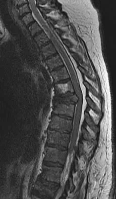
Figure 10 shows the MRI scan of a 56-year-old woman with metastatic breast cancer who now reports progressive paraparesis. Her general health remains good. Treatment should consist of


Explanation

If the patient's medical condition and prognosis remain good in the presence of significant and progressive neurologic deficit from cord compression, then the most reliable means of restoring function is via surgical decompression and fusion. Decompression should be directed toward the compressing structure (eg, anteriorly if the compression is from the anterior side). This procedure can be done via a posterolateral technique, such as costotransversectomy in some cases.

Question 2

When 6 weeks of noninvasive nonsurgical management fails to provide relief for a lumbar disk herniation, a trial of epidural steroid injections is likely to yield which of the following results?


Explanation

Lumbar epidural steroid injections appear to play a role in management of a lumbar disk herniation that has failed to respond to at least 6 weeks of nonsurgical treatment. Approximately 42% to 56% of patients report significant pain relief compared with 92% to 98% of those patients treated with diskectomy. Patients with extruded or sequestered herniations report the greatest and most rapid relief. Similarly, those with well-hydrated disk fragments report rapid relief of symptoms. A smaller percentage of patients report symptom relief compared with those having surgery, but the degree of improvement is similar for both groups and the improvement lasts up to 3 years. Butterman GR: Treatment of lumbar disc herniation: Epidural steroid injection compares with discectomy: A prospective, randomized study. J Bone Joint Surg Am 2004;86:670-679.


Question 3

Which of the following anatomic changes is observed as part of the normal aging process of the adult spine?


Explanation

The primary change that takes place in the aging spine is degeneration of the lumbar disks and loss of the overall lumbar lordosis. This also may be associated with osteopenic-related compression fractures. With these changes, the sagittal vertical line moves anteriorly relative to the sacrum; cervical scoliosis is uncommon and not part of the normal aging process. Overall kyphosis in the thoracic spine gradually increases, but the coronal balance remains essentially the same unless scoliosis develops. Gelb DE, Lenke LG, Bridwell KH, et al: An analysis of sagittal spinal alignment in 100 asymptomatic middle and older aged volunteers. Spine 1995;20:1351-1358.


Question 4

A previously healthy 30-year-old woman has neck pain and bilateral hand and lower extremity tingling with weakness after falling down stairs. She is alert and oriented. Examination reveals incomplete quadriplegia at the C6 level that remains unchanged throughout her evaluation and initial treatment. Radiographs show a bilateral facet dislocation of C6 on C7 without fracture. Attempts at reduction with halo cervical traction up to her body weight are unsuccessful. What is the next most appropriate step?


Explanation

A facet dislocation that cannot be reduced in an alert, awake patient with some preservation of cord function requires MRI to evaluate the disk prior to a reduction under anesthesia. The presence or absence of a disk herniation must be assessed, as this factor may influence the method of reduction. Vaccaro AR, Falatyn SP, Flanders AE, et al: Magnetic resonance evaluation of the intervertebral disc, spinal ligaments, and spinal cord before and after closed traction reduction of cervical spine dislocations. Spine 1999;24:1210-1217. Fardon DF, Garfin SR, Abitbol J (eds): Orthopaedic Knowledge Update: Spine 2. Rosemont, IL, American Academy of Orthopaedic Surgeons, 2002, pp 247-262. Eismont FJ, Arena MJ, Green BA: Extrusion of an intervertebral disc associated with traumatic subluxation or dislocation of cervical facets. J Bone Joint Surg Am 1991;73:1555-1560.


Question 5

Which of the following findings is the best radiographic indicator of segmental instability at L4-L5?


Explanation

Motion segments that demonstrate more than 4 mm of translation or 10 degrees of angulation compared with adjacent motion segments on flexion-extension radiographs have excessive motion and instability. Anterior marginal osteophytes form at the insertion of the annulus from increased forces but do not indicate increased motion. A spondylolisthesis or lateral listhesis is often static without increased motion. More than 3.5 mm of translation or 11 degrees of angulation is considered instability criteria for the cervical spine. Internal disk disruption does not denote instability. Boden SD, Wiesel SW: Lumbosacral segmental motion in normal individuals. Have we been measuring instability properly? Spine 1990;15:571-576.


Question 6

In a patient who has undergone fusion with instrumentation from T4 to the sacrum for adult scoliosis, at which site is a pseudarthrosis most likely to be discovered?


Explanation

Although pseudarthrosis can be found anywhere within the spine that has been fused using long multisegmental fixation to the sacrum, it most commonly occurs at the lumbosacral junction. The thoracolumbar junction is another common site of potential pseudarthrosis. In this location, the anatomy changes from lumbar transverse processes to thoracic through the transition zone, and overlying instrumentation often makes it difficult to obtain enough sound bone on decorticated bone to achieve a successful fusion. Saer EH III, Winter RB, Lonstein JE: Long scoliosis fusion to the sacrum in adults with nonparalytic scoliosis: An improved method. Spine 1990;15;650-653. Kostuik JP, Hall BB: Spinal fusions to the sacrum in adults with scoliosis. Spine 1983;8:489-500.

Question 7

The afferent pain innervation of the L3-L4 facet joint arises from the medial branch nerve of


Explanation

Afferent pain fibers to the lumbar facet joints arise from the medial branch nerves originating from the next two cephalad levels. Therefore, innervation of the L3-L4 facet joint arises from the L2 and L3 medial branch nerves. This effect should be taken into account when considering a medial branch block or facet denervation. The medial branch nerve arises from the dorsal ramus of the exiting nerve root. Nade SL, Bell E, Wyke BD: The innervation of the lumbar spinal joint and its significance. J Bone Joint Surg Br 1980;62:255-261

Question 8

When posterior fusion with instrumentation to the sacrum is used to treat adult scoliosis, what instrumentation technique best increases the chance of a successful lumbosacral fusion?


Explanation

As the chance of success of lumbosacral fusion increases with the stiffness and rigidity of the construct, fixation and stiffness improve with fixation into both the upper sacrum and the ilium. In a review of individuals treated with long constructs to the pelvis for adult scoliosis, Islam and associates reported that the rate of pseudarthrosis was significantly lower with sacral and iliac fixation compared with sacral fixation alone or iliac fixation alone. Iliac screws provide significant fixation anterior to the instantaneous axis of rotation for flexion and extension, as well as provides resistance to lateral bending and rotational forces. Numerous biomechanical studies support the concept of increasing biomechanical stabilization with increased fixation from the sacrum to the ilium. Islam NC, Wood KB, Transfeldt EE, et al: Extension of fusions to the pelvis in idiopathic scoliosis. Spine 2001;26:166-173. O'Brien N, et al: Sacral pelvic fixation and spinal deformity, in DeWald RL (ed): Spinal Deformities: A Comprehensive Text. New York, NY, Thieme, 2003, pp 601-614.

Question 9

Which of the following structures runs through the site indicated by the arrow in Figure 11?


Explanation

The vertebral artery traverses through the arcuate foramen after exiting the lateral aspect of C1 and before entering the skull. The foramen usually is not fully formed, but a complete foramen such as this one has been reported in up to 18% of patients. Stubbs DM: The arcuate foramen: Variability in distribution related to race and sex. Spine 1992;17:1502-1504.


Question 10

A 42-year-old man has had left lower extremity pain in an L5 radicular pattern for the past 6 weeks. He denies significant axial low back pain. History reveals that he underwent an L4-5 diskectomy with successful relief of similar pain 5 years ago. Which of the following imaging studies would offer the greatest amount of information?


Explanation

MRI with gadolinium will best identify recurrent herniated nucleus pulposus or other root compression and distinguish scar from recurrent disk. CT is unable to distinguish scar from recurrent disk density, and the addition of myelogram dye can reveal compromise of the thecal sac but cannot distinguish the scar from recurrent disk as the source of compression. Although lateral flexion-extension radiographs may be important to rule out any instability, much of that information can be inferred from the associated disk and adjacent bony changes on MRI. Bone scan techniques may identify subtle stress fractures resulting from previous aggressive facet resection, but low back pain also would be expected. Mirowitz SA, Shady KL: Gadopentetate dimeglumine-enhanced MR imaging of the postoperative lumbar spine: Comparison of fat-suppressed and conventional T1-weighted images. Am J Roentgenol 1992;159:385-389.

Question 11

Figure 12 shows the radiograph of an 80-year-old woman who has had an 8-month history of back pain after a fall. What is the most likely diagnosis based on the radiographic findings at the fractured vertebrae?


Explanation

An intravertebral vacuum cleft suggests nonunion of the vertebral fracture with osteonecrosis and is not seen in routine healing fractures. MRI characteristically shows a high T2 signal in the cleft. The cleft is not indicative of an infectious or neoplastic lesion. A vacuum disk phenomenon is associated with end-stage degenerative disk disease, but those findings are not found in the vertebral body. Murakami H, Kawahara N, Gabata T, et al: Vertebral body osteonecrosis without vertebral collapse. Spine 2003;28:E323-E328.


Question 12

Which of the following complications is uniquely associated with an anterior approach to the lumbosacral junction?


Explanation

Retrograde ejaculation is a sequela of injury to the superior hypogastric plexus. The structure needs protection, especially during anterior exposure of the lumbosacral junction. The use of monopolar electrocautery should be avoided in this region. The ideal exposure starts with blunt dissection just to the medial aspect of the left common iliac vein, sweeping the prevertebral tissues toward the patient's right side. Although erectile dysfunction can be seen after spinal surgery, it is not typically related to the surgical exposure because erectile function is regulated by parasympathetic fibers derived from the second, third, and fourth sacral segments that are deep in the pelvis and are not at risk with the anterior approach. The other choices are complications of spinal surgery but are not uniquely associated with an anterior L5-S1 exposure. Flynn JC, Price CT: Sexual complications of anterior fusion of the lumbar spine. Spine 1984;9:489-492. Watkins RG (ed): Surgical Approaches to the Spine, ed 1. New York, NY, Springer-Verlag, 1983, p 107.

Question 13

A 68-year-old woman with a history of rheumatoid arthritis has had neck pain and weakness in all four extremities that has become worse in the past 6 months. She has gone from a community to a household ambulator and uses a wheelchair outside of the home. Examination of the extremities reveals poor coordination, diffuse weakness, hyperactive reflexes, and bilateral sustained clonus. She has a broad-based and unsteady gait. The posterior atlanto-dens interval is 12 mm. Based on these findings and the radiograph and MRI scan shown in Figures 13a and 13b, the treatment of choice is surgical decompression and stabilization. However, the patient inquires about the prognosis with surgery compared to nonsurgical management. Assuming there are no complications from surgery, the patient should be informed that, with surgery, she will most likely


Explanation

The patient has a cervical myelopathy with more than 10 mm of space available for the cord; therefore, she has a reasonable chance of improved neurologic function following surgery. If not treated with surgery, however, her neurologic condition likely will worsen and she will die earlier than if she had surgery. Matsunaga S, Sakou T, Onishi T, et al: Prognosis of patients with upper cervical lesions caused by rheumatoid arthritis: Comparison of occipitocervical fusion between C1 laminectomy and nonsurgical management. Spine 2003;28:1581-1587.


Question 14

Five weeks after undergoing a successful L4-L5 diskectomy, with complete relief of his preoperative sciatica, a 36-year-old man has severe, relentless back and buttock pain. Examination and laboratory studies are unremarkable with the exception of an erythrocyte sedimentation rate (ESR) of 90 mm/h. What is the next most appropriate step in management?


Explanation

The patient's history, including the timing and type of symptoms, is typical for postoperative diskitis. The elevated ESR, 5 weeks after surgery, is also consistent with infection; a normal WBC count is not unusual. Management should consist of MRI with gadolinium; if positive, this should be followed by percutaneous biopsy to confirm the organism. Open biopsy may be considered if the percutaneous biopsy is unsuccessful. Anterior debridement and interbody fusion is reserved for the occasional patient that fails to respond to intravenous antibiotics, bed rest, and immobilization. Garfin SR, Vaccaro AR (eds): Orthopaedic Knowledge Update: Spine. Rosemont, IL, American Academy of Orthopaedic Surgeons, 1997, pp 257-271.

Question 15

An 18-year-old man sustained a knife injury to his midback, with the entry wound 2 cm to the left of the midline. He has been diagnosed with a hemicord transection. Neurologic examination will most likely reveal left-sided loss of


Explanation

Brown-Sequard syndrome results from an injury to one half of the spinal cord and is characteristically seen in penetrating injuries. The spinothalamic fibers cross the midline below the level of the lesion, resulting in contralateral loss of pain and temperature sensation. The posterior columns and corticospinal tracts carry vibratory, position, and light touch sensation, as well as motor function from the ipsilateral side of the body. This results in the characteristic neurologic findings seen with Brown-Sequard syndrome. Northrup BE, Evaluation and early treatment of acute injuries to the spine and spinal cord, in Clark CR (ed): The Cervical Spine, ed 3. Philadelphia, PA, Lippincott Raven, 1998, pp 541-549.

Question 16

When using surgery extending to the pelvis to treat long spinal deformity in adults, the addition of anterior interbody structural support at the lumbosacral junction serves what biomechanical function?


Explanation

Shufflebarger and others have reported that the placement of anterior interbody structural support at the lumbosacral junction increases the overall construct stiffness and reduces the strain on posterior instrumentation, thereby reducing the risk of screw pull-out or fracture. The stiffness of the posterior instrumentation actually increases, whereas the actual strength of the instrumentation remains the same. Actual strain measured at an adjacent intervertebral disk to a fusion construct is expected to increase. Shufflebarger HL: Moss-Miami spinal instrumentation system: Methods of fixation of the spondylopelvic junction, in Margulies JI, Floman Y, Farcy JPC, et al (eds): Lumbosacral and Spinal Pelvic Fixation. Philadelphia, PA, Lippincott-Raven, 1996, pp 381-393. Cunningham BW: A biomechanical approach to posterior spinal instrumentation: principles and applications, in DeWald RL (ed): Spinal Deformities: A Comprehensive Text. New York, NY, Thieme, 2003, pp 588-600.

Question 17

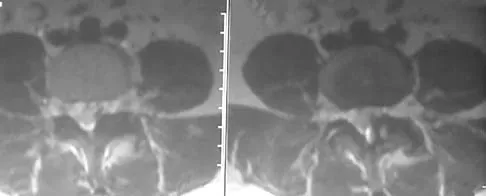
A 40-year-old woman has had sciatic pain on the left side for the past 8 weeks. She reports that the pain radiates to her posterior thigh, lateral calf, and into the dorsum of her left foot. Neurologic examination shows weakness of the left extensor hallucis longus. Axial T2-weighted MRI scans through L4-L5 are shown in Figure 14. Management should consist of


Explanation

The MRI scans show hypertrophy of the left L4-L5 facet joint and ligamentum flavum, with a synovial cyst. Appropriate surgical management consists of a hemilaminectomy and direct decompression of the neural elements. Fusion, in addition to the decompression, may be considered, particularly in patients with an associated spondylolisthesis. Epstein NE: Lumbar laminectomy for the resection of synovial cysts and coexisting lumbar spinal stenosis or degenerative spondylolisthesis: An outcome study. Spine 2004;29:1049-1055.


Question 18

During C1-C2 transarticular screw fixation, screw misplacement is most likely to result in injury to the


Explanation

With C1-C2 transarticular screw fixation, the following structures are potentially at risk: vertebral artery, spinal cord, occiput-C1 joint, and hypoglossal nerve. The vertebral artery is most vulnerable to injury with drill misdirection or anatomic variations in the vertebral foramen. The hypoglossal nerve may be injured if the drill, tap, or screw passes too far anterior to the lateral mass of C1. This complication is extremely rare. The occiput-C1 joint may be injured if the screw trajectory is too cephalad or cranially directed; however,this scenario is very unlikely because the exposure tends to direct the screw into a caudally inclined direction. This caudal orientation has the potential to cause vertebral artery injury, especially in patients who have a large vertebral foramen in the lateral mass of C2 because of erosions (rheumatoid arthritis) or anatomic variation. CT of the vertebral foramen is recommended when C1-C2 transarticular fixation is being considered. Spinal cord injury is extremely unlikely because of the very large size of the spinal canal in the upper cervical spine; the spinal cord lies far away from the lateral masses of C1 and C2. Mueller ME, Allgower M, et al: Manual of Internal Fixation, ed 3. New York, NY, Springer-Verlag, 1991, pp 634-636.

Question 19

A 27-year-old professional soccer player sustained an injury to his cervical spine in a collision with another player. Initially he was diagnosed with a right C6 radiculopathy that resolved with rest, anti-inflammatory medications, and physical therapy. Following a fall in a game, he noted a recurrence of neck pain without radicular signs or symptoms. Additional nonsurgical management over the past few months has failed to provide relief. A cervical MRI scan shows a right-sided C5-6 herniation without any evidence of disk disease at other cervical levels. The patient desires to continue his career as a professional soccer player. What treatment offers the best long-term option for return to play?


Explanation

The patient has chronic neck pain that is affecting his career as a professional soccer player. Although he had signs and symptoms of a right C6 radiculopathy, neck pain is his only current symptom. Therefore, procedures to address the relief of radiculopathy (keyhole foraminotomy and transforaminal epidural steroid injection) are likely to be ineffective. Although Watkins and others have described continuing nonsurgical management for symptomatic herniated disks and return to play only when asymptomatic, the patient has not found relief with these modalities. A single-level cervical fusion (either postoperative or congenital) generally is not considered a contraindication for return to play in collision or contact sports. Therefore, anterior cervical fusion at C5-6 offers the best long-term option for return to play. Watkins RG: Cervical spine injuries in athletes, in Clark CR (ed): The Cervical Spine, ed 3. Philadelphia, PA, Lippincott Raven, 1998, pp 373-386. Watkins RG: Neck injuries in football players. Clin Sports Med 1986;5:215-246. Morganti C, Sweeney CA, Albanese SA, et al: Return to play after cervical spine injury. Spine 2001;26:1131-1136.

Question 20

A collegiate football player who sustained an injury to his neck has significant neck pain and weakness in his extremities. Following immobilization, which of the following steps should be taken prior to transport?


Explanation

Prior to transport, the face mask should be removed so that the airway can be easily accessible. If serious injury is suspected, the helmet and shoulder pads should be left in place until he is assessed at the hospital and radiographs are obtained. Leaving the helmet and shoulder pads in place helps to keep the spine in the most neutral alignment. Removal of the helmet will result in extension of the neck, whereas removal of the shoulder pads will most likely result in flexion of the neck. Clark CR (ed): The Cervical Spine, ed 3. Philadelphia, PA, Lippincott Williams & Wilkins, 1998, p 376. Thomas B, McCullen GM, Yuan HA: Cervical spine injuries in football players. J Am Acad Orthop Surg 1999;7:338-347. Waninger KN, Richards JG, Pan WT, et al: An evaluation of head movement in backboard-immobilized helmeted football, lacrosse, and ice hockey players. Clin J Sport Med 2001;11:82-86. Donaldson WF III, Lauerman WC, Heil B, et al: Helmet and shoulder pad removal from a player with suspected cervical spine injury: A cadaveric model. Spine 1998;23:1729-1732.

Question 21

What is the most common complication following total disk arthroplasty in the lumbar spine?


Explanation

In a midterm (7 to 11 years) follow-up study of lumbar total disk arthroplasty, 5 of 55 patients had transient radicular leg pain without evidence of nerve root compression. Implant migration is rare. Deep venous thrombosis, incisional hernia, and retrograde ejaculation are less common complications of disk arthroplasty.

Question 22

A 42-year-old woman has cervical stenosis and radicular deficits at the C5-6 and C6-7 levels. History reveals that she has smoked one pack of cigarettes a day for 25 years. Because nonsurgical management has failed to provide relief, she is now seeking surgical treatment. After preoperative counseling, it becomes clear that she is not likely to stop smoking. Which of the following surgical procedures should be used?


Explanation

In a review of 190 anterior cervical fusions, Hilibrand and associates reported that only 20 of 40 patients who smoked had solid fusion at all levels, whereas 64 of 91 nonsmokers had solid fusions at all levels when treated with multilevel interbody technique (Smith-Robinson). When fused with strut grafts, 14 of 15 smokers and 41 of 44 nonsmokers had solid fusions with a fusion rate of 93% in the same series. Multilevel allografts have a lower fusion rate than autografts, and diskectomy without fusion has an increased rate of residual neck pain. Hilibrand AS, Fye MA, Emery SE, et al: Impact of smoking on the outcome of anterior cervical arthrodesis with interbody or strut-grafting. J Bone Joint Surg Am 2001;83:668-673.

Question 23

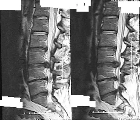
An otherwise healthy 54-year-old man who underwent a successful multilevel lumbar decompression and fusion 4 years ago now reports increasingly severe bilateral thigh claudication with paresthesia and severe back pain for the past 12 months. Physical therapy, bracing, and epidural steroids have failed to provide relief. A radiograph and MRI scans are shown in Figures 15a through 15c. He is afebrile, and laboratory studies show an erythrocyte sedimentation rate of 5 mm/h and a normal WBC count. What is the best course of action?


Explanation

The patient has degeneration of an adjacent segment with resultant kyphosis and stenosis. Because he is healthy, has responded well to previous surgery, and has a potentially correctable lesion, he is not a good candidate for an end-stage failed back procedure such as a morphine pump. The stenosis is exacerbated by the deformity; therefore, a simple decompression will contribute to instability. Because of the kyphosis and the patient's relatively young age, the treatment of choice is restoration of sagittal alignment and posterior decompression.


Question 24

Which of the following is considered a risk factor for the development of low back pain?


Explanation

Risk factors associated with low back pain include poor physical fitness, smoking, a history of repetitive bending or stooping on the job, or whole body vibration exposure. Some radiographic factors such as stenosis, spondyloarthropathy, severe deformity, or instability are also associated with low back pain. Gender, weight, transitional anatomy, or facet trophism are not associated with low back pain.

Question 25

A corset-type brace may help reduce symptoms during an episode of acute low back pain as the result of


Explanation

Although there is no significant alteration in motion with a corset, studies have shown a decrease in intradiskal pressure. Nachemson A, Morris JM: In vivo measurements of intradiscal pressure: Discometry, a method for determination of pressure in the low lumbar disc. J Bone Joint Surg Am 1964;46:1077-1092.

Question 26

A 68-year-old man falls and strikes his chin, sustaining a hyperextension injury to his neck. He has severe weakness in his hands but can walk with assistance. Which of the following is the most likely diagnosis?


Explanation

Central cord syndrome typically occurs after a hyperextension injury in a patient with pre-existing cervical spondylosis. It classically presents with upper extremity weakness that is proportionally greater than lower extremity weakness.

Question 27

In evaluating a patient with a suspected L4-L5 far lateral (extraforaminal) disc herniation, which of the following physical examination findings is most likely expected?


Explanation

A far lateral disc herniation at L4-L5 compresses the exiting L4 nerve root, unlike a paracentral herniation which compresses the traversing L5 root. L4 radiculopathy presents with quadriceps weakness (knee extension), a decreased patellar reflex, and medial leg numbness.

Question 28

A 72-year-old man presents with an acute Type II odontoid fracture following a fall. His fracture is posteriorly displaced by 6 mm.

Which of the following factors places him at the highest risk for nonunion with nonoperative management?


Explanation

Risk factors for nonunion in Type II odontoid fractures include displacement > 5 mm, angulation > 10 degrees, and age > 50 years. Displacement > 5 mm is generally considered the strongest independent predictor of nonunion.

Question 29

Which of the following represents the most common source of neurogenic claudication in patients older than 60 years?


Explanation

Degenerative spondylolisthesis, most commonly occurring at L4-L5, is the leading cause of acquired lumbar spinal stenosis and neurogenic claudication in older adults. It is primarily driven by degenerative changes in the facet joints and intervertebral discs.

Question 30

A patient involved in a high-speed collision sustains a burst fracture of L1. On examination, he has loss of motor function, pain, and temperature sensation below the umbilicus, but retains proprioception and vibratory sense. This presentation is characteristic of which spinal cord syndrome?


Explanation

Anterior cord syndrome results from injury to the anterior two-thirds of the spinal cord, often due to flexion injuries. It causes bilateral loss of motor function, pain, and temperature sensation, while dorsal column functions (proprioception, vibration) are preserved.

Question 31

According to the Thoracolumbar Injury Classification and Severity (TLICS) score, which of the following injury characteristics assigns the highest independent point value toward the decision for surgical intervention?


Explanation

In the TLICS system, an injured (disrupted) PLC is assigned 3 points, which is the highest individual score for a single category, alongside an incomplete neurologic deficit. A total score of 5 or more generally indicates surgical management.

Question 32

A 45-year-old man has a large sequestered (free fragment) disc herniation at L5-S1 causing severe S1 radiculopathy without motor deficit. What is the expected natural history of this specific type of disc herniation?


Explanation

Sequestered disc herniations have the highest rate of spontaneous resorption among all disc herniation types. The free fragment is exposed to the systemic circulation in the epidural space, triggering a robust macrophage-mediated inflammatory response.

Question 33

A 25-year-old man is brought to the ED after a lap-belt restrained motor vehicle collision. X-rays reveal a flexion-distraction injury (Chance fracture) at L2. Which of the following associated injuries must be actively ruled out?


Explanation

Chance fractures (flexion-distraction injuries) are highly associated with intra-abdominal injuries, particularly hollow viscus injuries like bowel perforations (up to 40-50% incidence). Prompt general surgery evaluation and abdominal CT are mandated.

Question 34

An 80-year-old man with long-standing Ankylosing Spondylitis falls backwards, hitting his head. He reports severe neck pain but has a normal neurological examination. Initial cross-table lateral cervical X-rays are read as normal. What is the most appropriate next step in management?


Explanation

Patients with Ankylosing Spondylitis have rigid, osteopenic spines that are highly susceptible to unstable "chalk-stick" fractures even from minor trauma. Standard X-rays are inadequate; a CT scan of the entire cervical spine down to T1 is mandatory to rule out occult fractures.

Question 35

A 55-year-old woman presents with progressive clumsiness in her hands and difficulty walking. Examination reveals a positive Hoffmann sign bilaterally, hyperreflexia in the lower extremities, and an inverted radial reflex. What is the most likely diagnosis?


Explanation

Cervical spondylotic myelopathy classically presents with upper motor neuron signs (hyperreflexia, Hoffmann sign) and gait instability. The inverted radial reflex is highly specific for spinal cord compression at the C5-C6 level.

Question 36

A patient with a T4 spinal cord injury presents to the ER with a blood pressure of 80/50 mmHg and a heart rate of 50 bpm. His extremities are warm and well-perfused. What is the primary pathophysiologic mechanism for his hemodynamic state?


Explanation

The patient is in neurogenic shock, characterized by hypotension and bradycardia due to the loss of sympathetic tone, which normally originates from the T1-L2 levels. It is classically seen in spinal cord injuries above T6.

Question 37

In a patient with traumatic spinal cord injury, the end of the "spinal shock" phase is clinically indicated by the return of which of the following?


Explanation

Spinal shock is a transient state of physiologic complete loss of spinal cord function below the level of injury. Its resolution is clinically marked by the return of the bulbocavernosus reflex, typically within 24 to 48 hours.

Question 38

A 45-year-old woman presents with severe, burning right anterior thigh pain and weakness in knee extension. Her symptoms began acutely after lifting a heavy box. Physical examination reveals a diminished right patellar reflex and a positive femoral stretch test. MRI of the lumbar spine demonstrates a far lateral (extraforaminal) disc herniation at the L3-L4 level. Which nerve root is most likely being compressed?


Explanation

A far lateral (extraforaminal) disc herniation in the lumbar spine compresses the exiting nerve root at the same level. Therefore, a far lateral disc herniation at L3-L4 compresses the exiting L3 nerve root, leading to anterior thigh pain, quadriceps weakness, and a diminished patellar reflex.

Question 39

A 65-year-old man with a history of cervical spondylosis falls forward, striking his forehead. He presents to the emergency department with profound bilateral upper extremity weakness and relatively preserved lower extremity function. Sensation is decreased in a cape-like distribution over his shoulders. Following initial ATLS protocol, which of the following is the most critical medical management parameter for his spinal cord injury?


Explanation

This patient's presentation is classic for central cord syndrome, typically occurring via a hyperextension injury in a stenotic cervical spine. Current guidelines strongly recommend avoiding hypotension and maintaining a MAP > 85 mmHg for 5 to 7 days to optimize spinal cord perfusion.

Question 40

A 25-year-old man is brought to the trauma bay after a diving accident. He is awake, alert, and cooperative. Neurologic examination is entirely normal. Plain radiographs and CT scans demonstrate a unilateral C5-C6 facet dislocation with approximately 25% anterior translation of C5 on C6. What is the most appropriate next step in management?


Explanation

In an awake, alert, and cooperative patient with a cervical facet dislocation, rapid awake closed reduction with cranial tongs is indicated and can be safely performed without a pre-reduction MRI. Pre-reduction MRI is reserved for patients who are unexaminable (e.g., comatose) or those who fail closed reduction.

Question 41

An 82-year-old man with multiple medical comorbidities including severe COPD and ischemic heart disease falls from a standing height. CT of the cervical spine reveals a Type II odontoid fracture with 2 mm of posterior displacement. He is neurologically intact. What is the most appropriate definitive management?


Explanation

While surgery typically yields higher union rates for Type II odontoid fractures, elderly patients with significant comorbidities suffer high morbidity and mortality with both surgery and halo vest immobilization. Recent evidence supports treatment with a rigid cervical collar, as fibrous nonunion is often asymptomatic and well-tolerated.

Question 42

An 18-year-old male is involved in a high-speed motor vehicle collision while wearing a lap belt. He complains of severe back pain. Radiographs demonstrate a horizontal fracture line passing through the spinous process, pedicles, and vertebral body of L2. Neurologic exam is normal. Which of the following is the most essential next step in his evaluation?


Explanation

The patient has sustained a Chance fracture (flexion-distraction injury), which is highly associated with the use of lap seatbelts. Up to 50% of these injuries are associated with intra-abdominal solid or hollow viscus injuries, making a CT of the abdomen and pelvis mandatory.

Question 43

A 68-year-old woman presents with worsening back and bilateral leg pain that increases with walking and is relieved by leaning over a shopping cart. She has failed 6 months of non-operative management including physical therapy and epidural steroid injections. Imaging reveals an L4-L5 Grade 1 degenerative spondylolisthesis with severe central canal stenosis. Dynamic radiographs show 4 mm of translation. What is the most effective surgical treatment?


Explanation

In the setting of degenerative spondylolisthesis with symptomatic spinal stenosis and dynamic instability, surgical decompression combined with instrumented posterolateral fusion provides superior clinical outcomes compared to decompression alone.

Question 44

A 24-year-old male arrives in the emergency department after a motorcycle crash. He has a palpable step-off at the upper thoracic spine and is completely flaccid and areflexic below the T4 level. His heart rate is 50 beats/minute and his blood pressure is 80/50 mmHg. His extremities are warm and well-perfused. Which of the following best explains his hemodynamic parameters?


Explanation

The patient is experiencing neurogenic shock, characterized by hypotension, bradycardia, and warm extremities. This results from a complete spinal cord injury above the level of T6, leading to a loss of descending sympathetic vascular tone and unopposed vagal (parasympathetic) activity.

Question 45

A 14-year-old female gymnast complains of insidious onset lower back pain that worsens with back extension. Neurologic examination is entirely normal. Radiographs reveal a pars interarticularis defect at L5 bilaterally with 15% translation of L5 on S1. What is the most appropriate initial management?


Explanation

The patient has a Grade I isthmic spondylolisthesis. The first line of treatment for a symptomatic, low-grade isthmic spondylolisthesis in a neurologically intact adolescent is non-operative, focusing on activity modification and physical therapy to strengthen the core and stretch the hamstrings.

Question 46

A 40-year-old man presents to the emergency department with severe lower back pain, bilateral lower extremity weakness, and new-onset urinary incontinence. Physical examination reveals perianal numbness and decreased bilateral Achilles reflexes. What is the most appropriate next step in management?


Explanation

This patient is exhibiting classic symptoms of Cauda Equina Syndrome. An emergent MRI of the lumbar spine is required to confirm the diagnosis and define the anatomy before proceeding to emergent surgical decompression, which should typically be performed within 24 to 48 hours to optimize functional recovery.

Question 47

When evaluating an MRI of the cervical spine in a patient with severe cervical spondylotic myelopathy, which of the following intrinsic cord signal changes is associated with the poorest prognosis for neurologic recovery following surgical decompression?


Explanation

Intrinsic cord signal changes can predict outcomes in cervical myelopathy. A focal T1-weighted hypointense signal indicates permanent cystic necrosis or myelomalacia and is strongly correlated with poor clinical recovery compared to isolated T2-weighted hyperintensity, which may represent reversible edema.

Question 48

A 55-year-old man with a known history of ankylosing spondylitis presents to the emergency department complaining of severe neck pain after a minor fall from a chair. He is neurologically intact. Standard anteroposterior and lateral cervical radiographs demonstrate osteopenia and syndesmophyte formation but no obvious fracture. What is the mandatory next step in his evaluation?


Explanation

Patients with ankylosing spondylitis have rigidly fused, osteopenic spines that fracture easily even with low-energy trauma. Because these fractures are highly unstable and often missed on plain radiographs, advanced imaging (CT or MRI) of the entire spine is mandatory to rule out an occult, highly destabilizing fracture.

Question 49

A traumatic spondylolisthesis of the axis, commonly known as a Hangman's fracture, involves bilateral fractures through which specific anatomic structure?


Explanation

A Hangman's fracture is defined as a bilateral fracture through the pars interarticularis (or sometimes the adjacent pedicles) of the C2 vertebra. It is typically caused by hyperextension and axial loading.

Question 50

The Thoracolumbar Injury Classification and Severity (TLICS) score is used to guide the surgical management of thoracolumbar trauma. Which of the following is NOT one of the three main categories evaluated in the TLICS system?


Explanation

The TLICS system determines the need for surgery based on three components: injury morphology (compression, burst, translation/rotation, distraction), integrity of the posterior ligamentous complex (PLC), and neurologic status. The degree of spinal canal compromise is not a distinct scoring category in this system.

Question 51

A 65-year-old man is evaluated for mild mid-back stiffness. Radiographs reveal flowing ossification along the anterolateral aspect of the thoracic vertebrae with preservation of disc height and no evidence of sacroiliac joint erosions. To meet the Resnick diagnostic criteria for Diffuse Idiopathic Skeletal Hyperostosis (DISH), this flowing ossification must bridge at least how many contiguous vertebral bodies?


Explanation

The diagnostic criteria for DISH established by Resnick include the presence of flowing ossification along the anterolateral aspect of at least four contiguous vertebral bodies, relative preservation of intervertebral disc height, and absence of apophyseal joint ankylosis or sacroiliac erosions.

Question 52

A 52-year-old woman presents with neck pain radiating down her left arm. Physical examination demonstrates weakness in elbow extension and wrist flexion, along with an absent triceps reflex. She reports numbness primarily in her long (middle) finger. Which cervical nerve root is most likely compressed?


Explanation

Compression of the C7 nerve root typically results in weakness of the triceps (elbow extension) and wrist flexors, an absent or diminished triceps reflex, and sensory deficits localized to the long (middle) finger. This most commonly occurs from a C6-C7 disc herniation.

Question 53

A 60-year-old man with end-stage renal disease on hemodialysis presents with severe, unrelenting mid-back pain and fever. Laboratory tests show an ESR of 100 mm/h and a CRP of 50 mg/L. MRI of the spine with gadolinium reveals a dorsal epidural abscess causing moderate cord compression. What is the most common causative organism for this condition?


Explanation

Staphylococcus aureus is the most common causative pathogen for spontaneous spinal epidural abscesses and pyogenic vertebral osteomyelitis, accounting for more than half of all cases. Hematogenous spread is a common etiology, especially in patients with indwelling catheters or those on hemodialysis.

Question 54


A 56-year-old woman with a history of metastatic breast cancer presents with progressive paraparesis and hyperreflexia in her lower extremities over the last 3 days. Her systemic disease is otherwise well-controlled, and her expected survival is greater than 1 year. MRI reveals a pathological fracture at T8 with high-grade epidural spinal cord compression caused by tumor mass. Based on the Patchell trial criteria, what is the most appropriate initial treatment?


Explanation

According to the landmark randomized trial by Patchell et al., patients with high-grade epidural spinal cord compression from solid tumors (like breast cancer), who have a life expectancy > 3 months and neurologic deficit, have significantly better functional outcomes with direct surgical decompression and stabilization followed by radiotherapy compared to radiotherapy alone.

Question 55

A 30-year-old male is intubated following a high-speed motor vehicle collision. A trauma CT scan of the head and neck demonstrates a significantly increased basion-dens interval (>12 mm) and a Powers ratio greater than 1, diagnostic of atlanto-occipital dissociation (AOD). What is the definitive treatment for this highly unstable injury?


Explanation

Atlanto-occipital dissociation (AOD) is a highly unstable, predominantly ligamentous injury with little capacity for spontaneous healing. Definitive management requires surgical occipitocervical fusion. Halo traction or immobilization is strictly contraindicated as it can cause fatal over-distraction of the spinal cord.

Question 56

A 70-year-old man presents with bilateral upper extremity weakness (hands greater than shoulders) and mild lower extremity clumsiness after a hyperextension injury during a fall.

What is the most appropriate initial hemodynamic management for this patient?


Explanation

This patient has Central Cord Syndrome, which often results from a hyperextension injury in a stenotic cervical spine. Current guidelines recommend maintaining a Mean Arterial Pressure (MAP) > 85 mmHg for 7 days to optimize spinal cord perfusion and limit secondary ischemic injury.

Question 57

A 35-year-old man falls from a roof, sustaining an L1 burst fracture. His neurological examination is completely intact. MRI demonstrates an intact posterior ligamentous complex (PLC). The fracture exhibits 15 degrees of local kyphosis. Based on the Thoracolumbar Injury Classification and Severity (TLICS) score, what is the recommended management?


Explanation

The TLICS score is 2 (burst fracture morphology = 2, intact PLC = 0, intact neurological status = 0). A TLICS score of less than 4 indicates that non-operative management, such as TLSO bracing and early mobilization, is appropriate.

Question 58

Which of the following preoperative factors is the most reliable negative prognostic indicator for postoperative neurological recovery in a patient undergoing surgical decompression for degenerative cervical spondylotic myelopathy?


Explanation

A prolonged duration of symptoms (typically greater than 12-18 months) prior to surgical decompression is a well-established negative predictor for neurological recovery in cervical myelopathy. Hypointense T1 signal changes are also a strong negative prognostic sign, whereas T2 changes alone are less definitive.

Question 59

An 82-year-old man sustains a Type II odontoid fracture after a ground-level fall. He is neurologically intact. Which of the following stabilization methods is associated with the highest risk of severe morbidity and mortality in this specific patient population?


Explanation

Halo vest immobilization in the elderly (typically defined as over 65-70 years) is associated with unacceptably high rates of morbidity and mortality, primarily due to respiratory complications, dysphagia, and falls. If surgery is indicated, posterior C1-C2 fusion is often preferred.

Question 60

A 65-year-old woman presents with severe neurogenic claudication and low back pain. Radiographs demonstrate a Grade I L4-L5 degenerative spondylolisthesis that increases to Grade II on dynamic flexion films. What is the most appropriate surgical intervention if a 6-month trial of conservative management fails?


Explanation

In the presence of a mobile or unstable degenerative spondylolisthesis associated with spinal stenosis, performing decompression combined with instrumented fusion provides superior long-term clinical outcomes and prevents progressive slip compared to decompression alone.

Question 61

A 22-year-old restrained passenger in a high-speed motor vehicle collision sustains a flexion-distraction (Chance) fracture of L2. Due to the mechanism of this injury, which of the following associated injuries must be aggressively ruled out?


Explanation

Chance fractures (flexion-distraction injuries) result from a seatbelt fulcrum effect acting on the abdominal wall and spine. They have a high association (up to 40-50%) with intra-abdominal injuries, particularly hollow viscus injuries like bowel perforations.

Question 62

A patient sustains a traumatic spondylolisthesis of the axis (Hangman's fracture). Imaging demonstrates severe angulation with minimal translation, consistent with a Levine-Edwards Type IIA fracture. Which of the following management steps is strictly contraindicated?


Explanation

Type IIA Hangman's fractures result from a flexion-distraction mechanism. Application of longitudinal cervical traction is strictly contraindicated as it exacerbates the injury, increases displacement, and stretches the spinal cord by widening the posterior disc space.

Question 63

A 30-year-old man presents neurologically intact but with severe neck pain following a football tackle. CT demonstrates a unilateral C5-C6 facet dislocation. He is awake, cooperative, and able to follow commands perfectly. What is the next most appropriate step in management?


Explanation

In an awake, cooperative, and neurologically examinable patient with a cervical facet dislocation, rapid awake closed reduction via skeletal traction is safely indicated. Pre-reduction MRI is mandatory if the patient is unexaminable (e.g., obtunded) or fails closed reduction, to rule out a compressive herniated disc.

Question 64

A 60-year-old man with a 30-year history of ankylosing spondylitis presents with new-onset mechanical neck pain after a minor fall from a chair. Initial plain radiographs of the cervical spine are read as normal. His neurological exam is intact. What is the most appropriate next step?


Explanation

Patients with ankylosing spondylitis have brittle, fused spines and are at extremely high risk for unstable, through-and-through occult spinal fractures even after trivial trauma. If plain films are negative or obscured, a CT scan (or MRI) of the spine is mandatory to definitively rule out a fracture.

Question 65

A 45-year-old man presents with right arm pain and weakness following a weightlifting injury. Examination reveals a diminished brachioradialis reflex, weakness in wrist extension, and numbness over the dorsal web space of the thumb and index finger. Which cervical nerve root is most likely compressed?


Explanation

A C6 radiculopathy typically presents with weakness in wrist extension and elbow flexion, a diminished brachioradialis reflex, and sensory changes in the thumb and index finger. This is most commonly caused by a herniated disc at the C5-C6 level.

Question 66

A 50-year-old man presents with severe left-sided anterior thigh pain. Examination reveals profound weakness in left knee extension and a diminished left patellar reflex. Sensation is decreased over the medial aspect of the leg. MRI demonstrates a far lateral (extraforaminal) disc herniation. At which level is this herniation most likely located?


Explanation

A far lateral (extraforaminal) disc herniation at the L4-L5 level compresses the exiting L4 nerve root, leading to L4 radiculopathy symptoms (quadriceps weakness, decreased patellar reflex, medial leg numbness). A paracentral disc at the same level would compress the traversing L5 root.

Question 67

According to the Denis classification of sacral fractures, fractures involving Zone III are most highly associated with which of the following clinical complications?


Explanation

Denis Zone III sacral fractures involve the central sacral canal. Because they directly disrupt the bilateral sacral neural elements, they carry the highest risk (greater than 50%) of neurological deficits, specifically saddle anesthesia and bowel, bladder, or sexual dysfunction.

Question 68

An 82-year-old woman falls from a standing height and presents with severe neck pain. Radiographs reveal a Type II odontoid fracture with 3 mm of posterior displacement. She has a history of severe COPD and ischemic heart disease. What is the most appropriate non-operative management for this patient?


Explanation

In elderly patients with Type II odontoid fractures, halo vest immobilization is associated with high morbidity and a mortality rate approaching 40%. A hard cervical collar is the preferred non-operative treatment, despite a higher risk of nonunion, because it is much better tolerated and has significantly lower complication rates.

Question 69

A 65-year-old man sustains a hyperextension injury to his cervical spine during a motor vehicle collision. On examination, he has motor weakness in all four extremities, but his upper extremities (especially the hands) are significantly weaker than his lower extremities. What is the typical pattern of functional recovery in this syndrome?


Explanation

This patient has Central Cord Syndrome. The typical pattern of neurological recovery proceeds from the lower extremities to bowel/bladder function, then to the proximal upper extremities, with fine motor function of the hands being the last and least likely to recover fully.

Question 70

A 70-year-old man presents with bilateral leg pain and fatigue that worsens after walking two blocks. Which of the following clinical findings most reliably differentiates neurogenic claudication from vascular claudication?


Explanation

Neurogenic claudication due to lumbar spinal stenosis is classically relieved by sitting or forward lumbar flexion (the "shopping cart sign"), which increases the cross-sectional area of the spinal canal. Vascular claudication is typically relieved by simply standing still and is exacerbated by the increased metabolic demand of walking uphill.

Question 71

A 55-year-old man presents with difficulty buttoning his shirts and a wide-based, unsteady gait. Physical examination reveals a positive Hoffmann's sign. Which of the following additional physical examination findings is most specific for this patient's underlying condition?


Explanation

The patient has symptoms of cervical spondylotic myelopathy, characterized by upper motor neuron signs. The inverted supinator reflex (brachioradialis reflex testing causing finger flexion) is a highly specific upper motor neuron sign, whereas Spurling's indicates radiculopathy and Lhermitte's is non-specific.

Question 72

A 25-year-old woman involved in a high-speed motor vehicle collision presents with a transverse ecchymosis across her abdomen (seatbelt sign). Radiographs reveal an osseous flexion-distraction injury (Chance fracture) of L2. What associated injury must be most urgently and specifically ruled out?


Explanation

Flexion-distraction injuries (Chance fractures) are highly associated with intra-abdominal injuries, particularly hollow viscus injuries like bowel rupture, which occur in up to 40-50% of cases. A thorough abdominal evaluation, usually with CT, is strictly required.

Question 73

A 65-year-old woman with L4-L5 degenerative spondylolisthesis and severe spinal stenosis has failed six months of conservative management. She complains of severe neurogenic claudication and mechanical back pain. What is the most appropriate surgical treatment?


Explanation

For symptomatic degenerative spondylolisthesis with spinal stenosis, laminectomy with instrumented posterolateral fusion provides superior long-term clinical outcomes compared to laminectomy alone. Decompression alone without stabilization often leads to progressive instability and recurrent symptoms.

Question 74

A 30-year-old man falls from a ladder and sustains a traumatic fracture of the L1 vertebra. According to the Denis three-column classification system, which of the following features defines a burst fracture and differentiates it from a simple compression fracture?


Explanation

Under the Denis classification, a burst fracture involves failure of both the anterior and middle columns under axial loading. A simple compression fracture involves only the anterior column, leaving the middle column intact and protecting the spinal canal.

Question 75

A 22-year-old man presents with severe neck pain after a rugby tackle. Radiographs reveal a unilateral facet dislocation at C5-C6. What is the classic mechanism of injury responsible for this specific pathology?


Explanation

Unilateral facet dislocations of the cervical spine typically result from a flexion-rotation mechanism, causing asymmetric disruption of the posterior ligamentous complex and facet joint capsule. In contrast, bilateral facet dislocations are typically caused by pure, severe flexion-distraction forces.

Question 76

A 60-year-old man with a known history of advanced ankylosing spondylitis presents to the emergency department complaining of new, severe neck pain after tripping and falling onto a carpeted floor. Initial plain radiographs of the cervical spine are interpreted as negative. What is the most appropriate next step in management?


Explanation

Patients with ankylosing spondylitis possess a rigid, osteopenic spine that acts like a long bone, making them highly susceptible to unstable fractures even from trivial trauma. Occult fractures are common and can lead to devastating epidural hematomas or cord injury, making advanced imaging (CT or MRI) mandatory when plain films are negative.

Question 77

A 45-year-old man presents with acute onset right leg pain that radiates down the lateral aspect of his calf to the dorsum of his foot. MRI reveals a large paracentral disc herniation at the L4-L5 level. Which nerve root is most likely compressed, and what is the expected motor deficit?


Explanation

In the lumbar spine, a paracentral disc herniation typically compresses the traversing nerve root at that level. An L4-L5 herniation compresses the L5 nerve root, which classically causes weakness in the extensor hallucis longus (great toe extension) and altered sensation over the dorsum of the foot.

Question 78

An 8-year-old boy is brought to the ED after a motor vehicle collision. He had transient numbness and weakness in both legs that has since resolved. Plain radiographs and a non-contrast CT of the spine are completely normal. What is the diagnostic modality of choice to fully evaluate this clinical picture?


Explanation

The patient is exhibiting signs of Spinal Cord Injury Without Radiographic Abnormality (SCIWORA), which is most common in pediatric patients due to inherent ligamentous laxity. MRI is the gold standard for detecting spinal cord edema, hemorrhage, or subtle ligamentous injury not visible on CT or plain films.

Question 79

A 62-year-old woman underwent an L4-S1 posterior spinal instrumented fusion 5 years ago. She now presents with new-onset radicular leg pain and progressive low back pain. Radiographs show significant degenerative changes and stenosis at the L3-L4 level. Which biomechanical factor is the primary contributor to this new pathology?


Explanation

Adjacent segment disease occurs as a result of the loss of motion at the surgically fused levels. This leads to compensatory hypermobility and increased biomechanical shear stresses at the adjacent unfused segments, thereby accelerating their degeneration.

Question 80

A 45-year-old man with a history of chronic low back pain suddenly develops severe bilateral sciatica, saddle anesthesia, and urinary retention. Physical examination reveals decreased rectal tone. What is the most critical and definitive step in his management?


Explanation

This patient presents with classic signs of Cauda Equina Syndrome, an absolute orthopedic emergency. The definitive management is an urgent MRI to confirm the diagnosis followed by emergent surgical decompression (typically within 24-48 hours) to prevent permanent neurological deficits.

Question 81

A 28-year-old unrestrained driver is involved in a head-on collision. Radiographs demonstrate bilateral fractures through the pars interarticularis of the C2 vertebra with mild anterior subluxation of C2 on C3. What is the classic mechanism of injury for this specific fracture pattern?


Explanation

Traumatic spondylolisthesis of the axis (Hangman's fracture) classically results from sudden hyperextension and axial loading, commonly seen in unrestrained motor vehicle accidents when the chin strikes the dashboard. This leads to bilateral fractures through the pars interarticularis.

Question 82

A 35-year-old pedestrian is struck by a vehicle and sustains a severe traumatic brain injury. Lateral cervical spine radiographs demonstrate a basion-dental interval (BDI) of 14 mm. Which of the following initial stabilization techniques is strictly contraindicated in this patient?


Explanation

A basion-dental interval (BDI) > 12 mm indicates an atlanto-occipital dissociation, a highly unstable, life-threatening ligamentous disruption. Cervical traction is absolutely contraindicated because it can cause severe over-distraction, leading to catastrophic stretching of the brainstem and spinal cord.

Question 83

A 68-year-old man presents with a 2-year history of bilateral buttock and leg pain that worsens with walking and standing. He reports that leaning forward on a shopping cart relieves his symptoms. Which of the following physical examination findings is most likely to be present in this patient?


Explanation

Neurogenic claudication from lumbar spinal stenosis is typically exacerbated by lumbar extension, which further narrows the spinal canal and neural foramina. Flexion increases the canal diameter, thereby providing symptom relief.

Question 84

A 72-year-old man presents with severe neck pain following a ground-level fall. CT imaging reveals a Type II odontoid fracture with 6 mm of posterior displacement. He is neurologically intact. Which of the following factors is most strongly predictive of nonunion if this patient is treated conservatively with a halo vest?


Explanation

In Type II odontoid fractures, patient age greater than 65 years, displacement greater than 5 mm, and a fracture gap greater than 1 mm are significant risk factors for nonunion with conservative management.

Question 85

A 45-year-old construction worker falls 15 feet, sustaining a T12 burst fracture. He remains neurologically intact. Which of the following radiographic findings is the most reliable indicator of a concurrent posterior ligamentous complex (PLC) injury?


Explanation

Splaying or widening of the interspinous distance on an AP or lateral radiograph indicates disruption of the posterior ligamentous complex (PLC). This finding upgrades the injury classification, often making it highly unstable and an indication for surgical stabilization.

Question 86

A 65-year-old man with known advanced cervical spondylosis is involved in a motor vehicle collision. He presents with severe motor weakness in his upper extremities but is able to move his lower extremities against resistance. He also has decreased sensation over the cape-like distribution of his shoulders. What is the most likely diagnosis?


Explanation

Central cord syndrome classically occurs in older patients with preexisting cervical spondylosis following a hyperextension injury. It presents with disproportionately greater motor impairment in the upper extremities compared to the lower extremities.

None

Clinic OS
Medically Verified Content by
Prof. Clinic OS
Consultant Orthopedic & Spine Surgeon
Chapter Index